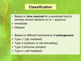
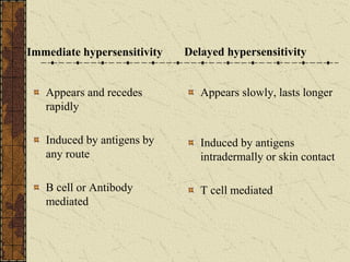
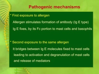
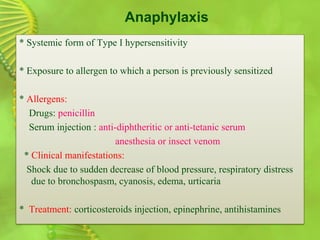
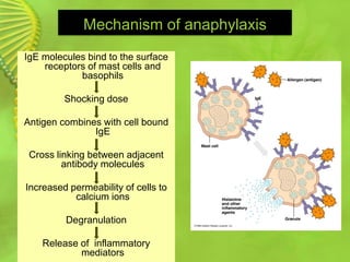
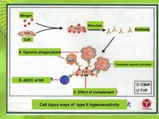
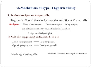
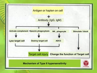
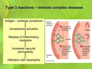
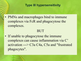
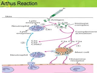
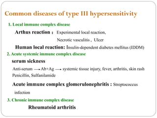
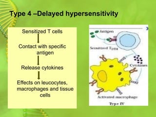
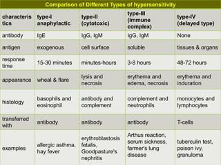
Dr. PULIPATI SOWJANYA
Professor & Head
Dept. of Pharmaceutical Biotechnology
Vignan Pharmacy College
Vadlamudi, Guntur (Dt)
Immunopathology
Exaggerated immune response may lead to different
forms of tissue damage
1) An overactive immune response:
produce more damage than it prevents
e.g. hypersensitivity reactions and graft rejection
2) Failure of appropriate recognition:
as in autoimmune diseases
Hypersensitivity Reaction
Hypersensitivity or allergy
* An immune response results in exaggerated reactions
harmful to the host
* There are four types of hypersensitivity reactions:
Type I, Type II, Type III, Type IV
* Types I, II and III are antibody mediated
* Type IV is cell mediated
Classification
• Based on time required for a sensitized host to
develop clinical reactions on re – exposure
➢ Immediate
➢ Delayed
• Based on different mechanisms of pathogenesis
➢ Type 1 ( IgE mediated)
➢ Type 2 (cytotoxic or cell stimulating)
➢ Type 3 (immune complex)
➢ Type 4 ( cell mediated)
Immediate hypersensitivity
Appears and recedes
rapidly
Induced by antigens by
any route
B cell or Antibody
mediated
Delayed hypersensitivity
Appears slowly, lasts longer
Induced by antigens
intradermally or skin contact
T cell mediated
Type I: Immediate hypersensitivity
* An antigen reacts with cell fixed antibody (Ig E)
leading to release of soluble molecules
An antigen (allergen)
soluble molecules (mediators)
* Soluble molecules cause the manifestation of disease
* Systemic life threatening; anaphylactic shock
* Local atopic allergies; bronchial asthma, hay fever
and food allergies
Pathogenic mechanisms
* First exposure to allergen
Allergen stimulates formation of antibody (Ig E type)
Ig E fixes, by its Fc portion to mast cells and basophils
* Second exposure to the same allergen
It bridges between Ig E molecules fixed to mast cells
leading to activation and degranulation of mast cells
and release of mediators
Pathogenic mechanisms
• Three classes of mediators derived from mast cells:
1) Preformed mediators stored in granules (histamine)
2) Newly sensitized mediators:
leukotrienes, prostaglandins, platelets activating factor
3) Cytokines produced by activated mast cells, basophils
e.g. TNF, IL3, IL-4, IL-5 IL-13, chemotactic factors
* These mediators cause: smooth muscle contraction,
mucous secretion and bronchial spasm, vasodilatation,
vascular permeability and edema
Anaphylaxis
* Systemic form of Type I hypersensitivity
* Exposure to allergen to which a person is previously sensitized
* Allergens:
Drugs: penicillin
Serum injection : anti-diphtheritic or anti-tetanic serum
anesthesia or insect venom
* Clinical manifestations:
Shock due to sudden decrease of blood pressure, respiratory distress
due to bronchospasm, cyanosis, edema, urticaria
* Treatment: corticosteroids injection, epinephrine, antihistamines
Mechanism of anaphylaxis
IgE molecules bind to the surface
receptors of mast cells and
basophils
Shocking dose
Antigen combines with cell bound
IgE
Cross linking between adjacent
antibody molecules
Increased permeability of cells to
calcium ions
Degranulation
Release of inflammatory
mediators
Atopy
• Local form of type I hypersensitivity
• Exposure to certain allergens that induce production of specific Ig E
* Allergens :
Inhalants: dust, tree or pollens, mould spur.
Ingestants: milk, egg, fish, chocolate
Contactants: wool, nylon, animal fur
Drugs: penicillin, salicylates, anaesthesia, insect venom
• There is a strong familial predisposition to atopic allergy
• The predisposition is genetically determined
4. Skin allergy:
4. Common disease of type I hypersensitivity
1. Systemic anaphylaxis: a very dangerous syndrome
1) Anaphylactic drug allergy :penicillin
2) Anaphylactic serum allergy :
2. Respiratory allergic diseases :
1) Allergic asthma:acute response, chronic response
2) Allergic rhinitis
3. Gastrointestinal allergic diseases :
The lack of SIgA protein
hydrolase
Undigested
protein
Allergen
Type II Hypersensitivity
Primed IgG or IgM + Antigen or hapten on membrane
Injury and dysfunction of target cells
Type 2 Reaction: cytolytic and cytotoxic
Ig G and rarely Ig M antibodies
Bind to antigenic cell
Lysis of cell
➢ Cell surface antigens
(autoimmune anaemia's)
➢ Adsorption of antigens on
cell surface (drugs)
➢ Cell surface receptor and
disrupts normal function
(Graves disease)
Allergen
Stimulate
Antibody
A. Opsonic phagocytosis
D. ADCC of NK
C. Effect of complement
Combined opsonic activities
Cell injury ways of type II hypersensitivity
Cell
2. Mechanism of Type II hypersentivity
1. Surface antigen on target cells
Target cells: Normal tissue cell, changed or modified self tissue cells
2.Antibody, complement and modified self-cell
Antigen : Blood group antigen, Common antigen,
Self-antigen modified by physical factors or infection
Drug antigen,
Antigen-antibody complex
Activate complement Lyses target cells
Opsonic phagocytosis Destroy target cells
Stimulating or blocking effect Promote /suppress the target cell function
Antigen or hapten on cell
Antibody (IgG, IgM)
Activate complement
Lyse target cell
Opsonic phagocytosis NK , phagocyte Stimulate / block
Destroy target cell ADCC
Target cell injury Change the function of Target cell
Mechanism of Type II hypersensitivity
Free Ag + Primed Ab Larger immune complex
Deposit in tissue or blood vessel wall
Inflammation
Type III hypersensitivity
Type 3 reactions – immune complex diseases
Antigen – antibody complexes
Complement activation
Release of inflammatory
mediators
Increased vascular
permeability
Infiltration with neutrophils
• The reaction may be general (e.g., serum sickness) or
may involve individual organs including skin (e.g.,
systemic lupus erythematosus, Arthus reaction), kidneys
(e.g., lupus nephritis), lungs (e.g., aspergillosis), joints
(e.g., rheumatoid arthritis) or other organs.
• This reaction may be the pathogenic mechanism of
diseases caused by many microorganisms.
Type III hypersensitivity
• The reaction may take 3 - 10 hours after exposure to the
antigen .
• It is mediated by soluble immune complexes.
• They are mostly of the IgG class, although IgM may also
be involved.
• The antigen may be exogenous (chronic bacterial, viral
or parasitic infections), or endogenous (non-organ
specific autoimmunity: e.g., systemic lupus
erythematosus, SLE).
• The antigen is soluble and not attached to the organ
involved
Type III hypersensitivity
• PMNs and macrophages bind to immune
complexes via FcR and phagocytose the
complexes.
BUT
• If unable to phagocytose the immune
complexes can cause inflammation via C’
activation ---> C3a C4a, C5a and "frustrated
phagocytes".
Type III hypersensitivity
Arthus Reaction
Mechanism of type III hypersensitivity
Formation of the intermediate immune complex
Deposition of the intermediate immune complex
Tissue injury by the immune complex
Common diseases of type III hypersensitivity
1. Local immune complex disease
Arthus reaction :Experimental local reaction,
Necrotic vasculitis , Ulcer
Human local reaction: Insulin-dependent diabetes mellitus (IDDM)
2. Acute systemic immune complex disease
serum sickness
Anti-serum Ab+Ag systemic tissue injury, fever, arthritis, skin rash
Penicillin, Sulfanilamide
Acute immune complex glomerulonephritis : Streptococcus
infection
3. Chronic immune complex disease
Rheumatoid arthritis
Interaction of primed T cells and associated antigen
Infiltration of Mononuclear Cells, Inflammatory response
Type IV hypersensitivity (Delayed)
Type 4 –Delayed hypersensitivity
Sensitized T cells
Contact with specific
antigen
Release cytokines
Effects on leucocytes,
macrophages and tissue
cells
Mechanism of type IV hypersensitivity
Formation of effector and memory T cells
Inflammation and cytotoxicity caused by effector T cells
1) Inflammation and tissue injury mediated by CD4+Th1
Release chemokines and cytokines
Immune injury mainly caused by infiltration of mononuclear cells and
lymphocytes
2) Cytotoxicity of CD8+CTL
Antigen T cell
(CD4+,CD8+)
Secondary
contact
Induce
PrimedT cell
CD4+
T cell
CD8+
T cell
Release
Cytokines
IL-2
TNF-b
INF-g
TF
MCF
MIF
MAF
SRF
Directly kill target cells
Infiltration of
monocyte and Mf
Proliferation ofT cell
Exudation and edema
Cytotoxicity
Inflammation characterized by infiltration of Mf , monocyte,
and tissue injury
Mechanism of type IV hypersensitivity
Delayed hypersensitivity reactions
Type Reaction time
Clinical
appearance
Histology
Antigen and
site
Contact
dermatitis
48-72 hr eczema
lymphocytes,
followed by
macrophages;
edema of
epidermis
epidermal
(organic
chemicals,
poison ivy,
heavy
metals, etc.)
tuberculin 48-72 hr
local
induration
lymphocytes,
monocytes,
macrophages
intradermal
(tuberculin,
lepromin, etc.)
granuloma 21-28 days hardening
macrophages,
epitheloid and
giant cells,
fibrosis
persistent
antigen or
foreign body
presence
(tuberculosis,
leprosy, etc.)
DIAGNOSIS AND TREATMENT
• Diagnostic tests in vivo include delayed cutaneous
reaction (e.g. Mantoux test and patch test (for contact
dermatitis).
• In vitro tests for delayed hypersensitivity include
mitogenic response, lympho-cytotoxicity and IL-2
production.
• Corticosteroids and other immunosuppressive agents
are used in treatment.
Common disease of type IV hypersensitivity
1) Infectious delayed type hypersensitivity
OT( Old Tuberculin ) test
2) Contact dermatitis :
Paint, drugs, water blister, dermatitis
Comparison of Different Types of hypersensitivity
characteris
tics
type-I
anaphylactic
type-II
(cytotoxic)
type-III
(immune
complex)
type-IV
(delayed type)
antibody IgE IgG, IgM IgG, IgM None
antigen exogenous cell surface soluble tissues & organs
response
time
15-30 minutes minutes-hours 3-8 hours 48-72 hours
appearance wheal & flare
lysis and
necrosis
erythema and
edema, necrosis
erythema and
induration
histology
basophils and
eosinophil
antibody and
complement
complement and
neutrophils
monocytes and
lymphocytes
transferred
with
antibody antibody antibody T-cells
examples
allergic asthma,
hay fever
erythroblastosis
fetalis,
Goodpasture's
nephritis
Arthus reaction,
serum sickness,
farmer's lung
disease
tuberculin test,
poison ivy,
granuloma
Hypersensitivity reactions

Hypersensitivity reactions

  • 1.
    Dr. PULIPATI SOWJANYA Professor& Head Dept. of Pharmaceutical Biotechnology Vignan Pharmacy College Vadlamudi, Guntur (Dt)
  • 2.
    Immunopathology Exaggerated immune responsemay lead to different forms of tissue damage 1) An overactive immune response: produce more damage than it prevents e.g. hypersensitivity reactions and graft rejection 2) Failure of appropriate recognition: as in autoimmune diseases
  • 3.
    Hypersensitivity Reaction Hypersensitivity orallergy * An immune response results in exaggerated reactions harmful to the host * There are four types of hypersensitivity reactions: Type I, Type II, Type III, Type IV * Types I, II and III are antibody mediated * Type IV is cell mediated
  • 4.
    Classification • Based ontime required for a sensitized host to develop clinical reactions on re – exposure ➢ Immediate ➢ Delayed • Based on different mechanisms of pathogenesis ➢ Type 1 ( IgE mediated) ➢ Type 2 (cytotoxic or cell stimulating) ➢ Type 3 (immune complex) ➢ Type 4 ( cell mediated)
  • 5.
    Immediate hypersensitivity Appears andrecedes rapidly Induced by antigens by any route B cell or Antibody mediated Delayed hypersensitivity Appears slowly, lasts longer Induced by antigens intradermally or skin contact T cell mediated
  • 6.
    Type I: Immediatehypersensitivity * An antigen reacts with cell fixed antibody (Ig E) leading to release of soluble molecules An antigen (allergen) soluble molecules (mediators) * Soluble molecules cause the manifestation of disease * Systemic life threatening; anaphylactic shock * Local atopic allergies; bronchial asthma, hay fever and food allergies
  • 7.
    Pathogenic mechanisms * Firstexposure to allergen Allergen stimulates formation of antibody (Ig E type) Ig E fixes, by its Fc portion to mast cells and basophils * Second exposure to the same allergen It bridges between Ig E molecules fixed to mast cells leading to activation and degranulation of mast cells and release of mediators
  • 8.
    Pathogenic mechanisms • Threeclasses of mediators derived from mast cells: 1) Preformed mediators stored in granules (histamine) 2) Newly sensitized mediators: leukotrienes, prostaglandins, platelets activating factor 3) Cytokines produced by activated mast cells, basophils e.g. TNF, IL3, IL-4, IL-5 IL-13, chemotactic factors * These mediators cause: smooth muscle contraction, mucous secretion and bronchial spasm, vasodilatation, vascular permeability and edema
  • 9.
    Anaphylaxis * Systemic formof Type I hypersensitivity * Exposure to allergen to which a person is previously sensitized * Allergens: Drugs: penicillin Serum injection : anti-diphtheritic or anti-tetanic serum anesthesia or insect venom * Clinical manifestations: Shock due to sudden decrease of blood pressure, respiratory distress due to bronchospasm, cyanosis, edema, urticaria * Treatment: corticosteroids injection, epinephrine, antihistamines
  • 10.
    Mechanism of anaphylaxis IgEmolecules bind to the surface receptors of mast cells and basophils Shocking dose Antigen combines with cell bound IgE Cross linking between adjacent antibody molecules Increased permeability of cells to calcium ions Degranulation Release of inflammatory mediators
  • 11.
    Atopy • Local formof type I hypersensitivity • Exposure to certain allergens that induce production of specific Ig E * Allergens : Inhalants: dust, tree or pollens, mould spur. Ingestants: milk, egg, fish, chocolate Contactants: wool, nylon, animal fur Drugs: penicillin, salicylates, anaesthesia, insect venom • There is a strong familial predisposition to atopic allergy • The predisposition is genetically determined
  • 12.
    4. Skin allergy: 4.Common disease of type I hypersensitivity 1. Systemic anaphylaxis: a very dangerous syndrome 1) Anaphylactic drug allergy :penicillin 2) Anaphylactic serum allergy : 2. Respiratory allergic diseases : 1) Allergic asthma:acute response, chronic response 2) Allergic rhinitis 3. Gastrointestinal allergic diseases : The lack of SIgA protein hydrolase Undigested protein Allergen
  • 13.
    Type II Hypersensitivity PrimedIgG or IgM + Antigen or hapten on membrane Injury and dysfunction of target cells
  • 14.
    Type 2 Reaction:cytolytic and cytotoxic Ig G and rarely Ig M antibodies Bind to antigenic cell Lysis of cell ➢ Cell surface antigens (autoimmune anaemia's) ➢ Adsorption of antigens on cell surface (drugs) ➢ Cell surface receptor and disrupts normal function (Graves disease)
  • 15.
    Allergen Stimulate Antibody A. Opsonic phagocytosis D.ADCC of NK C. Effect of complement Combined opsonic activities Cell injury ways of type II hypersensitivity Cell
  • 16.
    2. Mechanism ofType II hypersentivity 1. Surface antigen on target cells Target cells: Normal tissue cell, changed or modified self tissue cells 2.Antibody, complement and modified self-cell Antigen : Blood group antigen, Common antigen, Self-antigen modified by physical factors or infection Drug antigen, Antigen-antibody complex Activate complement Lyses target cells Opsonic phagocytosis Destroy target cells Stimulating or blocking effect Promote /suppress the target cell function
  • 17.
    Antigen or haptenon cell Antibody (IgG, IgM) Activate complement Lyse target cell Opsonic phagocytosis NK , phagocyte Stimulate / block Destroy target cell ADCC Target cell injury Change the function of Target cell Mechanism of Type II hypersensitivity
  • 18.
    Free Ag +Primed Ab Larger immune complex Deposit in tissue or blood vessel wall Inflammation Type III hypersensitivity
  • 19.
    Type 3 reactions– immune complex diseases Antigen – antibody complexes Complement activation Release of inflammatory mediators Increased vascular permeability Infiltration with neutrophils
  • 20.
    • The reactionmay be general (e.g., serum sickness) or may involve individual organs including skin (e.g., systemic lupus erythematosus, Arthus reaction), kidneys (e.g., lupus nephritis), lungs (e.g., aspergillosis), joints (e.g., rheumatoid arthritis) or other organs. • This reaction may be the pathogenic mechanism of diseases caused by many microorganisms. Type III hypersensitivity
  • 21.
    • The reactionmay take 3 - 10 hours after exposure to the antigen . • It is mediated by soluble immune complexes. • They are mostly of the IgG class, although IgM may also be involved. • The antigen may be exogenous (chronic bacterial, viral or parasitic infections), or endogenous (non-organ specific autoimmunity: e.g., systemic lupus erythematosus, SLE). • The antigen is soluble and not attached to the organ involved Type III hypersensitivity
  • 22.
    • PMNs andmacrophages bind to immune complexes via FcR and phagocytose the complexes. BUT • If unable to phagocytose the immune complexes can cause inflammation via C’ activation ---> C3a C4a, C5a and "frustrated phagocytes". Type III hypersensitivity
  • 23.
  • 24.
    Mechanism of typeIII hypersensitivity Formation of the intermediate immune complex Deposition of the intermediate immune complex Tissue injury by the immune complex
  • 25.
    Common diseases oftype III hypersensitivity 1. Local immune complex disease Arthus reaction :Experimental local reaction, Necrotic vasculitis , Ulcer Human local reaction: Insulin-dependent diabetes mellitus (IDDM) 2. Acute systemic immune complex disease serum sickness Anti-serum Ab+Ag systemic tissue injury, fever, arthritis, skin rash Penicillin, Sulfanilamide Acute immune complex glomerulonephritis : Streptococcus infection 3. Chronic immune complex disease Rheumatoid arthritis
  • 26.
    Interaction of primedT cells and associated antigen Infiltration of Mononuclear Cells, Inflammatory response Type IV hypersensitivity (Delayed)
  • 27.
    Type 4 –Delayedhypersensitivity Sensitized T cells Contact with specific antigen Release cytokines Effects on leucocytes, macrophages and tissue cells
  • 28.
    Mechanism of typeIV hypersensitivity Formation of effector and memory T cells Inflammation and cytotoxicity caused by effector T cells 1) Inflammation and tissue injury mediated by CD4+Th1 Release chemokines and cytokines Immune injury mainly caused by infiltration of mononuclear cells and lymphocytes 2) Cytotoxicity of CD8+CTL
  • 29.
    Antigen T cell (CD4+,CD8+) Secondary contact Induce PrimedTcell CD4+ T cell CD8+ T cell Release Cytokines IL-2 TNF-b INF-g TF MCF MIF MAF SRF Directly kill target cells Infiltration of monocyte and Mf Proliferation ofT cell Exudation and edema Cytotoxicity Inflammation characterized by infiltration of Mf , monocyte, and tissue injury Mechanism of type IV hypersensitivity
  • 30.
    Delayed hypersensitivity reactions TypeReaction time Clinical appearance Histology Antigen and site Contact dermatitis 48-72 hr eczema lymphocytes, followed by macrophages; edema of epidermis epidermal (organic chemicals, poison ivy, heavy metals, etc.) tuberculin 48-72 hr local induration lymphocytes, monocytes, macrophages intradermal (tuberculin, lepromin, etc.) granuloma 21-28 days hardening macrophages, epitheloid and giant cells, fibrosis persistent antigen or foreign body presence (tuberculosis, leprosy, etc.)
  • 31.
    DIAGNOSIS AND TREATMENT •Diagnostic tests in vivo include delayed cutaneous reaction (e.g. Mantoux test and patch test (for contact dermatitis). • In vitro tests for delayed hypersensitivity include mitogenic response, lympho-cytotoxicity and IL-2 production. • Corticosteroids and other immunosuppressive agents are used in treatment.
  • 32.
    Common disease oftype IV hypersensitivity 1) Infectious delayed type hypersensitivity OT( Old Tuberculin ) test 2) Contact dermatitis : Paint, drugs, water blister, dermatitis
  • 33.
    Comparison of DifferentTypes of hypersensitivity characteris tics type-I anaphylactic type-II (cytotoxic) type-III (immune complex) type-IV (delayed type) antibody IgE IgG, IgM IgG, IgM None antigen exogenous cell surface soluble tissues & organs response time 15-30 minutes minutes-hours 3-8 hours 48-72 hours appearance wheal & flare lysis and necrosis erythema and edema, necrosis erythema and induration histology basophils and eosinophil antibody and complement complement and neutrophils monocytes and lymphocytes transferred with antibody antibody antibody T-cells examples allergic asthma, hay fever erythroblastosis fetalis, Goodpasture's nephritis Arthus reaction, serum sickness, farmer's lung disease tuberculin test, poison ivy, granuloma