Downloaded 191 times
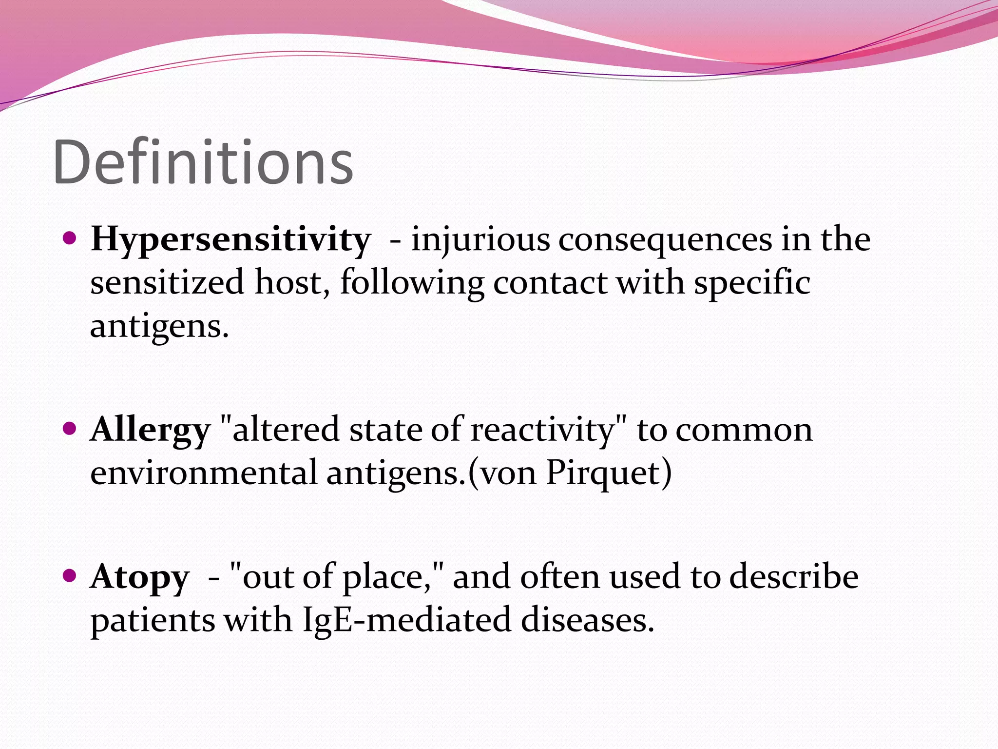

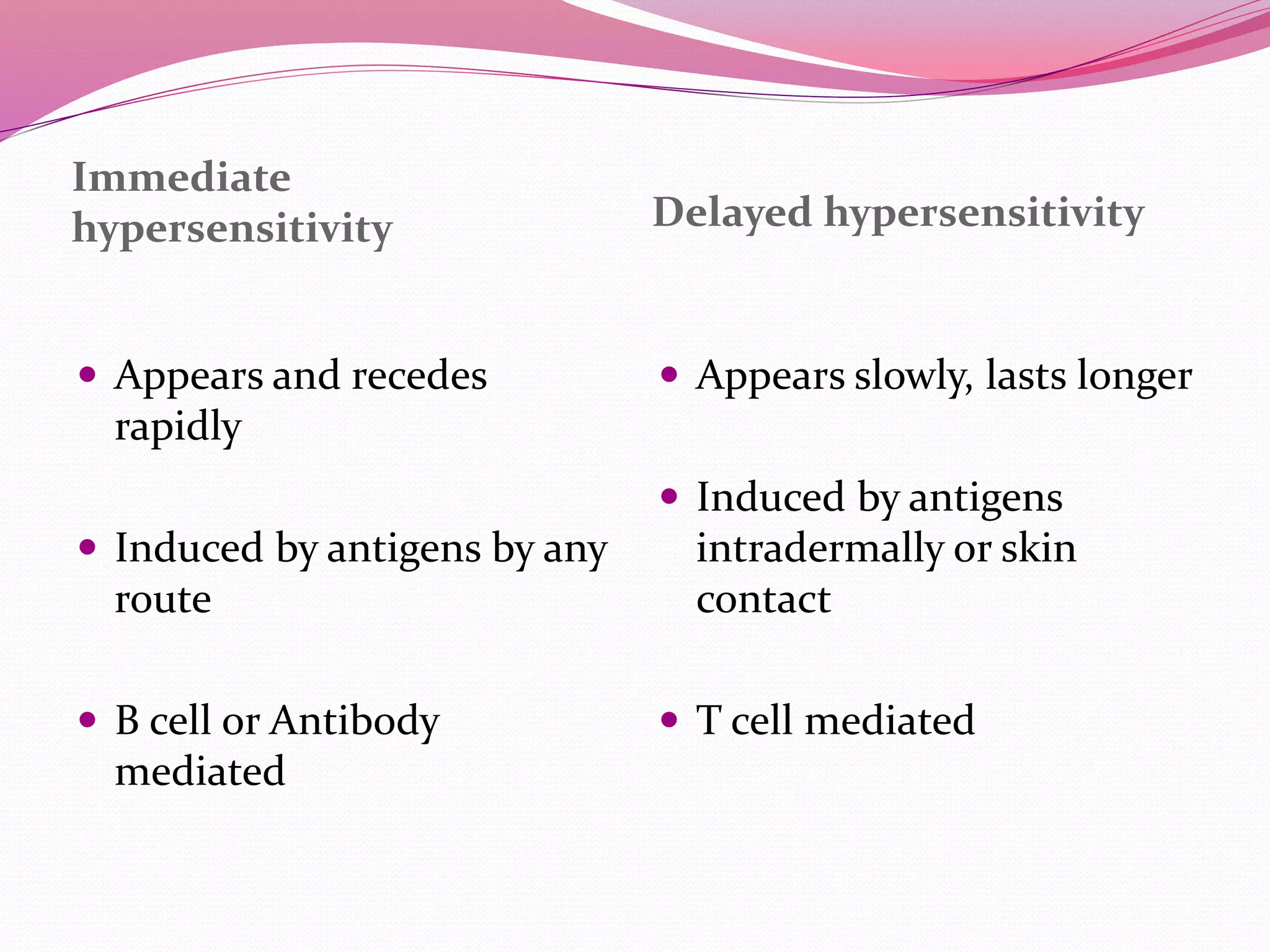
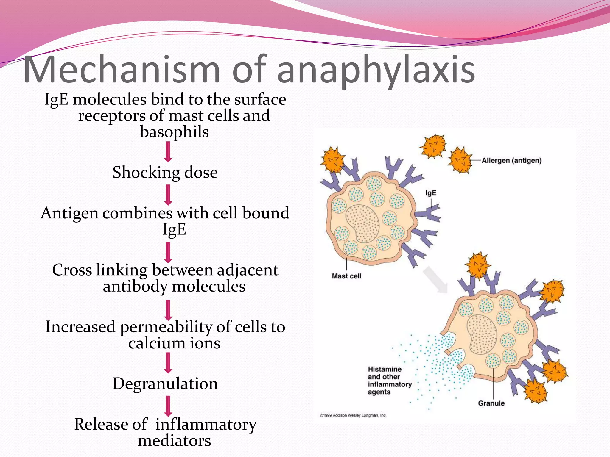

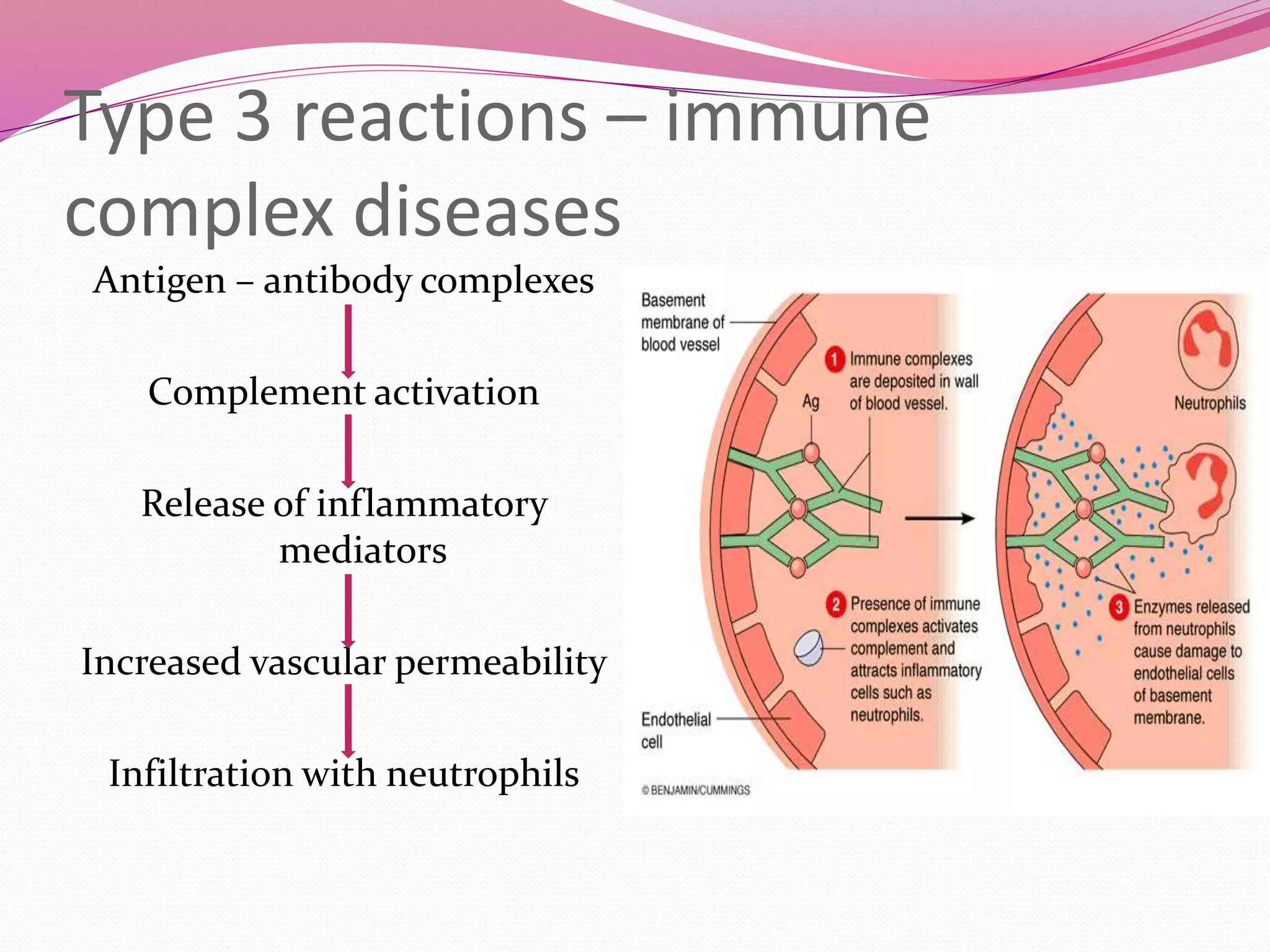

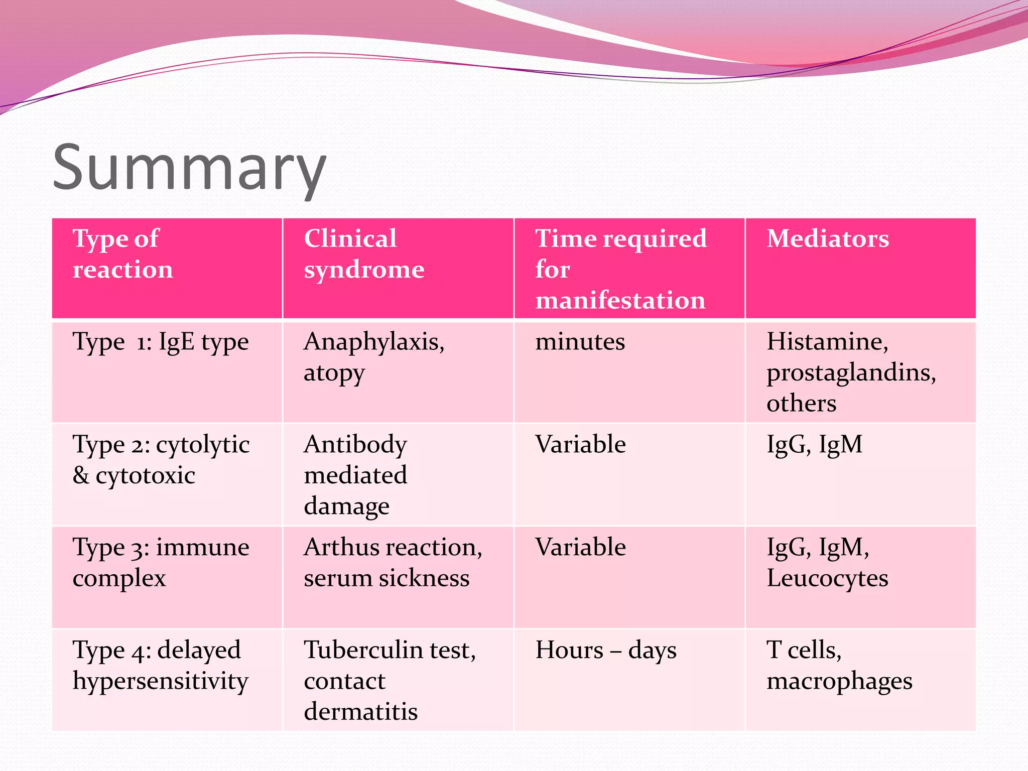

This document defines types of hypersensitivity reactions and allergies. It describes four main types of reactions: Type 1 IgE-mediated reactions like anaphylaxis occur rapidly via histamine release. Type 2 cytotoxic reactions damage cells via IgG/IgM antibodies. Type 3 immune complex reactions involve antigen-antibody complexes activating complement and attracting inflammatory cells. Type 4 delayed hypersensitivity reactions occur via T cell activation and cytokine release over hours to days.