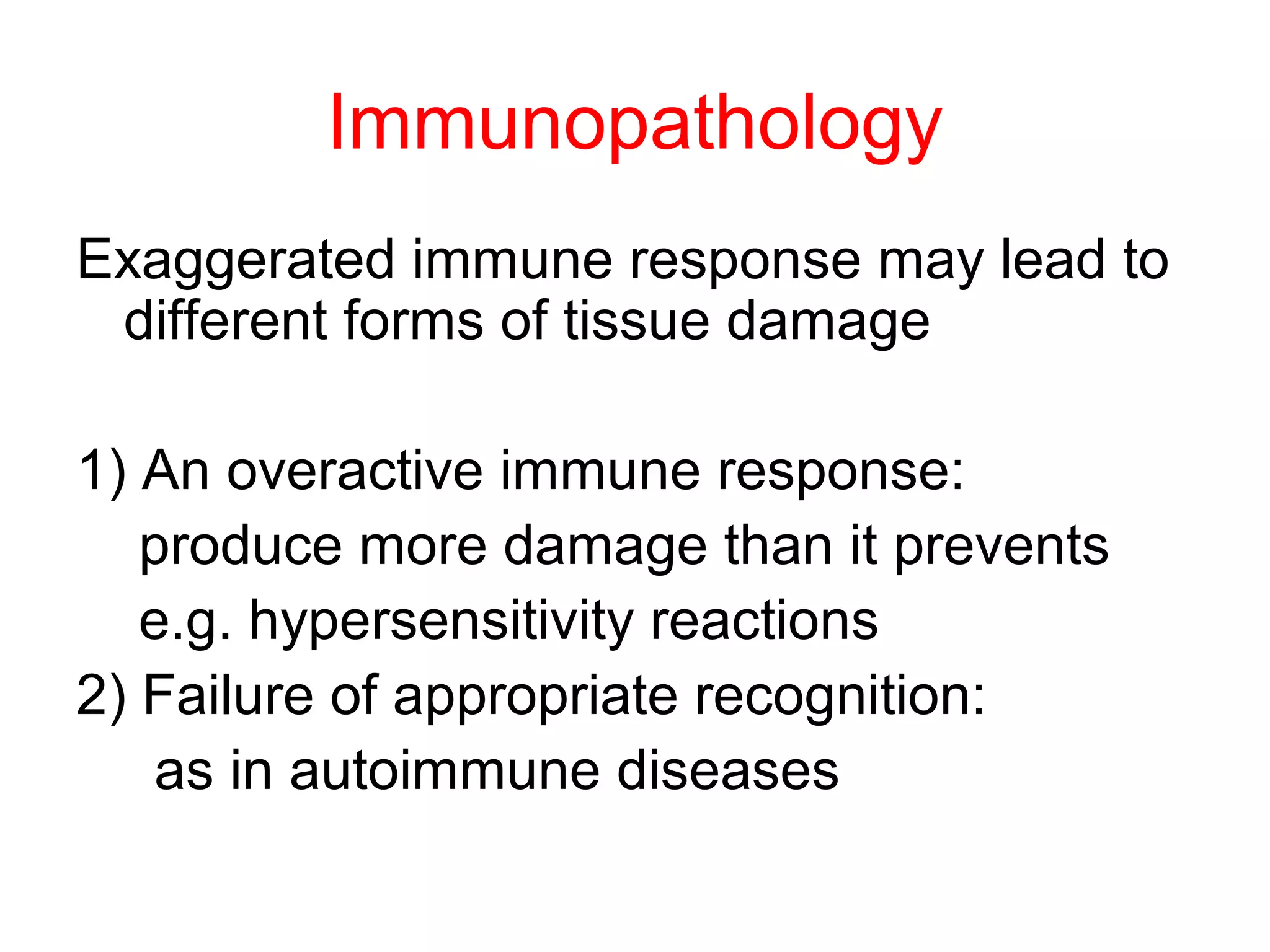
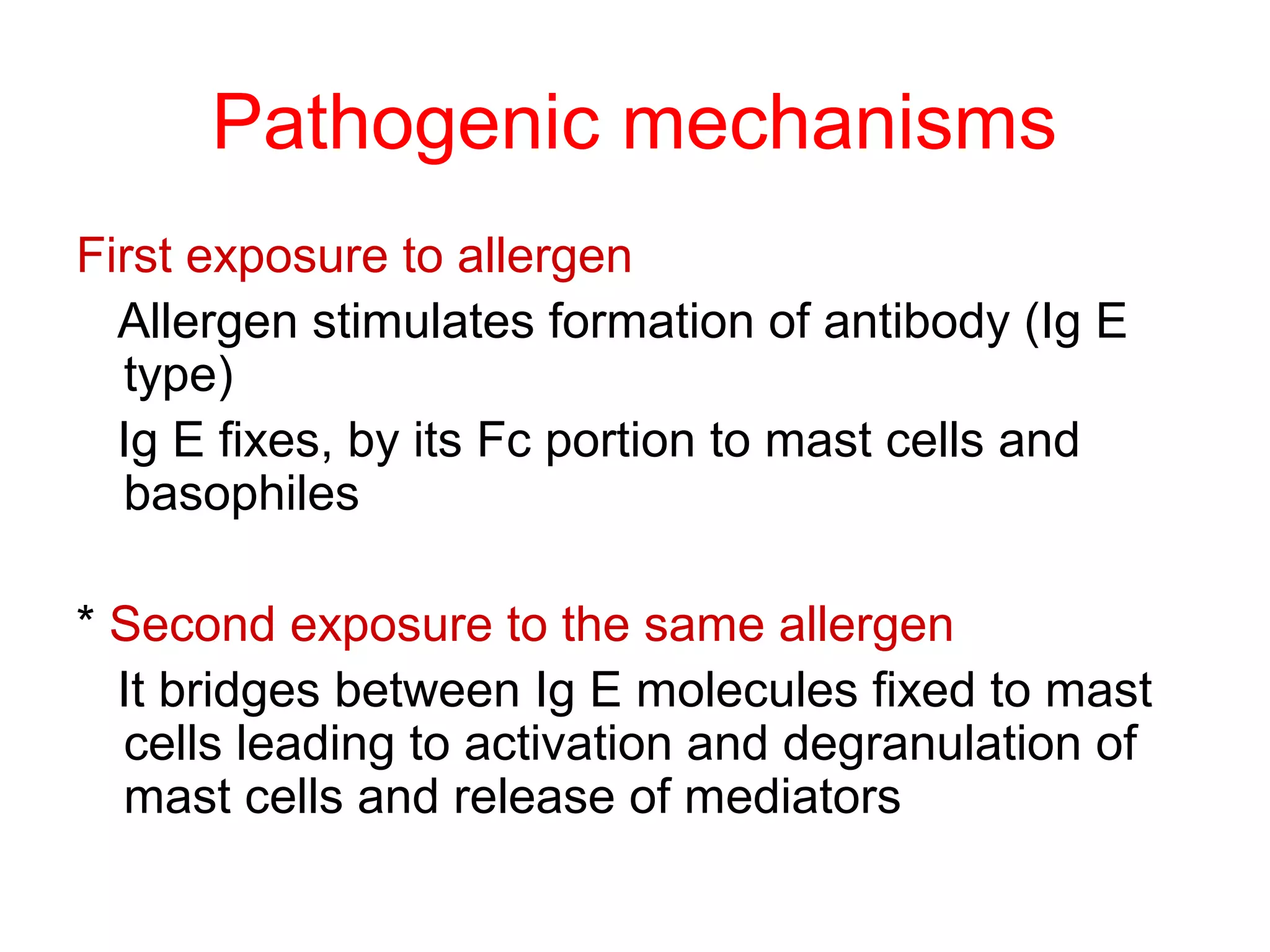
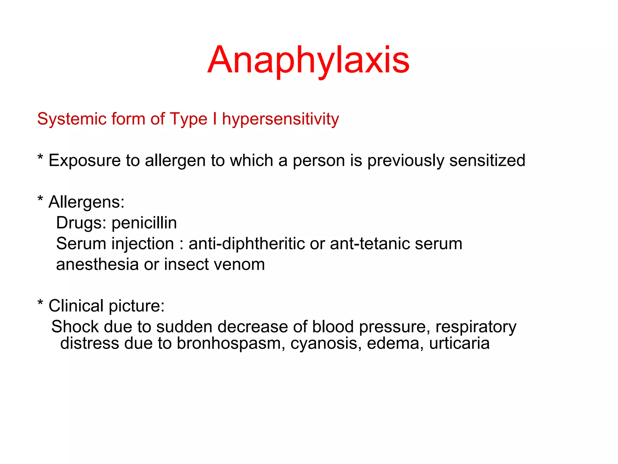
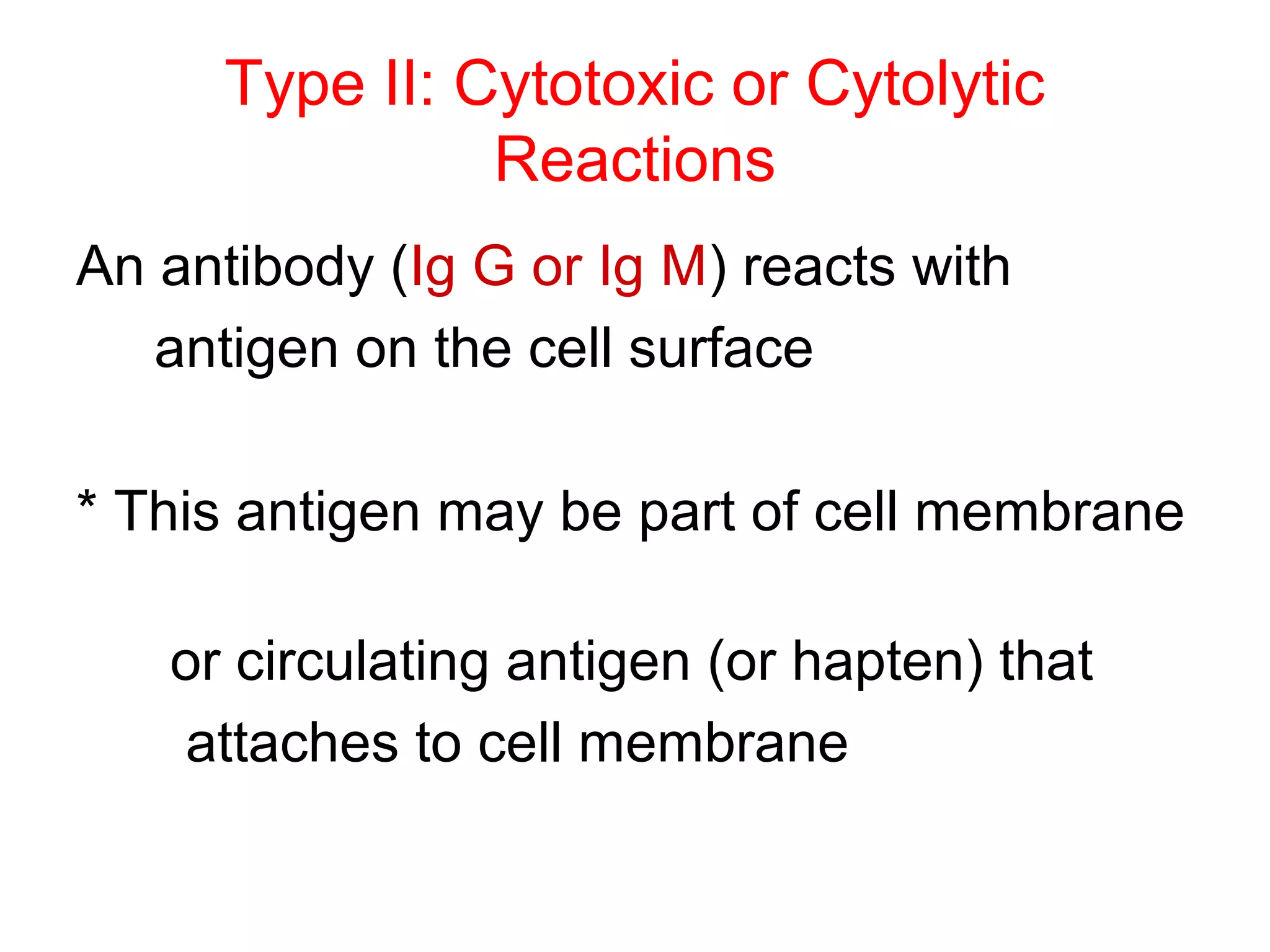
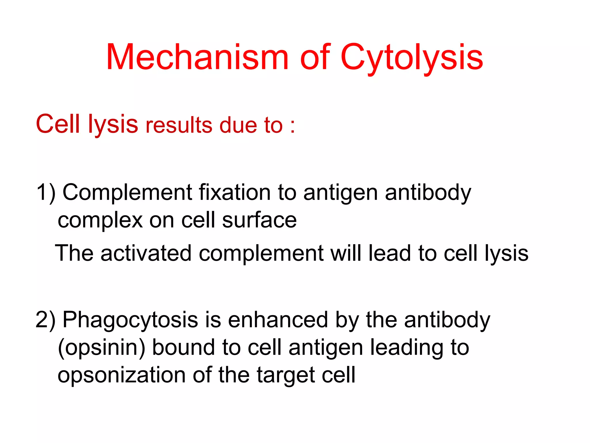
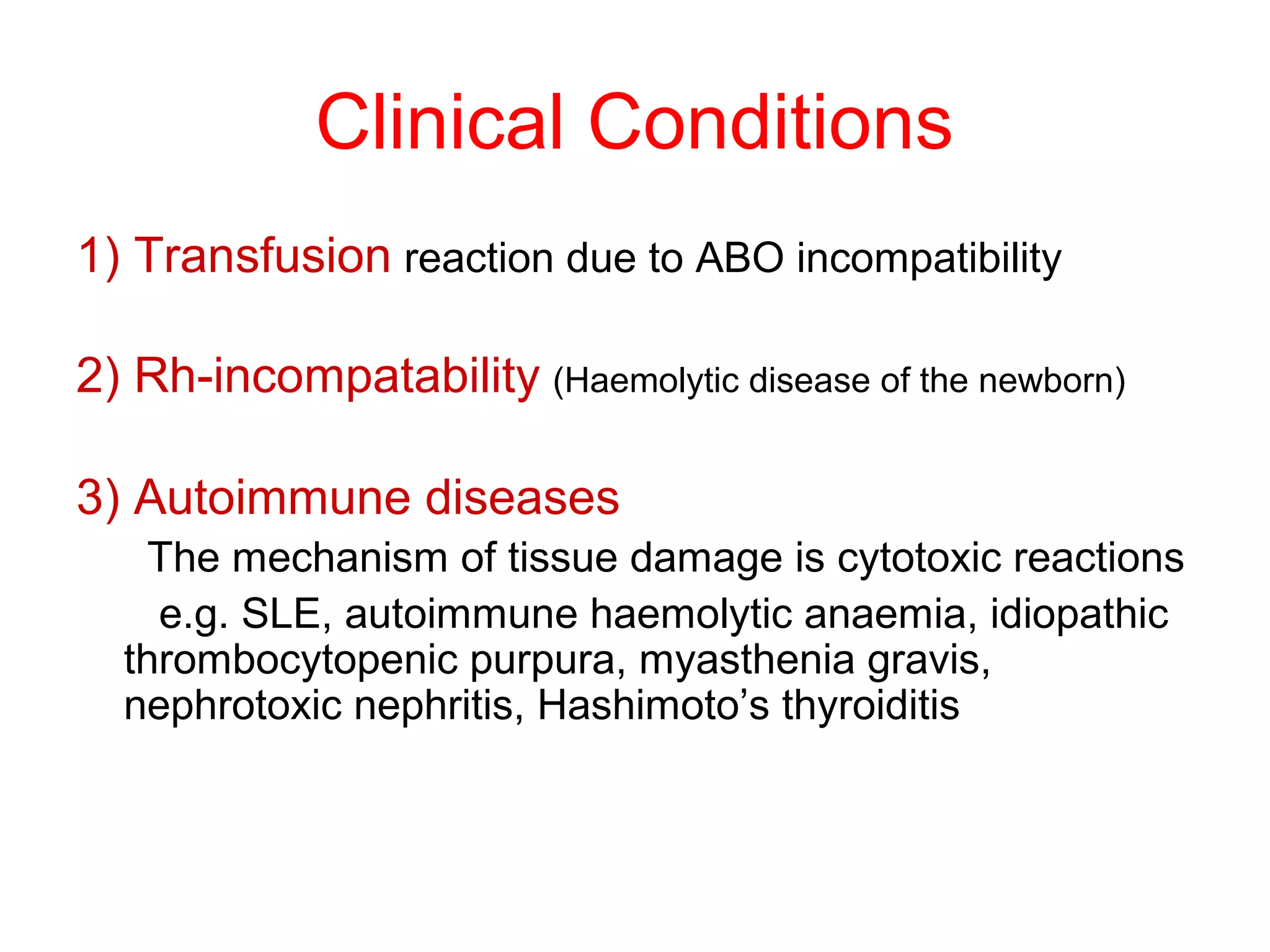
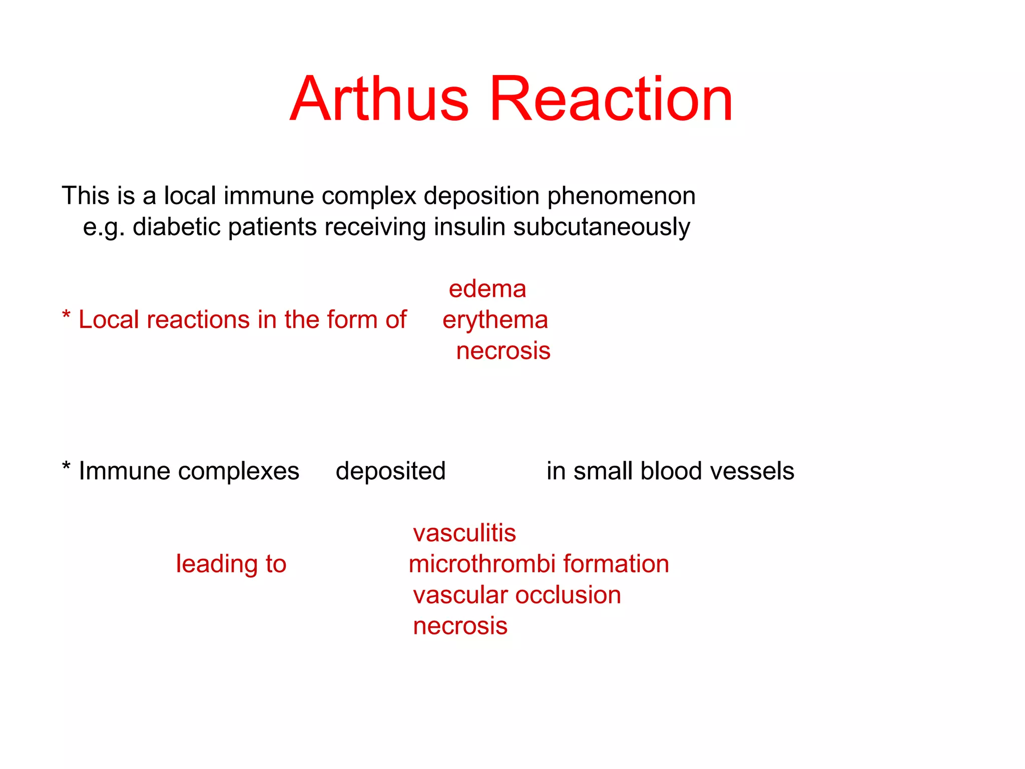
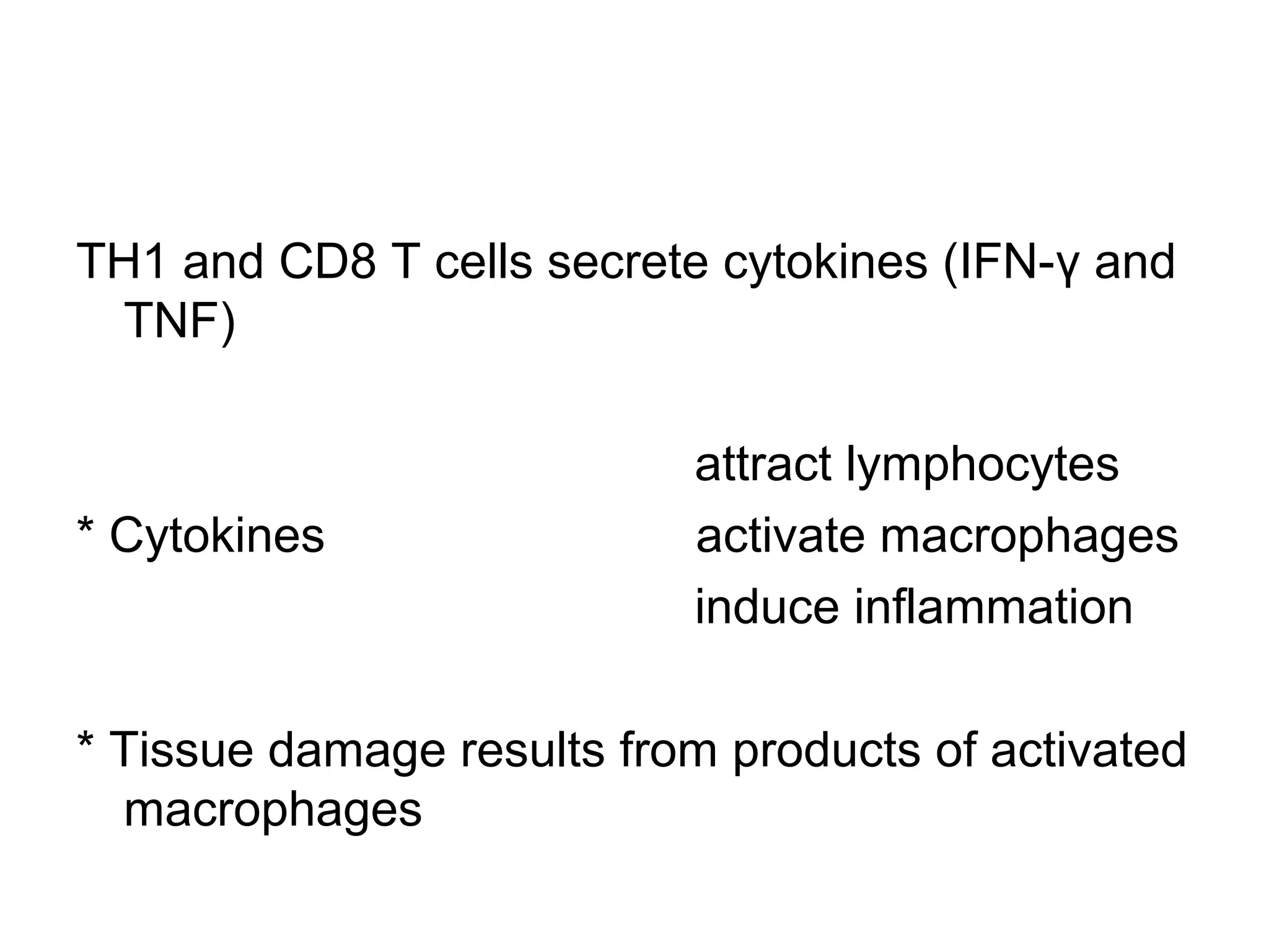
HYPERSENSITIVITY
Immunopathology
Exaggerated immune response may lead to
different forms of tissue damage
1) An overactive immune response:
produce more damage than it prevents
e.g. hypersensitivity reactions
2) Failure of appropriate recognition:
as in autoimmune diseases
Hypersensitivity Reaction
Hypersensitivity or allergy
* An immune response results in exaggerated reactions
harmful to the host
* There are four types of hypersensitivity reactions:
Type I, Type II, Type III, Type IV
* Types I, II and III are antibody mediated
* Type IV is cell mediated
Type I: Immediate hypersensitivity
An antigen reacts with cell fixed antibody (Ig E)
leading to release of soluble molecules
An antigen (allergen)
soluble molecules (mediators)
* Soluble molecules cause the manifestation of disease
* Systemic life threatening; anaphylactic shock
* Local atopic allergies; bronchial asthma,
and food allergies
Pathogenic mechanisms
First exposure to allergen
Allergen stimulates formation of antibody (Ig E
type)
Ig E fixes, by its Fc portion to mast cells and
basophiles
* Second exposure to the same allergen
It bridges between Ig E molecules fixed to mast
cells leading to activation and degranulation of
mast cells and release of mediators
Three classes of mediators derived from mast cells:
1) Preformed mediators stored in granules (histamine)
2) Newly sensitized mediators:
leukotrienes, prostaglandins, platelets activating factor
3) Cytokines produced by activated mast cells, basophils
e.g. TNF, IL3, IL-4, IL-5 IL-13
These mediators cause: smooth muscle
contraction, mucous secretion and
bronchial spasm, vasodilatation, vascular
permeability and edema
Anaphylaxis
Systemic form of Type I hypersensitivity
* Exposure to allergen to which a person is previously sensitized
* Allergens:
Drugs: penicillin
Serum injection : anti-diphtheritic or ant-tetanic serum
anesthesia or insect venom
* Clinical picture:
Shock due to sudden decrease of blood pressure, respiratory
distress due to bronhospasm, cyanosis, edema, urticaria
Atopy
Local form of type I hypersensitivity
* Exposure to certain allergens that induce production of specific Ig E
* Allergens :
Inhalants:dust mite faeces, tree or pollens, mould spor.
Ingestants: milk, egg, fish, choclate
Contactants: wool, nylon, animal fur
Drugs: penicillin, salicylates, anesthesia insect venom
* There is a strong familial predisposition to atopic allergy
* The predisposition is genetically determined
Type II: Cytotoxic or Cytolytic
Reactions
An antibody (Ig G or Ig M) reacts with
antigen on the cell surface
* This antigen may be part of cell membrane
or circulating antigen (or hapten) that
attaches to cell membrane
Mechanism of Cytolysis
Cell lysis results due to :
1) Complement fixation to antigen antibody
complex on cell surface
The activated complement will lead to cell lysis
2) Phagocytosis is enhanced by the antibody
(opsinin) bound to cell antigen leading to
opsonization of the target cell
3) Antibody depended cellular cytotoxicity (ADCC):
- Antibody coated cells
e.g. tumour cells, graft cells or infected cells
can be killed by cells possess Fc receptors
- - Cells most active in ADCC are:
NK, macrophages, neutrophils and eosinophils
Clinical Conditions
1) Transfusion reaction due to ABO incompatibility
2) Rh-incompatability (Haemolytic disease of the newborn)
3) Autoimmune diseases
The mechanism of tissue damage is cytotoxic reactions
e.g. SLE, autoimmune haemolytic anaemia, idiopathic
thrombocytopenic purpura, myasthenia gravis,
nephrotoxic nephritis, Hashimoto’s thyroiditis
• Type III Hypersensitivity
Immune Complex Mediated Reaction
Type III: Immune Complex Mediated
Reaction
When antibodies (Ig G or Ig M) and antigen coexist
immune complexes are formed
*Immune complexes are removed by reticuloendoth. syst.
*Some immune complexes escape phagocytosis
*Immune complexes deposited in tissues on the basement
membrane of blood vessels and cause tissue injury
Diseases produced by immune complexes are those in
which antigens persists without being eliminated as:
a- Repeated exposure to extrinsic antigen
b- injection of large amounts of antigens
c- Persistent infections
d- Autoimmunity to self components
Arthus Reaction
This is a local immune complex deposition phenomenon
e.g. diabetic patients receiving insulin subcutaneously
edema
* Local reactions in the form of erythema
necrosis
* Immune complexes deposited in small blood vessels
vasculitis
leading to microthrombi formation
vascular occlusion
necrosis
Serum Sickness
A systemic immune complex phenomenon
* Injection of large doses of foreign serum
* Antigen is slowly cleared from circulation
* Immune complexes are deposited in various sites
fever
urticaria
* 10 days after injection arthralgia
lymphadenopathy
splenomegaly
glomerulonephritis
Post-streptococcal glomerulonephritis
glomerulitis associated with infective
endocarditis
Hypersensitive pneumonitis (farmer lung)
immune complexes depositition in lung after
repeated inhalation of dust , mould spores
Endogenous antigen antibody complexes involved
in autoimmune diseases
e.g. SLE, rheumatoid arthritis
Type IV
Cell Mediated
Delayed Type Hypersensitivity
TH1 and CD8 T cells secrete cytokines (IFN-γ and
TNF)
attract lymphocytes
* Cytokines activate macrophages
induce inflammation
* Tissue damage results from products of activated
macrophages
Tuberculin –Type Hypersensitivity
• The classical example of this hypersensitivity is
tuberculin (Montoux) reaction (figure 5) which
peaks 48 hours after the injection of antigen
(PPD or old tuberculin).
• The lesion is characterized by induration and
erythema.
In chronic diseases : T.B., Leprosy, schistosomiases
* Intracellular organisms resist destruction by macrophag.
* Persistent antigen in tissues stimulate local DTH reaction
* Continuous release of cytokines leads to accumulation of
macrophages which give rise to epitheloidal and giant
cell granuloma
Hypersensitivity made easy

Hypersensitivity made easy

  • 1.
  • 3.
    Immunopathology Exaggerated immune responsemay lead to different forms of tissue damage 1) An overactive immune response: produce more damage than it prevents e.g. hypersensitivity reactions 2) Failure of appropriate recognition: as in autoimmune diseases
  • 4.
    Hypersensitivity Reaction Hypersensitivity orallergy * An immune response results in exaggerated reactions harmful to the host * There are four types of hypersensitivity reactions: Type I, Type II, Type III, Type IV * Types I, II and III are antibody mediated * Type IV is cell mediated
  • 5.
    Type I: Immediatehypersensitivity An antigen reacts with cell fixed antibody (Ig E) leading to release of soluble molecules An antigen (allergen) soluble molecules (mediators) * Soluble molecules cause the manifestation of disease * Systemic life threatening; anaphylactic shock * Local atopic allergies; bronchial asthma, and food allergies
  • 6.
    Pathogenic mechanisms First exposureto allergen Allergen stimulates formation of antibody (Ig E type) Ig E fixes, by its Fc portion to mast cells and basophiles * Second exposure to the same allergen It bridges between Ig E molecules fixed to mast cells leading to activation and degranulation of mast cells and release of mediators
  • 7.
    Three classes ofmediators derived from mast cells: 1) Preformed mediators stored in granules (histamine) 2) Newly sensitized mediators: leukotrienes, prostaglandins, platelets activating factor 3) Cytokines produced by activated mast cells, basophils e.g. TNF, IL3, IL-4, IL-5 IL-13
  • 8.
    These mediators cause:smooth muscle contraction, mucous secretion and bronchial spasm, vasodilatation, vascular permeability and edema
  • 9.
    Anaphylaxis Systemic form ofType I hypersensitivity * Exposure to allergen to which a person is previously sensitized * Allergens: Drugs: penicillin Serum injection : anti-diphtheritic or ant-tetanic serum anesthesia or insect venom * Clinical picture: Shock due to sudden decrease of blood pressure, respiratory distress due to bronhospasm, cyanosis, edema, urticaria
  • 10.
    Atopy Local form oftype I hypersensitivity * Exposure to certain allergens that induce production of specific Ig E * Allergens : Inhalants:dust mite faeces, tree or pollens, mould spor. Ingestants: milk, egg, fish, choclate Contactants: wool, nylon, animal fur Drugs: penicillin, salicylates, anesthesia insect venom * There is a strong familial predisposition to atopic allergy * The predisposition is genetically determined
  • 11.
    Type II: Cytotoxicor Cytolytic Reactions An antibody (Ig G or Ig M) reacts with antigen on the cell surface * This antigen may be part of cell membrane or circulating antigen (or hapten) that attaches to cell membrane
  • 12.
    Mechanism of Cytolysis Celllysis results due to : 1) Complement fixation to antigen antibody complex on cell surface The activated complement will lead to cell lysis 2) Phagocytosis is enhanced by the antibody (opsinin) bound to cell antigen leading to opsonization of the target cell
  • 13.
    3) Antibody dependedcellular cytotoxicity (ADCC): - Antibody coated cells e.g. tumour cells, graft cells or infected cells can be killed by cells possess Fc receptors - - Cells most active in ADCC are: NK, macrophages, neutrophils and eosinophils
  • 14.
    Clinical Conditions 1) Transfusionreaction due to ABO incompatibility 2) Rh-incompatability (Haemolytic disease of the newborn) 3) Autoimmune diseases The mechanism of tissue damage is cytotoxic reactions e.g. SLE, autoimmune haemolytic anaemia, idiopathic thrombocytopenic purpura, myasthenia gravis, nephrotoxic nephritis, Hashimoto’s thyroiditis
  • 15.
    • Type IIIHypersensitivity Immune Complex Mediated Reaction
  • 16.
    Type III: ImmuneComplex Mediated Reaction When antibodies (Ig G or Ig M) and antigen coexist immune complexes are formed *Immune complexes are removed by reticuloendoth. syst. *Some immune complexes escape phagocytosis *Immune complexes deposited in tissues on the basement membrane of blood vessels and cause tissue injury
  • 17.
    Diseases produced byimmune complexes are those in which antigens persists without being eliminated as: a- Repeated exposure to extrinsic antigen b- injection of large amounts of antigens c- Persistent infections d- Autoimmunity to self components
  • 18.
    Arthus Reaction This isa local immune complex deposition phenomenon e.g. diabetic patients receiving insulin subcutaneously edema * Local reactions in the form of erythema necrosis * Immune complexes deposited in small blood vessels vasculitis leading to microthrombi formation vascular occlusion necrosis
  • 19.
    Serum Sickness A systemicimmune complex phenomenon * Injection of large doses of foreign serum * Antigen is slowly cleared from circulation * Immune complexes are deposited in various sites fever urticaria * 10 days after injection arthralgia lymphadenopathy splenomegaly glomerulonephritis
  • 20.
    Post-streptococcal glomerulonephritis glomerulitis associatedwith infective endocarditis Hypersensitive pneumonitis (farmer lung) immune complexes depositition in lung after repeated inhalation of dust , mould spores Endogenous antigen antibody complexes involved in autoimmune diseases e.g. SLE, rheumatoid arthritis
  • 21.
    Type IV Cell Mediated DelayedType Hypersensitivity
  • 22.
    TH1 and CD8T cells secrete cytokines (IFN-γ and TNF) attract lymphocytes * Cytokines activate macrophages induce inflammation * Tissue damage results from products of activated macrophages
  • 24.
    Tuberculin –Type Hypersensitivity •The classical example of this hypersensitivity is tuberculin (Montoux) reaction (figure 5) which peaks 48 hours after the injection of antigen (PPD or old tuberculin). • The lesion is characterized by induration and erythema.
  • 25.
    In chronic diseases: T.B., Leprosy, schistosomiases * Intracellular organisms resist destruction by macrophag. * Persistent antigen in tissues stimulate local DTH reaction * Continuous release of cytokines leads to accumulation of macrophages which give rise to epitheloidal and giant cell granuloma