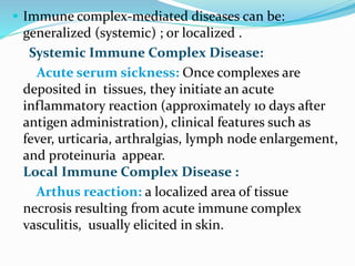
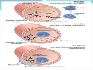
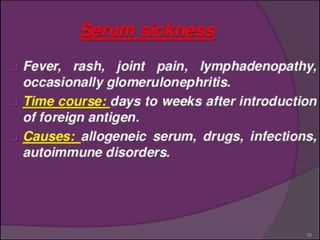
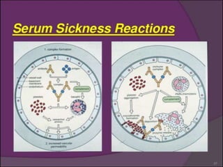
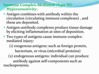
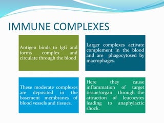
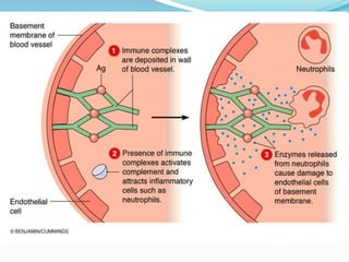
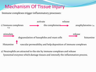
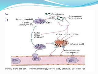
Immune Complex-Mediated (Type III)
Hypersensitivity:
 Antigen combines with antibody within the
circulation (circulating immune complexes) , and
these are deposited.
 Antigen-antibody complexes produce tissue damage
by eliciting inflammation at sites of deposition.
 Two types of antigens cause immune complex-
mediated injury:
(1) exogenous antigens: such as foreign protein,
bacterium, or virus.(microbial proteins)
(2) endogenous antigens: individual can produce
antibody against self-components such as
nucleoproteins.
IMMUNE COMPLEXES
Antigen binds to IgG and
forms complex and
circulate through the blood
Larger complexes activate
complement in the blood
and are phagocytosed by
macrophages.
These moderate complexes
are deposited in the
basement membranes of
blood vessels and tissues.
Here they cause
inflammation of target
tissue/organ through the
attraction of leucocytes
leading to anaphylactic
shock.
Mechanism Of Tissue Injury
Immune complexes trigger inflammatory processes:
activate release
1) Immune complexes the complement anaphylatoxins C3a,
C5a
stimulate release
degranulation of basophiles and mast cells histamine
Histamine vascular permeability and help deposition of immune complexes
2) Neutrophils are attracted to the site by immune complexes and release
lysosomal enzymes which damage tissues and intensify the inflammation process.
 Examples of Immune Complex-Mediated Diseases: [
diseases and antigens ]
 Systemic lupus erythematosus : DNA, and
nucleoproteins antigens.
 Polyarteritis nodosa : Hepatitis B virus surface antigen
 Poststreptococcal glomerulonephritis : Streptococcal cell
wall antigen.
 Acute glomerulonephritis : Bacterial antigens
(Treponema); parasite antigens (malaria, schistosomes);
and tumor antigens.
 Reactive arthritis : Bacterial antigens (Yersinia).
 Immune complex-mediated diseases can be:
generalized (systemic) ; or localized .
Systemic Immune Complex Disease:
Acute serum sickness: Once complexes are
deposited in tissues, they initiate an acute
inflammatory reaction (approximately 10 days after
antigen administration), clinical features such as
fever, urticaria, arthralgias, lymph node enlargement,
and proteinuria appear.
Local Immune Complex Disease :
Arthus reaction: a localized area of tissue
necrosis resulting from acute immune complex
vasculitis, usually elicited in skin.
ARTHUS REACTION
 Local inflammatory reaction with Necrosis
 Few hours after Ag inoculation
Inoculated Animal is previously immunized by same Ag
 immunized animal has titers of precipitating IgG Abs
 Arthus lesions evolve over a few hours and reach a
peak 4 to 10 hrs after injection
 When injection site develops visible edema with severe
hemorrhage occasionally followed by ulceration
General
hypersensitivity e.g.
serum toxicity.
Specific Reactions like
kidney lupus nephritis,
Joints rheumatoid
arthritis.
Anaphylaxis
Non human
proteins given
therapeutical
y bind to IgG.
These
complexes get
deposited in
the alveoli of
the lungs.
Inflammation
leads to
anaphylaxis.
Hypersensitivity type -3 reactions

Hypersensitivity type -3 reactions

  • 2.
    Immune Complex-Mediated (TypeIII) Hypersensitivity:  Antigen combines with antibody within the circulation (circulating immune complexes) , and these are deposited.  Antigen-antibody complexes produce tissue damage by eliciting inflammation at sites of deposition.  Two types of antigens cause immune complex- mediated injury: (1) exogenous antigens: such as foreign protein, bacterium, or virus.(microbial proteins) (2) endogenous antigens: individual can produce antibody against self-components such as nucleoproteins.
  • 3.
    IMMUNE COMPLEXES Antigen bindsto IgG and forms complex and circulate through the blood Larger complexes activate complement in the blood and are phagocytosed by macrophages. These moderate complexes are deposited in the basement membranes of blood vessels and tissues. Here they cause inflammation of target tissue/organ through the attraction of leucocytes leading to anaphylactic shock.
  • 5.
    Mechanism Of TissueInjury Immune complexes trigger inflammatory processes: activate release 1) Immune complexes the complement anaphylatoxins C3a, C5a stimulate release degranulation of basophiles and mast cells histamine Histamine vascular permeability and help deposition of immune complexes 2) Neutrophils are attracted to the site by immune complexes and release lysosomal enzymes which damage tissues and intensify the inflammation process.
  • 7.
     Examples ofImmune Complex-Mediated Diseases: [ diseases and antigens ]  Systemic lupus erythematosus : DNA, and nucleoproteins antigens.  Polyarteritis nodosa : Hepatitis B virus surface antigen  Poststreptococcal glomerulonephritis : Streptococcal cell wall antigen.  Acute glomerulonephritis : Bacterial antigens (Treponema); parasite antigens (malaria, schistosomes); and tumor antigens.  Reactive arthritis : Bacterial antigens (Yersinia).
  • 8.
     Immune complex-mediateddiseases can be: generalized (systemic) ; or localized . Systemic Immune Complex Disease: Acute serum sickness: Once complexes are deposited in tissues, they initiate an acute inflammatory reaction (approximately 10 days after antigen administration), clinical features such as fever, urticaria, arthralgias, lymph node enlargement, and proteinuria appear. Local Immune Complex Disease : Arthus reaction: a localized area of tissue necrosis resulting from acute immune complex vasculitis, usually elicited in skin.
  • 12.
    ARTHUS REACTION  Localinflammatory reaction with Necrosis  Few hours after Ag inoculation Inoculated Animal is previously immunized by same Ag  immunized animal has titers of precipitating IgG Abs  Arthus lesions evolve over a few hours and reach a peak 4 to 10 hrs after injection  When injection site develops visible edema with severe hemorrhage occasionally followed by ulceration
  • 13.
    General hypersensitivity e.g. serum toxicity. SpecificReactions like kidney lupus nephritis, Joints rheumatoid arthritis.
  • 14.
    Anaphylaxis Non human proteins given therapeutical ybind to IgG. These complexes get deposited in the alveoli of the lungs. Inflammation leads to anaphylaxis.