HEMODYNAMICS In the CRITICAL CARE UNIT Sherry L. Knowles, RN, CCRN, CRNI
DEFINITION PURPOSE DEFINITION HEMODYNAMIC MONITORING Measuring and monitoring the factors that influence the force and flow of blood.  To aid in diagnosing, monitoring and managing critically ill patients.
OBJECTIVES Define Basic Hemodynamics Understand The Risks and Benefits Identify PA Catheter Components Analyze Cardiac Profiles Interpret Shock States Learn Appropriate Interventions Describe How To Optimize Cardiac Output Recognize Potential Complications Upon completion of this program the student will be able to:
OBJECTIVES Collect appropriate equipment/supplies necessary to set-up a transducer pressure system.  State the correct solutions/medications used at Kaiser Vallejo for the flush bags.  Correctly level and zero the transducer. Correctly identify the location and purpose of each port/ lumen of the PA catheter  Identify in sequence the normal waveforms observed during PA catheter insertion, and state the corresponding pressure.  Correctly obtain the following pressures:  Pulmonary artery systolic, diastolic and mean Pulmonary capillary wedge. Central venous pressure. Briefly describe the indications, limitations and complications of PA catheterization, guidelines for accurate monitoring, and troubleshooting techniques. Upon completion of this program the student will be able to:
INDICATIONS To diagnose shock states  To determine fluid volume status To measure cardiac output To monitor and manage unstable patients  To assess hemodynamic response to therapies  To diagnose primary pulmonary hypertension, valvular disease, intracardiac shunts, cardiac tamponade, and pulmonary embolus
CONTRAINDICATIONS Tricuspid or pulmonary valve  mechanical prosthesis  Right heart mass  (thrombus and/or tumor) Tricuspid or pulmonary  valve endocarditis
SWAN-GANZ CATHETERS The Cordis Offers A Large Bore Infusion Port There Are Ten Types Of Swan-Ganz Catheters VIP Catheter Has Three Other Infusion Ports Large Markers = 50cm, Small Markers = 10cm
SWAN GANZ CATHETER
SWAN GANZ COMPONENTS
SWAN GANZ PLACEMENT
SWAN GANZ PLACEMENT
Bleeding Infection Dysrhythmias Pulmonary Artery Rupture Pneumothorax Hemothorax Valvular Damage Embolization Balloon Rupture Catheter Migration RISKS WITH SWAN GANZ
INSERTION EQUIPMENT EQUIPMENT NECESSARY FOR INSERTION Flush solution for transducer system Flush solution for cardiac output system Arterial access line Disposable triple pressure transducer system Pulmonary artery catheter                                 Monitor, module, electrodes, cables Central line kit                            Transducer holder, I.V. pole, pressure bag Emergency resuscitation equipment     Prepackaged Introducer Kit; sutures Sterile gowns, gloves, and masks
RA  WAVEFORM Normal Value 0-8 mmHg RAP = CVP Wave Fluctuations Due To Contractions
RV  WAVEFORM Normal Value 15-25/0-8 mmHg Catheter In RV May Cause Ventricular Ectopy Swan Tip May Drift From PA to RV
PA  WAVEFORM Normal Value 15-25/8-15 mmHg Dicrotic Notch Represents PV Closure PAD Approximates  PAWP (LVEDP)  (in absence of lung or MV disease)
PAWP WAVEFORM Normal Value 8-12 mmHg Balloon Floats and Wedges in Pulmonary Artery  PAWP = LAP = LVEDP  Wedging Can Cause Capillary Rupture
PA INSERTION WAVEFORMS A =  RA (CVP) Waveform B  =  RV Waveform C  =  PA Waveform D  =  PAWP Waveform B C D A
PA CATHETER WAVEFORMS A wave  - due to atrial contraction. Absent in atrial fibrillation. Enlarged in  tricuspid  stenosis, pulmonary stenosis and pulmonary hypertension. C wave  - due to bulging of tricuspid valve into the right atrium or possibly  transmitted pulsations from the carotid artery. X descent  - due to atrial relaxation. V wave  - due to the rise in atrial pressure before the tricuspid valve opens.  Enlarged in tricuspid regurgitation. Y descent  - due to atrial emptying as blood enters the ventricle. Canon waves  - large waves not corresponding to a, v or c waves. Due to    complete heart block or junctional arrhythmias.
PA INSERTION SEQUENCE
POST INSERTION Assess ECG for dysrhythmias. Assess for signs and symptoms of respiratory distress. Ascertain sterile dressing is in place. Obtain PCXR to check placement. Zero and level transducer(s) at the phlebostatic axis.  Assess quality of waveforms (i.e., proper configuration, dampening, catheter whip).  Obtain opening pressures and wave form tracings for each waveform. Assess length at insertion site.  Ensure that all open ends of stopcocks are covered with sterile dead-end caps (red dead-end caps, injection caps, or male Luer lock caps).  Update physician of abnormalities.
ZEROING & REFERENCING Zeroing is performed by opening the system to air to establish atmospheric pressure as zero.  Referencing is accomplished by placing the air-fluid interface of the catheter (the transducer) at the phlebostatic axis.
PHLEBOSTATIC AXIS
RESPIRATORY VARIATION Intrathoracic pressure     decreases   during spontaneous inspiration     (ventilation)  This presents a  negative (  )  deflection on a PAWP tracing Intrathoracic pressure     increases during spontaneous expiration   This present a  positive (  )  deflection on a PAWP tracing SPONTANUOUS VENTILATION :
RESPIRATORY VARIATION Intrathoracic pressure     increases   during positive pressure ventilation    (ventilator breaths) This presents a  positive (  )  deflection on a PAWP tracing Intrathoracic pressure     decreases during positive pressure expiration   This present a  negative (  )  deflection on a PAWP tracing POSITIVE PRESSURE VENTILATION :
RESPIRATORY VARIATION Spontaneous Breathing
RESPIRATORY VARIATION
END EXPIRATION
RAP WAVEFORM RAP WAVEFORM
(CVP) RA WAVEFORM & ECG
(CVP) RA WAVEFORM (CVP) RA WAVEFORM
WEDGING THE CATHETER
PAWP TRACING PAWP WAVEFORM
PAWP WAVEFORM PAWP WAVEFORM
PA vs PAWP WAVEFORM PA vs PAWP WAVEFORM
PAWP WAVEFORM & ECG PAWP WAVEFORM
PAWP WAVEFORM PAWP WAVEFORM
V  WAVES PAWP WAVEFORM
PAWP WITH V WAVES
SVO 2  MONITORING
SVO2 MONITORING Normal Values:  60-75% Decreased   (  )   SVO 2  Values Indicate    Increased      Extraction   From Decreased Oxygen Delivery   or   From Increased Oxygen Demands
POTENTIAL COMPLICATIONS POTENTIAL COMPLICATIONS Same as arterial pressure monitoring plus the following: Cardiac arrest Air emboli PA hemorrhage or infarction Pulmonary artery extravasation Altered skin integrity Loss of balloon integrity Frank Hemorrhage Pneumothorax/Hemothorax Pulmonary artery rupture Equipment malfunction Electromicroshock Inaccurate pressures Lung ischemia Infection Balloon rupture Catheter displacement Dysrhythmias Thromboembolism Cardiac tamponade Air emboli
Wedging Can Cause Capillary Rupture Catheter In RV Can Cause Ventricular Ectopy Swan Tip Can Drift From  PA to RV POTENTIAL COMPLICATIONS
PERICARDIAL TAMPONADE Hemodynamic monitoring can diagnose tamponade: Pericardial tamponade presents with equalization of the diastolic pressures on the left and right side of the heart Other PAP signs of pericardial tamponade include: Elevated right atrial pressure Kussmaul sign (increase in right atrial pressure with inspiration) Pulsus Paradoxus Elevated right atrial pressure (RAP)  Pulmonary artery diastolic pressure (PAD) = mean right atrial pressure(RA) = right ventricular (RV) diastolic pressure = mean wedge pressure
PRECAUTIONS Always set alarms, approximately 20 mmHg above and below the patient’s readings.  If balloon is down and you find PA catheter tracing in wedge position, you may ask the patient to deep breathe and cough, or reposition patient in bed to dislodge it.  If unable to dislodge catheter from wedge position by above measures notify physician immediately to reposition catheter by pulling back gently; then, get chest x-ray to confirm proper placement.  If patient coughs up blood or it is suctioned via endotracheal tube, suspect PA rupture and notify physician immediately.
TROUBLESHOOTING
= 0-8 mm Hg = 15-25 / 8-15 mm Hg = 8-12 mm Hg = 8-12mm Hg = 50-100 ml/beat = 4-8 L/min = 2.5-4.0 L/min M 2 = 0.60-0.75 NORMAL VALUES Right Atrial Pressure (CVP) Pulmonary Artery Pressure  Pulmonary Artery Wedge Pressure Left Ventricular Diastolic Pressure  Stroke Volume  Cardiac Output Cardiac Index SVO 2
DAMPENED PA WAVEFORM PAWP WAVEFORM
ALTERATIONS IN SVO 2
Optimize  HR  and  SV  (Stroke Volume) Stroke Volume =  PRELOAD AFTERLOAD  CONTRACTILITY Chronotropic Medications  Diuretics / Volume  Vasodilators / Vasoconstrictors Inotropic Medications (Positive or Negative) IABP OPTIMIZING CARDIAC OUTPUT
Shock States Cardiogenic Shock Hypovolemic Shock Septic Shock Anaphylactic Shock Cardiac Tamponade Left Ventricular Failure Right Ventricular Failure Pulmonary Hypertension CARDIAC PROFILES
SHOCK PARAMETERS Cardiogenic Shock is the only shock with    PAWP. Early (Hyperdynamic) Shock is the only shock with     CO and     SVR. Neurogenic Shock is the only shock with     bradycardia. Anaphylactic Shock has the definitive characteristic of wheezing due to bronchospasm.
TREATMENTS
SAMPLE MEASUREMENTS
MEASUREMENTS
SAMPLE MEASUREMENTS
SAMPLE MEASUREMENTS
SAMPLE MEASUREMENTS
SAMPLE MEASUREMENTS
SAMPLE MEASUREMENTS
SAMPLE MEASUREMENTS
SAMPLE MEASUREMENTS
SAMPLE MEASUREMENTS
SAMPLE MEASUREMENTS
SAMPLE MEASUREMENTS
SAMPLE MEASUREMENTS
SAMPLE MEASUREMENTS
SAMPLE MEASUREMENTS
SAMPLE MEASUREMENTS
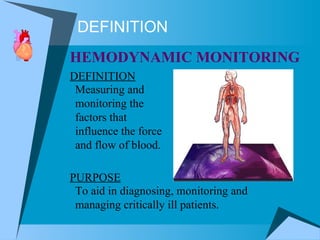
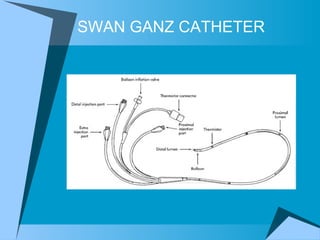
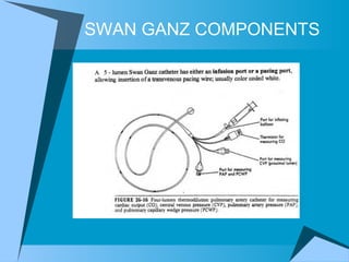
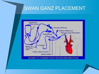
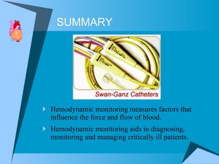
Hemodynamic monitoring measures factors that influence the force and flow of blood.  Hemodynamic monitoring aids in diagnosing, monitoring and managing critically ill patients. SUMMARY
THE END
REFERENCES AACN (American Association of Critical Care Nurses). Clinical Care References. 2002 SCCM (Society of Critical Care Medicine. PACEP (Pulmonary Artery Catheter Education Project). 701 Lee Street, Suite 200, Des Plaines, Illinoise 60016. 2000. Bridges, EJ, and Woods, SL. Pulmonary artery pressure measurement: State of the art. Heart Lung 1993; 22:99.  Mirini, JJ. Pulmonary artery occlusion pressure: Clinical physiology, measurement and interpretation. Am Rev Respir Dis 1983; 128:319.  Putterman, C. The Swan-Ganz catheter: A decade of hemodynamic monitoring. J Crit Care 1989; 4:127. Nemens, EJ, Woods, SL. Normal Fluctuations in pulmonary artery and pulmonary capillary wedge pressures in acutely ill patients. Heart Lung 1982; 11:393.  Darovic, G.O., (1995) H emodynamic monitoring: invasive and noninvasive clinical application  (2d ed), New York: W. B. Saunders

Hemodynamics

  • 1.
    HEMODYNAMICS In theCRITICAL CARE UNIT Sherry L. Knowles, RN, CCRN, CRNI
  • 2.
    DEFINITION PURPOSE DEFINITIONHEMODYNAMIC MONITORING Measuring and monitoring the factors that influence the force and flow of blood. To aid in diagnosing, monitoring and managing critically ill patients.
  • 3.
    OBJECTIVES Define BasicHemodynamics Understand The Risks and Benefits Identify PA Catheter Components Analyze Cardiac Profiles Interpret Shock States Learn Appropriate Interventions Describe How To Optimize Cardiac Output Recognize Potential Complications Upon completion of this program the student will be able to:
  • 4.
    OBJECTIVES Collect appropriateequipment/supplies necessary to set-up a transducer pressure system. State the correct solutions/medications used at Kaiser Vallejo for the flush bags. Correctly level and zero the transducer. Correctly identify the location and purpose of each port/ lumen of the PA catheter Identify in sequence the normal waveforms observed during PA catheter insertion, and state the corresponding pressure. Correctly obtain the following pressures: Pulmonary artery systolic, diastolic and mean Pulmonary capillary wedge. Central venous pressure. Briefly describe the indications, limitations and complications of PA catheterization, guidelines for accurate monitoring, and troubleshooting techniques. Upon completion of this program the student will be able to:
  • 5.
    INDICATIONS To diagnoseshock states To determine fluid volume status To measure cardiac output To monitor and manage unstable patients To assess hemodynamic response to therapies To diagnose primary pulmonary hypertension, valvular disease, intracardiac shunts, cardiac tamponade, and pulmonary embolus
  • 6.
    CONTRAINDICATIONS Tricuspid orpulmonary valve mechanical prosthesis Right heart mass (thrombus and/or tumor) Tricuspid or pulmonary valve endocarditis
  • 7.
    SWAN-GANZ CATHETERS TheCordis Offers A Large Bore Infusion Port There Are Ten Types Of Swan-Ganz Catheters VIP Catheter Has Three Other Infusion Ports Large Markers = 50cm, Small Markers = 10cm
  • 8.
  • 9.
  • 10.
  • 11.
  • 12.
    Bleeding Infection DysrhythmiasPulmonary Artery Rupture Pneumothorax Hemothorax Valvular Damage Embolization Balloon Rupture Catheter Migration RISKS WITH SWAN GANZ
  • 13.
    INSERTION EQUIPMENT EQUIPMENTNECESSARY FOR INSERTION Flush solution for transducer system Flush solution for cardiac output system Arterial access line Disposable triple pressure transducer system Pulmonary artery catheter                                Monitor, module, electrodes, cables Central line kit                            Transducer holder, I.V. pole, pressure bag Emergency resuscitation equipment     Prepackaged Introducer Kit; sutures Sterile gowns, gloves, and masks
  • 14.
    RA WAVEFORMNormal Value 0-8 mmHg RAP = CVP Wave Fluctuations Due To Contractions
  • 15.
    RV WAVEFORMNormal Value 15-25/0-8 mmHg Catheter In RV May Cause Ventricular Ectopy Swan Tip May Drift From PA to RV
  • 16.
    PA WAVEFORMNormal Value 15-25/8-15 mmHg Dicrotic Notch Represents PV Closure PAD Approximates PAWP (LVEDP) (in absence of lung or MV disease)
  • 17.
    PAWP WAVEFORM NormalValue 8-12 mmHg Balloon Floats and Wedges in Pulmonary Artery PAWP = LAP = LVEDP Wedging Can Cause Capillary Rupture
  • 18.
    PA INSERTION WAVEFORMSA = RA (CVP) Waveform B = RV Waveform C = PA Waveform D = PAWP Waveform B C D A
  • 19.
    PA CATHETER WAVEFORMSA wave - due to atrial contraction. Absent in atrial fibrillation. Enlarged in tricuspid stenosis, pulmonary stenosis and pulmonary hypertension. C wave - due to bulging of tricuspid valve into the right atrium or possibly transmitted pulsations from the carotid artery. X descent - due to atrial relaxation. V wave - due to the rise in atrial pressure before the tricuspid valve opens. Enlarged in tricuspid regurgitation. Y descent - due to atrial emptying as blood enters the ventricle. Canon waves - large waves not corresponding to a, v or c waves. Due to complete heart block or junctional arrhythmias.
  • 20.
  • 21.
    POST INSERTION AssessECG for dysrhythmias. Assess for signs and symptoms of respiratory distress. Ascertain sterile dressing is in place. Obtain PCXR to check placement. Zero and level transducer(s) at the phlebostatic axis. Assess quality of waveforms (i.e., proper configuration, dampening, catheter whip). Obtain opening pressures and wave form tracings for each waveform. Assess length at insertion site. Ensure that all open ends of stopcocks are covered with sterile dead-end caps (red dead-end caps, injection caps, or male Luer lock caps). Update physician of abnormalities.
  • 22.
    ZEROING & REFERENCINGZeroing is performed by opening the system to air to establish atmospheric pressure as zero. Referencing is accomplished by placing the air-fluid interface of the catheter (the transducer) at the phlebostatic axis.
  • 23.
  • 24.
    RESPIRATORY VARIATION Intrathoracicpressure  decreases during spontaneous inspiration  (ventilation) This presents a negative (  ) deflection on a PAWP tracing Intrathoracic pressure  increases during spontaneous expiration  This present a positive (  ) deflection on a PAWP tracing SPONTANUOUS VENTILATION :
  • 25.
    RESPIRATORY VARIATION Intrathoracicpressure  increases during positive pressure ventilation  (ventilator breaths) This presents a positive (  ) deflection on a PAWP tracing Intrathoracic pressure  decreases during positive pressure expiration  This present a negative (  ) deflection on a PAWP tracing POSITIVE PRESSURE VENTILATION :
  • 26.
  • 27.
  • 28.
  • 29.
  • 30.
  • 31.
    (CVP) RA WAVEFORM(CVP) RA WAVEFORM
  • 32.
  • 33.
  • 34.
  • 35.
    PA vs PAWPWAVEFORM PA vs PAWP WAVEFORM
  • 36.
    PAWP WAVEFORM &ECG PAWP WAVEFORM
  • 37.
  • 38.
    V WAVESPAWP WAVEFORM
  • 39.
  • 40.
    SVO 2 MONITORING
  • 41.
    SVO2 MONITORING NormalValues: 60-75% Decreased (  ) SVO 2 Values Indicate  Increased  Extraction From Decreased Oxygen Delivery or From Increased Oxygen Demands
  • 42.
    POTENTIAL COMPLICATIONS POTENTIALCOMPLICATIONS Same as arterial pressure monitoring plus the following: Cardiac arrest Air emboli PA hemorrhage or infarction Pulmonary artery extravasation Altered skin integrity Loss of balloon integrity Frank Hemorrhage Pneumothorax/Hemothorax Pulmonary artery rupture Equipment malfunction Electromicroshock Inaccurate pressures Lung ischemia Infection Balloon rupture Catheter displacement Dysrhythmias Thromboembolism Cardiac tamponade Air emboli
  • 43.
    Wedging Can CauseCapillary Rupture Catheter In RV Can Cause Ventricular Ectopy Swan Tip Can Drift From PA to RV POTENTIAL COMPLICATIONS
  • 44.
    PERICARDIAL TAMPONADE Hemodynamicmonitoring can diagnose tamponade: Pericardial tamponade presents with equalization of the diastolic pressures on the left and right side of the heart Other PAP signs of pericardial tamponade include: Elevated right atrial pressure Kussmaul sign (increase in right atrial pressure with inspiration) Pulsus Paradoxus Elevated right atrial pressure (RAP) Pulmonary artery diastolic pressure (PAD) = mean right atrial pressure(RA) = right ventricular (RV) diastolic pressure = mean wedge pressure
  • 45.
    PRECAUTIONS Always setalarms, approximately 20 mmHg above and below the patient’s readings. If balloon is down and you find PA catheter tracing in wedge position, you may ask the patient to deep breathe and cough, or reposition patient in bed to dislodge it. If unable to dislodge catheter from wedge position by above measures notify physician immediately to reposition catheter by pulling back gently; then, get chest x-ray to confirm proper placement. If patient coughs up blood or it is suctioned via endotracheal tube, suspect PA rupture and notify physician immediately.
  • 46.
  • 47.
    = 0-8 mmHg = 15-25 / 8-15 mm Hg = 8-12 mm Hg = 8-12mm Hg = 50-100 ml/beat = 4-8 L/min = 2.5-4.0 L/min M 2 = 0.60-0.75 NORMAL VALUES Right Atrial Pressure (CVP) Pulmonary Artery Pressure Pulmonary Artery Wedge Pressure Left Ventricular Diastolic Pressure Stroke Volume Cardiac Output Cardiac Index SVO 2
  • 48.
    DAMPENED PA WAVEFORMPAWP WAVEFORM
  • 49.
  • 50.
  • 51.
    Optimize HR and SV (Stroke Volume) Stroke Volume = PRELOAD AFTERLOAD CONTRACTILITY Chronotropic Medications Diuretics / Volume Vasodilators / Vasoconstrictors Inotropic Medications (Positive or Negative) IABP OPTIMIZING CARDIAC OUTPUT
  • 52.
    Shock States CardiogenicShock Hypovolemic Shock Septic Shock Anaphylactic Shock Cardiac Tamponade Left Ventricular Failure Right Ventricular Failure Pulmonary Hypertension CARDIAC PROFILES
  • 53.
    SHOCK PARAMETERS CardiogenicShock is the only shock with  PAWP. Early (Hyperdynamic) Shock is the only shock with  CO and  SVR. Neurogenic Shock is the only shock with  bradycardia. Anaphylactic Shock has the definitive characteristic of wheezing due to bronchospasm.
  • 54.
  • 55.
  • 56.
  • 57.
  • 58.
  • 59.
  • 60.
  • 61.
  • 62.
  • 63.
  • 64.
  • 65.
  • 66.
  • 67.
  • 68.
  • 69.
  • 70.
  • 71.
    Hemodynamic monitoring measuresfactors that influence the force and flow of blood. Hemodynamic monitoring aids in diagnosing, monitoring and managing critically ill patients. SUMMARY
  • 72.
  • 73.
    REFERENCES AACN (AmericanAssociation of Critical Care Nurses). Clinical Care References. 2002 SCCM (Society of Critical Care Medicine. PACEP (Pulmonary Artery Catheter Education Project). 701 Lee Street, Suite 200, Des Plaines, Illinoise 60016. 2000. Bridges, EJ, and Woods, SL. Pulmonary artery pressure measurement: State of the art. Heart Lung 1993; 22:99. Mirini, JJ. Pulmonary artery occlusion pressure: Clinical physiology, measurement and interpretation. Am Rev Respir Dis 1983; 128:319. Putterman, C. The Swan-Ganz catheter: A decade of hemodynamic monitoring. J Crit Care 1989; 4:127. Nemens, EJ, Woods, SL. Normal Fluctuations in pulmonary artery and pulmonary capillary wedge pressures in acutely ill patients. Heart Lung 1982; 11:393. Darovic, G.O., (1995) H emodynamic monitoring: invasive and noninvasive clinical application (2d ed), New York: W. B. Saunders