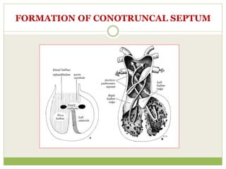
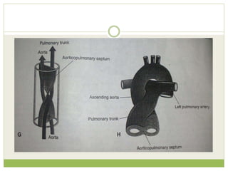
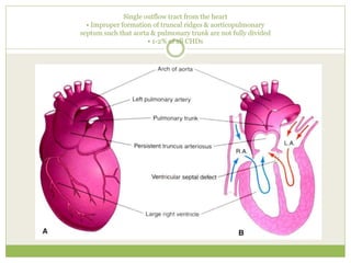
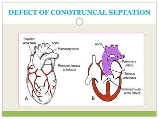
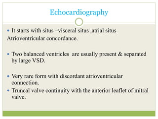
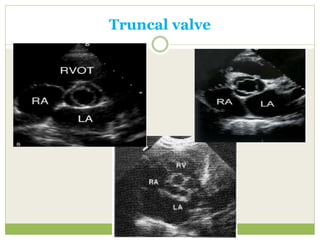
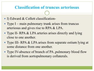
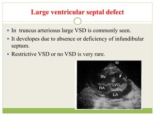
D R V I S H W A N A T H H E S A R U R
J N M C , B E L G A U M
TRUNCUS
ARTERIOSUS
FORMATION OF CONOTRUNCAL SEPTUM
Single outflow tract from the heart
• Improper formation of truncal ridges & aorticopulmonary
septum such that aorta & pulmonary trunk are not fully divided
• 1-2% of all CHDs
DEFECT OF CONOTRUNCAL SEPTATION
TRUNCUS ARTERIOSUS
 Truncus arteriosus is one of the least common cynotic
congenital heart disease.
 1-2.5 % in all CHD.
 Frequently syndromic as 22q 11 deletion syndrome.
 DiGeorge’s syndrome.
DiGeorge’s syndrome
Definition
 It is congenital cardiac malformation in which one great
artery arises from the base of the heart by a way of single
semilunar valve (truncal valve),it gives origin to systemic
arteries,coronary arteries and pulmonary arteries.
 History – Wilson – 1st described in 1798.
 Buchanan – clinical and autopsy reports in 1864.
 Collett and Edwards –classification in 1949.
 Van praagh- alternative classification in 1965.
 McGoon – 1st repair with homograft in 1967.
Echocardiography
 It starts with situs –visceral situs ,atrial situs
Atrioventricular concordance.
 Two balanced ventricles are usually present & separated
by large VSD.
 Very rare form with discordant atrioventricular
connection.
 Truncal valve continuity with the anterior leaflet of mitral
valve.
Truncal valve
 It is best seen in parasternal short axis view.
 It can be tricuspid – 60-67 %.
 Quadricuspid – 25- 31%
 Bicuspid – 8%
 Pentacuspid -0.3%
 Valve leaflet can be normal or may be stenotic or
regurgitant.
Truncal valve
Classification of truncus arteriosus
 I) Edward & Collett classification-
 Type I – main pulmonary trunk arises from truncus
arteriosus and gives rise to RPA & LPA.
 Type II- RPA & LPA arteries arises directly and lying
close to one another.
 Type III- RPA & LPA arises from separate ostium lying at
some distance from one another.
 Type IV-absence of branch of PA ,pulmonary blood flow
is derived from aortopulmonary collaterals.
EDWARD AND COLLETT CLASSIFICATION-1949
Van praagh classification-1965
 Type A1-corresponds to type I of collett & Edward.
 Type A2- corresponds to Type II and Type III of collett &
Edward.
 Type A3-Absence of truncus origin of one of the
pulmonary Artery.
 Type A4- Hypoplasia of Aorta.
Differential diagnosis
 A)Pulmonary atresia-pulmonary ateries are small with
continues flow being seen in branch PA either from PDA
or collateral flow.
 B) very rarely this large great vessel may be PA in
patient’s of aortic atresia.(Imp – identify Aorta).
 C) anomalous origin of RPA from posterior aspect of
aorta.
2D-Parastenal long axis view
 Common arterial trunk arising predominantly from LV –
4-6%
 Truncus arteriosus with biventricular origin – 69%
 Right ventricular origin – 11 – 29 %
 Truncal override –
 It is discontinuity between IVS and anterior truncal wall.
Large ventricular septal defect
 In truncus arteriosus large VSD is commonly seen.
 It developes due to absence or deficiency of infundibular
septum.
 Restrictive VSD or no VSD is very rare.
Short MPA with RPA &
LPA arising frm left lateral
aspect of TR
NO MPA –direct RPA &
LPA arising from
posterior aspect of TR
Parasternal short axis view
Apical view
Suprasternal long axis view
Regurgitant truncal valve
 Causes-
 1) Thickening , dysplastic cusps.
 2) Prolapsed cusps.
 3) unequal cusps.
 4) truncal root dilation.
Stenotic truncal valve
 By using colour and continues doppler gradient across
pulmonary arteries and stenotic truncal valve can be
recorded.
 Stenosis and regurgitant gradient is frequently
overestimated across truncal valve bz both the ventricular
output has to pass across it.
 Gradient up to 40 – 50 mmHg have been documented.
 Significant stenosis of origin of branch of pulmonary
artery is the presence of diastolic spill on doppler.
Coronary artery anomalies
Associated anomalies
 Right aortic arch- 25 to 35 %
 Coarction of aorta.-10 to 15 %
 Patent ductus arteriosus-10%
 Interrupted aortic arch- 10 to 15 %
 Ostium secundum ASD-10 %
 Left SVC draining into coronary sinus- 10%
 TAPVC / PAPVC
Surgical correction

Truncus Arteriosus

  • 1.
    D R VI S H W A N A T H H E S A R U R J N M C , B E L G A U M TRUNCUS ARTERIOSUS
  • 2.
  • 4.
    Single outflow tractfrom the heart • Improper formation of truncal ridges & aorticopulmonary septum such that aorta & pulmonary trunk are not fully divided • 1-2% of all CHDs
  • 5.
  • 6.
    TRUNCUS ARTERIOSUS  Truncusarteriosus is one of the least common cynotic congenital heart disease.  1-2.5 % in all CHD.  Frequently syndromic as 22q 11 deletion syndrome.  DiGeorge’s syndrome.
  • 7.
  • 8.
    Definition  It iscongenital cardiac malformation in which one great artery arises from the base of the heart by a way of single semilunar valve (truncal valve),it gives origin to systemic arteries,coronary arteries and pulmonary arteries.  History – Wilson – 1st described in 1798.  Buchanan – clinical and autopsy reports in 1864.  Collett and Edwards –classification in 1949.  Van praagh- alternative classification in 1965.  McGoon – 1st repair with homograft in 1967.
  • 9.
    Echocardiography  It startswith situs –visceral situs ,atrial situs Atrioventricular concordance.  Two balanced ventricles are usually present & separated by large VSD.  Very rare form with discordant atrioventricular connection.  Truncal valve continuity with the anterior leaflet of mitral valve.
  • 12.
    Truncal valve  Itis best seen in parasternal short axis view.  It can be tricuspid – 60-67 %.  Quadricuspid – 25- 31%  Bicuspid – 8%  Pentacuspid -0.3%  Valve leaflet can be normal or may be stenotic or regurgitant.
  • 13.
  • 14.
    Classification of truncusarteriosus  I) Edward & Collett classification-  Type I – main pulmonary trunk arises from truncus arteriosus and gives rise to RPA & LPA.  Type II- RPA & LPA arteries arises directly and lying close to one another.  Type III- RPA & LPA arises from separate ostium lying at some distance from one another.  Type IV-absence of branch of PA ,pulmonary blood flow is derived from aortopulmonary collaterals.
  • 15.
    EDWARD AND COLLETTCLASSIFICATION-1949
  • 16.
    Van praagh classification-1965 Type A1-corresponds to type I of collett & Edward.  Type A2- corresponds to Type II and Type III of collett & Edward.  Type A3-Absence of truncus origin of one of the pulmonary Artery.  Type A4- Hypoplasia of Aorta.
  • 18.
    Differential diagnosis  A)Pulmonaryatresia-pulmonary ateries are small with continues flow being seen in branch PA either from PDA or collateral flow.  B) very rarely this large great vessel may be PA in patient’s of aortic atresia.(Imp – identify Aorta).  C) anomalous origin of RPA from posterior aspect of aorta.
  • 19.
    2D-Parastenal long axisview  Common arterial trunk arising predominantly from LV – 4-6%  Truncus arteriosus with biventricular origin – 69%  Right ventricular origin – 11 – 29 %  Truncal override –  It is discontinuity between IVS and anterior truncal wall.
  • 21.
    Large ventricular septaldefect  In truncus arteriosus large VSD is commonly seen.  It developes due to absence or deficiency of infundibular septum.  Restrictive VSD or no VSD is very rare.
  • 23.
    Short MPA withRPA & LPA arising frm left lateral aspect of TR NO MPA –direct RPA & LPA arising from posterior aspect of TR Parasternal short axis view
  • 24.
  • 26.
  • 27.
    Regurgitant truncal valve Causes-  1) Thickening , dysplastic cusps.  2) Prolapsed cusps.  3) unequal cusps.  4) truncal root dilation.
  • 28.
    Stenotic truncal valve By using colour and continues doppler gradient across pulmonary arteries and stenotic truncal valve can be recorded.  Stenosis and regurgitant gradient is frequently overestimated across truncal valve bz both the ventricular output has to pass across it.  Gradient up to 40 – 50 mmHg have been documented.  Significant stenosis of origin of branch of pulmonary artery is the presence of diastolic spill on doppler.
  • 30.
  • 31.
    Associated anomalies  Rightaortic arch- 25 to 35 %  Coarction of aorta.-10 to 15 %  Patent ductus arteriosus-10%  Interrupted aortic arch- 10 to 15 %  Ostium secundum ASD-10 %  Left SVC draining into coronary sinus- 10%  TAPVC / PAPVC
  • 32.