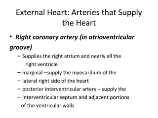
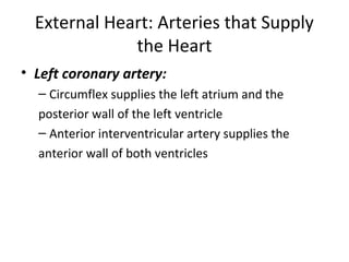
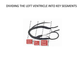
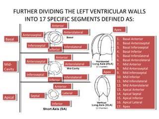
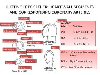
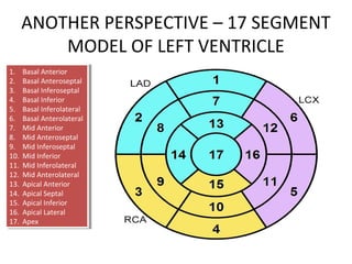
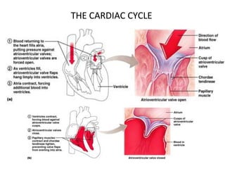
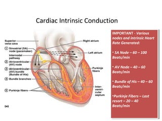
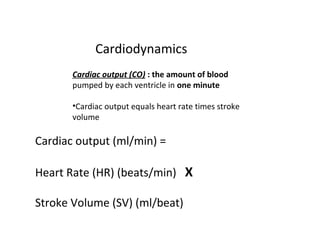
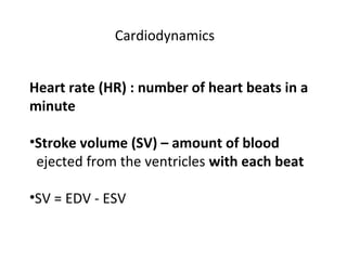
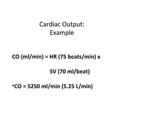
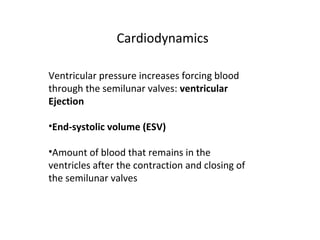
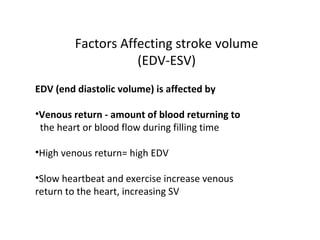
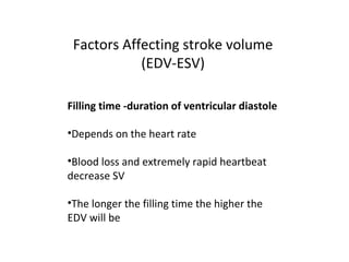
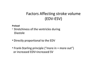
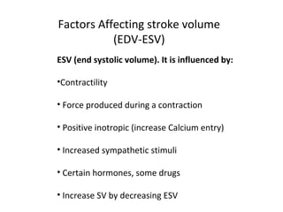
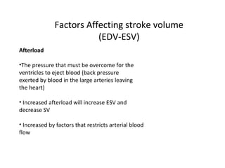
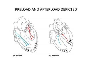
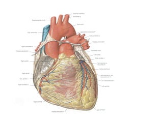
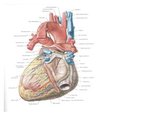
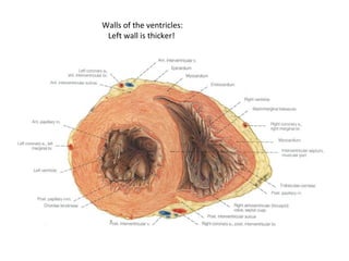
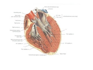
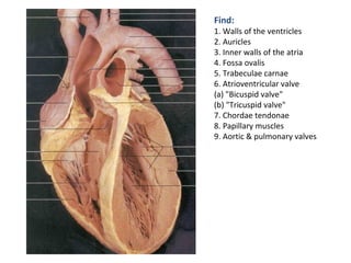
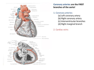
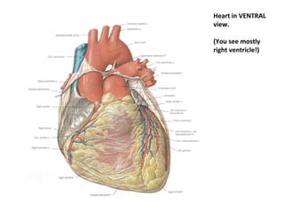
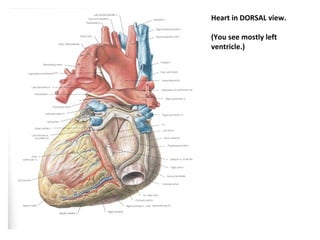
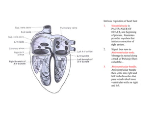
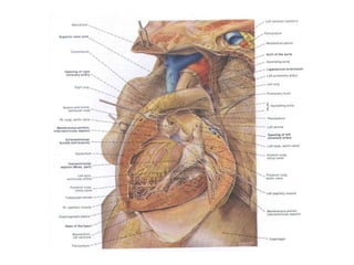
SlideShare a Scribd company logo
External Heart: Arteries that Supply
the Heart
• Coronary circulation is the functional blood
supply to the heart muscle itself (i.e. the fuel
for the heart)
• Collateral routes ensure blood delivery to
heart
• even if major vessels are occluded/clogged
Different parts of the heart are supplied by
different coronary artery segments
Heart chambers in geometric
models are subdivided into
perfusion zones using
anatomic knowledge in the
FMA ontology
CORONARY ARTERIES SUPPLYING THE
HEART WITH OXYGEN-RICH BLOOD
Right
Coronary
Artery
(RCA)
Right
Coronary
Artery
(RCA)
Left Main
Coronary
Artery (LMCA)
Left Main
Coronary
Artery (LMCA)
Left Anterior
Descending
Artery (LAD)
Left Anterior
Descending
Artery (LAD)
Posterior
Descending Artery
(PDA)
Posterior
Descending Artery
(PDA)
Left Circumflex
Coronary Artery
(LCx)
Left Circumflex
Coronary Artery
(LCx)
Arterial Coronary Circulation
External Heart: Arteries that Supply
the Heart
• Right coronary artery (in atrioventricular
groove)
– Supplies the right atrium and nearly all the
right ventricle
– marginal –supply the myocardium of the
– lateral right side of the heart
– posterior interventricular artery – supply the
– interventricular septum and adjacent portions
of the ventricular walls
External Heart: Arteries that Supply
the Heart
• Left coronary artery:
– Circumflex supplies the left atrium and the
posterior wall of the left ventricle
– Anterior interventricular artery supplies the
anterior wall of both ventricles
CERTAIN HEART WALL SEGMENTS ARE FED BY
CERTAIN CORONARY ARTERIES AS SPECIFIED BELOW:
BASE
MID
APEX
A4C
A2C
PARASTERNAL
LONG-AXIS
Key:
Red = LAD
Yellow = LCx
Green = RCA
LAD = Left Anterior Descending Coronary Artery, LCx = Left Circumflex Coronary
Artery, RCA = Right Coronary Artery
DIVIDING THE LEFT VENTRICLE INTO KEY SEGMENTS
Apical
Third
Apical
Third Mid
Third
Mid
Third
Basal
Third
Basal
Third
FURTHER DIVIDING THE LEFT VENTRICULAR WALLS
INTO 17 SPECIFIC SEGMENTS DEFINED AS:
AnteriorAnterior
AnterolateralAnterolateral
InferolateralInferolateral
InferiorInferior
InferoseptalInferoseptal
AnteroseptalAnteroseptal
ApexApex
AnteriorAnterior
AnteroseptalAnteroseptal
InferoseptalInferoseptal
AnterolateralAnterolateral
InferolateralInferolateral
InferiorInferior
AnteriorAnterior
SeptalSeptal
LateralLateral
InferiorInferior
ApexApex
BasalBasal
Mid-
Cavity
Mid-
Cavity
ApicalApical
1. Basal Anterior
2. Basal Anteroseptal
3. Basal Inferoseptal
4. Basal Inferior
5. Basal Inferolateral
6. Basal Anterolateral
7. Mid Anterior
8. Mid Anteroseptal
9. Mid Inferoseptal
10. Mid Inferior
11. Mid Inferolateral
12. Mid Anterolateral
13. Apical Anterior
14. Apical Septal
15. Apical Inferior
16. Apical Lateral
17. Apex
PUTTING IT TOGETHER: HEART WALL SEGMENTS
AND CORRESPONDING CORONARY ARTERIES
Coronary
Artery: Segments:
LAD 1, 2, 7, 8, 13, 14, 17
RCA 3, 4, 9, 10, 15
LCX 5, 6, 11, 12, 16
Coronary
Artery: Segments:
LAD 1, 2, 7, 8, 13, 14, 17
RCA 3, 4, 9, 10, 15
LCX 5, 6, 11, 12, 16
LAD = Left Anterior Descending
Artery
RCA = Right Coronary Artery
LCX = Left Circumflex Artery
LAD = Left Anterior Descending
Artery
RCA = Right Coronary Artery
LCX = Left Circumflex Artery
AnteriorAnterior
AnterolateralAnterolateral
InferolateralInferolateral
InferiorInferior
InferoseptalInferoseptal
AnteroseptalAnteroseptal
ApexApex
AnteriorAnterior
AnteroseptalAnteroseptal
InferoseptalInferoseptal
AnterolateralAnterolateral
InferolateralInferolateral
InferiorInferior
AnteriorAnterior
SeptalSeptal
LateralLateral
InferiorInferior
ApexApex
ANOTHER PERSPECTIVE – 17 SEGMENT
MODEL OF LEFT VENTRICLE
1. Basal Anterior
2. Basal Anteroseptal
3. Basal Inferoseptal
4. Basal Inferior
5. Basal Inferolateral
6. Basal Anterolateral
7. Mid Anterior
8. Mid Anteroseptal
9. Mid Inferoseptal
10. Mid Inferior
11. Mid Inferolateral
12. Mid Anterolateral
13. Apical Anterior
14. Apical Septal
15. Apical Inferior
16. Apical Lateral
17. Apex
SAMPLE – SEGMENTAL SCORING
Ortiz-Perez, J. T. et al. J Am Coll Cardiol Img 2008;1:282-293
Representative Segmentation and Pattern of Contrast
HE Over 17-Segments of the LAD, RCA, and LCX
Arteries
Ortiz-Perez, J. T. et al. J Am Coll Cardiol Img 2008;1:282-293
HEART ANATOMY – COVERINGS OF THE HEART
• Coverings of the Heart: Anatomy
• Pericardium – a double-walled sac around the
• heart composed of:
• A superficial fibrous pericardium
• A deep two-layer serous pericardium
• The parietal layer lines the internal surface
• of the fibrous pericardium
• The visceral layer or epicardium lines the
• surface of the heart
• They are separated by the fluid-filled
• pericardial cavity
• Coverings of the Heart: Physiology
• The pericardium:
• Protects the heart
COVERINGS OF THE HEART:
PHYSIOLOGY
• The pericardium:
– Protects and anchors the heart
– Prevents overfilling of the heart with blood
– Allows for the heart to work in a relatively
– friction-free environment
PERICARDIUM OF THE HEART
• Pericardium (3 layers)
• 1) Outer-fibrous pericardium
– Serous pericardium
• 2) parietal
• 3) visceral (epicardium)
• Pericardial Cavity
– between layers of serous pericardium
– serous fluid
– lubricate heart while beating
Pericardial Layers of the Heart
Heart Wall – Layers of the Heart
• Epicardium – visceral layer of the serous
• pericardium
• Myocardium – cardiac muscle layer forming
• the bulk of the heart
• Fibrous skeleton of the heart – crisscrossing,
• interlacing layer of connective tissue
• Endocardium – endothelial layer of the inner
• myocardial surface
• Cardiac Muscle Bundles
• External
Cardiac Muscle 1000X
intercalated disc
striations nucleus
Purkinje fibers 40X
EXTERNAL FEATURES OF THE HEART
• Interventricular sulcus
• Coronal/Coronary sulcus
• Auricles of atria
• Apex
• Base
• Coronary vessels
• Ligamentum Arteriosum
External Heart: Major Vessels of
the Heart (Anterior View)
• External Heart: Major Vessels of
the Heart (Anterior View)
• Vessels returning blood to the heart include:
– Superior and inferior vena cava
– Right and left pulmonary veins
• Vessels conveying blood away from the heart:
– Pulmonary trunk, which splits into right and
– left pulmonary arteries
– Ascending aorta (three branches) –
– brachiocephalic, left common carotid, and
– subclavian arteries
External Heart: Veins that Drain the
Heart
External Heart: Major Vessels of
the Heart (Posterior View)
Vessels returning blood to the heart include:
•Right and left pulmonary veins
• Superior and inferior vena cava
• Vessels conveying blood away from the heart
include:
• Aorta
• Right and left pulmonary arteries
FLOW OF BLOOD
• O2-poor blood (S+I VC, Coronary Sinus) enters Rt Atrium
• Travels through Tricuspid Valve into Rt Ventricle
• Pumped out through Pulmonary Semilunar Valve into
Pulmonary trunk (branches into Pulmonary Arteries) and to
lungs
•After circulating through lungs, O2-rich blood returns to the
heart through 4 Pulmonary veins
• The O2-rich blood enters the Left Atrium
• Travels through Bicuspid/Mitral Valve into Left Ventricle
• Pumped out through Aortic Semilunar Valve into Aorta to be
distributed to rest of body by descending aorta and branches
of aortic arch
Pathway of Blood Through the
Heart and Lungs
Right atrium > tricuspid valve > right ventricle >
pulmonary semilunar valve > pulmonary arteries >
Lungs > pulmonary veins > left atrium >
Mitral valve > left ventricle > Left ventricle >
aortic semilunar valve > aorta > systemic circulation
THE PATHWAY OF THE HEART
Cardiovascular Flow of Blood
• HeartArteries(conducting-distributing)
ArteriolesCapillaries of tissues
• At Capillaries O2 is delivered and CO2 picked
up
•CapillariesVenulesVeinsHeart
DETAILED IMAGES OF THE HEART’S
ANATOMY – CHORDAE TENDONAE
THE CARDIAC CYCLE
Semilunar Valve Function
Cardiac Intrinsic Conduction
IMPORTANT - Various
nodes and intrinsic Heart
Rate Generated:
• SA Node – 60 – 100
Beats/min
• AV Node – 40 – 60
Beats/min
• Bundle of His – 40 – 60
Beats/min
•Purkinje Fibers – Last
resort – 20 – 40
Beats/min
IMPORTANT - Various
nodes and intrinsic Heart
Rate Generated:
• SA Node – 60 – 100
Beats/min
• AV Node – 40 – 60
Beats/min
• Bundle of His – 40 – 60
Beats/min
•Purkinje Fibers – Last
resort – 20 – 40
Beats/min
Heart Excitation Related to ECG
atrial excitation begins
Impulse delayed
at AV node
Impulse passes to
heart apex; ventricular
excitation begins
Ventricular excitation
complete
SA node AV node Purkinje
fibers
Bundle
branches
Electrocardiography
Electrical activity is recorded by
electrocardiogram (ECG)
• P wave corresponds to depolarization of SA
•node
•QRS complex corresponds to ventricular
•depolarization
•T wave corresponds to ventricular
repolarization
•Atrial repolarization record is masked by the
•larger QRS complex
Electrocardiography
Electrocardiography
PR interval
•Atrial depolarization and contraction
•QT interval
•Ventricular depolarization, contraction and
repolarization
•PR segment
•Atrial contraction
•ST segment
•Ventricular contraction
•ECG Tracings
•Heart
ECG Tracings
Heart
Cardiac Cycle
Cardiac cycle refers to all
events associated
with blood flow through
the heart
•Systole – contraction of heart muscle
•Diastole – relaxation of heart muscle
Phases of the Cardiac Cycle
• Isovolumetric relaxation – early diastole
• Ventricles relax
• Backflow of blood in aorta and pulmonary trunk closes semilunar valves
• Dicrotic notch – brief rise in aortic pressure caused by backflow of blood. This
backflow
• causes the valve to close and creates a slight
pressure rebound
Phases of the Cardiac Cycle
THE CARDIAC CYCLE
Cardiodynamics
Cardiac output (CO) : the amount of blood
pumped by each ventricle in one minute
•Cardiac output equals heart rate times stroke
volume
Cardiac output (ml/min) =
Heart Rate (HR) (beats/min) X
Stroke Volume (SV) (ml/beat)
Cardiodynamics
Heart rate (HR) : number of heart beats in a
minute
•Stroke volume (SV) – amount of blood
ejected from the ventricles with each beat
•SV = EDV - ESV
Cardiac Output:
Example
CO (ml/min) = HR (75 beats/min) x
SV (70 ml/beat)
•CO = 5250 ml/min (5.25 L/min)
Cardiodynamics
Ventricular pressure increases forcing blood
through the semilunar valves: ventricular
Ejection
•End-systolic volume (ESV)
•Amount of blood that remains in the
ventricles after the contraction and closing of
the semilunar valves
Factors Affecting stroke volume
(EDV-ESV)
EDV (end diastolic volume) is affected by
•Venous return - amount of blood returning to
the heart or blood flow during filling time
•High venous return= high EDV
•Slow heartbeat and exercise increase venous
return to the heart, increasing SV
Factors Affecting stroke volume
(EDV-ESV)
Filling time -duration of ventricular diastole
•Depends on the heart rate
•Blood loss and extremely rapid heartbeat
decrease SV
•The longer the filling time the higher the
EDV will be
Factors Affecting stroke volume
(EDV-ESV)
Preload
• Stretchiness of the ventricles during
Diastole
• Directly proportional to the EDV
• Frank-Starling principle (“more in = more out”)
or increased EDV=increased SV
Factors Affecting stroke volume
(EDV-ESV)
ESV (end systolic volume). It is influenced by:
•Contractility
• Force produced during a contraction
• Positive inotropic (increase Calcium entry)
• Increased sympathetic stimuli
• Certain hormones, some drugs
• Increase SV by decreasing ESV
Factors Affecting stroke volume
(EDV-ESV)
Afterload
•The pressure that must be overcome for the
ventricles to eject blood (back pressure
exerted by blood in the large arteries leaving
the heart)
• Increased afterload will increase ESV and
decrease SV
• Increased by factors that restricts arterial blood
flow
PRELOAD AND AFTERLOAD DEPICTED
STRESS PERFUSION IMAGES
Stress-
Perfusion
Rest-
Perfusion
Baseline
LV contrast
uptake
RV contrast
uptake
Myocardial
contrast uptake
Viability and
coronary
angiography
Walls of the ventricles:
Left wall is thicker!
Find:
1. Walls of the ventricles
2. Auricles
3. Inner walls of the atria
4. Fossa ovalis
5. Trabeculae carnae
6. Atrioventricular valve
(a) "Bicuspid valve"
(b) "Tricuspid valve"
7. Chordae tendonae
8. Papillary muscles
9. Aortic & pulmonary valves
Coronary arteries are the FIRST
branches of the aorta!
1. Coronary arteries
(a) Left coronary artery
(b) Right coronary artery
(c) Interventricular branches
(d) Right marginal branch
2. Cardiac veins
Heart in VENTRAL
view.
(You see mostly
right ventricle!)
Heart in DORSAL view.
(You see mostly left
ventricle.)
Intrinsic regulation of heart beat
1. Sinoatrial node is
PACEMAKER OF
HEART, and beginning
of process. Geenrates
periodic impulses that
initiate contraction of
right atrium.
2. Signal then runs to
Atrioventricular node.
Message is passed along
a track of Purkinje fibers
called the...
3. Atrioventricular bundle.
Atrioventricular bundle
then splits into right and
left limbs/branches that
pass to individual inner
ventricular walls on right
and left.
Maximum Heartrate Calculation
(Suggested) MAXIMUM HEARTRATE can be calculated by the
formula:
208 - (0.7)(your age) = normal maximum
heartrate.
Heartrate
Resting heartrate average is variable
depending on ages, sex, weight, etc.
MAXIMUM HEARTRATE used to be
calculated by the formula:
220 - your age = normal maximum
heartrate.
(This is now known to be oversimplified
and incorrect.)

More Related Content

What's hot

Coronary angiograpgy basic n special views by Author- Dr Surg Capt Rajesh Pa...
Coronary angiograpgy basic n special views by  Author- Dr Surg Capt Rajesh Pa...Coronary angiograpgy basic n special views by  Author- Dr Surg Capt Rajesh Pa...
Coronary angiograpgy basic n special views by Author- Dr Surg Capt Rajesh Pa...
Rajesh Pandey
 
Echo assessment of mitral regurgitation
Echo assessment of mitral regurgitationEcho assessment of mitral regurgitation
Echo assessment of mitral regurgitation
Dr. Md. Ahasanul Kabir Shahin
 
Echo Mitral Stenosis
Echo Mitral StenosisEcho Mitral Stenosis
Echo Mitral Stenosis
Mashiul Alam
 
Echo assessment of aortic stenosis
Echo assessment of aortic stenosisEcho assessment of aortic stenosis
Echo assessment of aortic stenosis
Nizam Uddin
 
Echo assessment of Aortic Regurgitation
Echo assessment of Aortic RegurgitationEcho assessment of Aortic Regurgitation
Echo assessment of Aortic Regurgitation
Mashiul Alam
 
Diastolic dysfunction
Diastolic dysfunctionDiastolic dysfunction
Diastolic dysfunction
mahendra maske
 
MVP Mitral Valve Prolapse - Echocardiographic Evaluation
 MVP Mitral Valve  Prolapse - Echocardiographic Evaluation MVP Mitral Valve  Prolapse - Echocardiographic Evaluation
MVP Mitral Valve Prolapse - Echocardiographic Evaluation
Praveen Nagula
 
Transesophageal echocardiography
Transesophageal echocardiographyTransesophageal echocardiography
Transesophageal echocardiography
Sheersha Pramanik 🇮🇳
 
Contrast Echocardiography
Contrast EchocardiographyContrast Echocardiography
Contrast Echocardiography
Adhi Arya
 
Percutaneous Pulmonary Valve Interventions
Percutaneous Pulmonary Valve InterventionsPercutaneous Pulmonary Valve Interventions
Percutaneous Pulmonary Valve Interventions
Praveen Nagula
 
Right heart catheterization
 Right heart catheterization Right heart catheterization
Right heart catheterization
Toufiqur Rahman
 
ECHO ASSESSMENT OF ASD FOR DEVICE CLOSURE
ECHO ASSESSMENT OF ASD FOR DEVICE CLOSURE ECHO ASSESSMENT OF ASD FOR DEVICE CLOSURE
ECHO ASSESSMENT OF ASD FOR DEVICE CLOSURE
SR,CARDIOLOGY,JIPMER,PUDUCHERRY
 
Shunt quantification
Shunt quantificationShunt quantification
Shunt quantification
Ankur Gupta
 
Non infarction Q waves
Non infarction Q wavesNon infarction Q waves
Non infarction Q waves
Aby Thankachan
 
Basics of Electrophysiologic study, part 1 (2020)
Basics of Electrophysiologic study, part 1 (2020)Basics of Electrophysiologic study, part 1 (2020)
Basics of Electrophysiologic study, part 1 (2020)
salah_atta
 
Asd echo assessment
Asd echo assessmentAsd echo assessment
Asd echo assessment
Mashiul Alam
 
PTMC/PBMC
PTMC/PBMCPTMC/PBMC
Echo in cardiomyopathies part 1
Echo in cardiomyopathies part 1Echo in cardiomyopathies part 1
Echo in cardiomyopathies part 1
sruthiMeenaxshiSR
 
Echocardiographic Evaluation of Hypertrophic Cardiomyopathy
Echocardiographic Evaluation of Hypertrophic CardiomyopathyEchocardiographic Evaluation of Hypertrophic Cardiomyopathy
Echocardiographic Evaluation of Hypertrophic Cardiomyopathy
Hatem Soliman Aboumarie
 
Coronary angiography
Coronary angiographyCoronary angiography
Coronary angiography
Raja Lahiri
 

What's hot (20)

Coronary angiograpgy basic n special views by Author- Dr Surg Capt Rajesh Pa...
Coronary angiograpgy basic n special views by  Author- Dr Surg Capt Rajesh Pa...Coronary angiograpgy basic n special views by  Author- Dr Surg Capt Rajesh Pa...
Coronary angiograpgy basic n special views by Author- Dr Surg Capt Rajesh Pa...
 
Echo assessment of mitral regurgitation
Echo assessment of mitral regurgitationEcho assessment of mitral regurgitation
Echo assessment of mitral regurgitation
 
Echo Mitral Stenosis
Echo Mitral StenosisEcho Mitral Stenosis
Echo Mitral Stenosis
 
Echo assessment of aortic stenosis
Echo assessment of aortic stenosisEcho assessment of aortic stenosis
Echo assessment of aortic stenosis
 
Echo assessment of Aortic Regurgitation
Echo assessment of Aortic RegurgitationEcho assessment of Aortic Regurgitation
Echo assessment of Aortic Regurgitation
 
Diastolic dysfunction
Diastolic dysfunctionDiastolic dysfunction
Diastolic dysfunction
 
MVP Mitral Valve Prolapse - Echocardiographic Evaluation
 MVP Mitral Valve  Prolapse - Echocardiographic Evaluation MVP Mitral Valve  Prolapse - Echocardiographic Evaluation
MVP Mitral Valve Prolapse - Echocardiographic Evaluation
 
Transesophageal echocardiography
Transesophageal echocardiographyTransesophageal echocardiography
Transesophageal echocardiography
 
Contrast Echocardiography
Contrast EchocardiographyContrast Echocardiography
Contrast Echocardiography
 
Percutaneous Pulmonary Valve Interventions
Percutaneous Pulmonary Valve InterventionsPercutaneous Pulmonary Valve Interventions
Percutaneous Pulmonary Valve Interventions
 
Right heart catheterization
 Right heart catheterization Right heart catheterization
Right heart catheterization
 
ECHO ASSESSMENT OF ASD FOR DEVICE CLOSURE
ECHO ASSESSMENT OF ASD FOR DEVICE CLOSURE ECHO ASSESSMENT OF ASD FOR DEVICE CLOSURE
ECHO ASSESSMENT OF ASD FOR DEVICE CLOSURE
 
Shunt quantification
Shunt quantificationShunt quantification
Shunt quantification
 
Non infarction Q waves
Non infarction Q wavesNon infarction Q waves
Non infarction Q waves
 
Basics of Electrophysiologic study, part 1 (2020)
Basics of Electrophysiologic study, part 1 (2020)Basics of Electrophysiologic study, part 1 (2020)
Basics of Electrophysiologic study, part 1 (2020)
 
Asd echo assessment
Asd echo assessmentAsd echo assessment
Asd echo assessment
 
PTMC/PBMC
PTMC/PBMCPTMC/PBMC
PTMC/PBMC
 
Echo in cardiomyopathies part 1
Echo in cardiomyopathies part 1Echo in cardiomyopathies part 1
Echo in cardiomyopathies part 1
 
Echocardiographic Evaluation of Hypertrophic Cardiomyopathy
Echocardiographic Evaluation of Hypertrophic CardiomyopathyEchocardiographic Evaluation of Hypertrophic Cardiomyopathy
Echocardiographic Evaluation of Hypertrophic Cardiomyopathy
 
Coronary angiography
Coronary angiographyCoronary angiography
Coronary angiography
 

Viewers also liked

Physiology
PhysiologyPhysiology
Physiology
cerebro75
 
L7 circulatory routes
L7 circulatory routesL7 circulatory routes
L7 circulatory routes
Reach Na
 
33. cardiovascular 5-1-08-09
33. cardiovascular 5-1-08-0933. cardiovascular 5-1-08-09
33. cardiovascular 5-1-08-09
Nasir Koko
 
Cvs physio
Cvs physioCvs physio
Cvs physio
samiksha khanooja
 
Circulatory system
Circulatory systemCirculatory system
Circulatory system
Sayeed Ahmad
 
Blood circulation
Blood circulationBlood circulation
Blood circulation
roy bendijo
 
Amp Slideshow
Amp SlideshowAmp Slideshow
Amp Slideshow
Kell Baldwin
 
Coronary circuln 07 02-2012
Coronary  circuln 07 02-2012Coronary  circuln 07 02-2012
Coronary circuln 07 02-2012
Imran Sheikh
 
coronary circulation
coronary circulationcoronary circulation
coronary circulation
Aparna Appzz
 
Circulation through special regions 1
Circulation through special regions 1Circulation through special regions 1
Circulation through special regions 1
vajira54
 
Coronary arteries
Coronary arteriesCoronary arteries
Coronary arteries
University of Edinburgh
 
Coronary Circulation
Coronary CirculationCoronary Circulation
Coronary Circulation
Medicine Mcq
 
Regulation of Coronary Blood Flow
Regulation of Coronary Blood FlowRegulation of Coronary Blood Flow
Regulation of Coronary Blood Flow
Hikaoru Hitachiin
 
Coronary Arteries
Coronary ArteriesCoronary Arteries
Coronary Arteries
jmlafroscia
 
Coronary circulation
Coronary circulationCoronary circulation
Coronary circulation
Tehmina Naru
 
CORONARY BLOOD FLOW
CORONARY BLOOD FLOWCORONARY BLOOD FLOW
CORONARY BLOOD FLOW
Praveen Nagula
 
Coronary circulation 14 10-14
Coronary circulation 14 10-14Coronary circulation 14 10-14
Coronary circulation 14 10-14
Aftab Hussain
 
DRAW A NEAT DIAGRAM - EXCRETARY SYSTEM
DRAW A NEAT DIAGRAM - EXCRETARY SYSTEMDRAW A NEAT DIAGRAM - EXCRETARY SYSTEM
DRAW A NEAT DIAGRAM - EXCRETARY SYSTEM
Dr Nilesh Kate
 
Anatomy of the Cardiovascular system
Anatomy of the Cardiovascular systemAnatomy of the Cardiovascular system
Anatomy of the Cardiovascular system
romainsperera
 
CORONARY CIRCULATION
CORONARY CIRCULATIONCORONARY CIRCULATION
CORONARY CIRCULATION
Dr Nilesh Kate
 

Viewers also liked (20)

Physiology
PhysiologyPhysiology
Physiology
 
L7 circulatory routes
L7 circulatory routesL7 circulatory routes
L7 circulatory routes
 
33. cardiovascular 5-1-08-09
33. cardiovascular 5-1-08-0933. cardiovascular 5-1-08-09
33. cardiovascular 5-1-08-09
 
Cvs physio
Cvs physioCvs physio
Cvs physio
 
Circulatory system
Circulatory systemCirculatory system
Circulatory system
 
Blood circulation
Blood circulationBlood circulation
Blood circulation
 
Amp Slideshow
Amp SlideshowAmp Slideshow
Amp Slideshow
 
Coronary circuln 07 02-2012
Coronary  circuln 07 02-2012Coronary  circuln 07 02-2012
Coronary circuln 07 02-2012
 
coronary circulation
coronary circulationcoronary circulation
coronary circulation
 
Circulation through special regions 1
Circulation through special regions 1Circulation through special regions 1
Circulation through special regions 1
 
Coronary arteries
Coronary arteriesCoronary arteries
Coronary arteries
 
Coronary Circulation
Coronary CirculationCoronary Circulation
Coronary Circulation
 
Regulation of Coronary Blood Flow
Regulation of Coronary Blood FlowRegulation of Coronary Blood Flow
Regulation of Coronary Blood Flow
 
Coronary Arteries
Coronary ArteriesCoronary Arteries
Coronary Arteries
 
Coronary circulation
Coronary circulationCoronary circulation
Coronary circulation
 
CORONARY BLOOD FLOW
CORONARY BLOOD FLOWCORONARY BLOOD FLOW
CORONARY BLOOD FLOW
 
Coronary circulation 14 10-14
Coronary circulation 14 10-14Coronary circulation 14 10-14
Coronary circulation 14 10-14
 
DRAW A NEAT DIAGRAM - EXCRETARY SYSTEM
DRAW A NEAT DIAGRAM - EXCRETARY SYSTEMDRAW A NEAT DIAGRAM - EXCRETARY SYSTEM
DRAW A NEAT DIAGRAM - EXCRETARY SYSTEM
 
Anatomy of the Cardiovascular system
Anatomy of the Cardiovascular systemAnatomy of the Cardiovascular system
Anatomy of the Cardiovascular system
 
CORONARY CIRCULATION
CORONARY CIRCULATIONCORONARY CIRCULATION
CORONARY CIRCULATION
 

Similar to Echo class 2_05262015

Cardiac anatomy and physiology
Cardiac anatomy and physiologyCardiac anatomy and physiology
Cardiac anatomy and physiology
MitchellChapman5
 
CARDIOVASCULAR SYSTEM - ANATOMY & PHYSIOLOGY
CARDIOVASCULAR SYSTEM - ANATOMY & PHYSIOLOGYCARDIOVASCULAR SYSTEM - ANATOMY & PHYSIOLOGY
CARDIOVASCULAR SYSTEM - ANATOMY & PHYSIOLOGY
Kameshwaran Sugavanam
 
cardiovascular anatomy and physiology
cardiovascular anatomy and physiologycardiovascular anatomy and physiology
cardiovascular anatomy and physiology
TariqQazi7
 
Anatomy of the heart lecture.pptx
Anatomy of the heart lecture.pptxAnatomy of the heart lecture.pptx
Anatomy of the heart lecture.pptx
SayaliPatil790915
 
Cardiac anatomy and physiology
Cardiac anatomy and physiologyCardiac anatomy and physiology
Cardiac anatomy and physiology
kamalrajkumar1
 
Cardiovascularsystem 110221045748-phpapp02
Cardiovascularsystem 110221045748-phpapp02Cardiovascularsystem 110221045748-phpapp02
Cardiovascularsystem 110221045748-phpapp02
Tracy Phillips
 
Cardiovascular anatomy, nerves and other structures
Cardiovascular anatomy, nerves and other structuresCardiovascular anatomy, nerves and other structures
Cardiovascular anatomy, nerves and other structures
NomceboNyawo
 
1 GNM - Anatomy unit - 4 - CVS by thirumurugan.pptx
1 GNM - Anatomy unit - 4 - CVS by thirumurugan.pptx1 GNM - Anatomy unit - 4 - CVS by thirumurugan.pptx
1 GNM - Anatomy unit - 4 - CVS by thirumurugan.pptx
thiru murugan
 
THE HEART.pptx
THE HEART.pptxTHE HEART.pptx
THE HEART.pptx
JohnTacorda1
 
CARDIO VASCULAR SYSTEM THE HEART
CARDIO VASCULAR SYSTEM THE HEARTCARDIO VASCULAR SYSTEM THE HEART
CARDIO VASCULAR SYSTEM THE HEART
PARUL UNIVERSITY
 
Human heart
Human heartHuman heart
Human heart
Vijay Salvekar
 
cardiovascular disease.pptx
cardiovascular disease.pptxcardiovascular disease.pptx
cardiovascular disease.pptx
Ahmad Thanin
 
Presentation on anatomy of heart ,coronary circulation ,mi and ecg interpreta...
Presentation on anatomy of heart ,coronary circulation ,mi and ecg interpreta...Presentation on anatomy of heart ,coronary circulation ,mi and ecg interpreta...
Presentation on anatomy of heart ,coronary circulation ,mi and ecg interpreta...
febin rony
 
The heart
The heartThe heart
The heart
Akshmala Sharma
 
Cardiovasular system
Cardiovasular systemCardiovasular system
Cardiovasular system
DR .PALLAVI PATHANIA
 
Vascular structure head and neck Almas khan , Khorfakkhan hospital
Vascular structure head and neck      Almas khan , Khorfakkhan hospitalVascular structure head and neck      Almas khan , Khorfakkhan hospital
Vascular structure head and neck Almas khan , Khorfakkhan hospital
almasmkm
 
7. CARDIOVASCULAR PHYSIOLOGY.pdf bsc nursing
7. CARDIOVASCULAR  PHYSIOLOGY.pdf bsc nursing7. CARDIOVASCULAR  PHYSIOLOGY.pdf bsc nursing
7. CARDIOVASCULAR PHYSIOLOGY.pdf bsc nursing
Yasinmohammed3571@gmail.com
 
12_heart.ppt
12_heart.ppt12_heart.ppt
12_heart.ppt
PaulMarkPilar2
 
15 Cardiovascular
15 Cardiovascular15 Cardiovascular
15 Cardiovascular
Mrs. Taber
 
Cardiovascular system
Cardiovascular systemCardiovascular system
Cardiovascular system
Nisha Mhaske
 

Similar to Echo class 2_05262015 (20)

Cardiac anatomy and physiology
Cardiac anatomy and physiologyCardiac anatomy and physiology
Cardiac anatomy and physiology
 
CARDIOVASCULAR SYSTEM - ANATOMY & PHYSIOLOGY
CARDIOVASCULAR SYSTEM - ANATOMY & PHYSIOLOGYCARDIOVASCULAR SYSTEM - ANATOMY & PHYSIOLOGY
CARDIOVASCULAR SYSTEM - ANATOMY & PHYSIOLOGY
 
cardiovascular anatomy and physiology
cardiovascular anatomy and physiologycardiovascular anatomy and physiology
cardiovascular anatomy and physiology
 
Anatomy of the heart lecture.pptx
Anatomy of the heart lecture.pptxAnatomy of the heart lecture.pptx
Anatomy of the heart lecture.pptx
 
Cardiac anatomy and physiology
Cardiac anatomy and physiologyCardiac anatomy and physiology
Cardiac anatomy and physiology
 
Cardiovascularsystem 110221045748-phpapp02
Cardiovascularsystem 110221045748-phpapp02Cardiovascularsystem 110221045748-phpapp02
Cardiovascularsystem 110221045748-phpapp02
 
Cardiovascular anatomy, nerves and other structures
Cardiovascular anatomy, nerves and other structuresCardiovascular anatomy, nerves and other structures
Cardiovascular anatomy, nerves and other structures
 
1 GNM - Anatomy unit - 4 - CVS by thirumurugan.pptx
1 GNM - Anatomy unit - 4 - CVS by thirumurugan.pptx1 GNM - Anatomy unit - 4 - CVS by thirumurugan.pptx
1 GNM - Anatomy unit - 4 - CVS by thirumurugan.pptx
 
THE HEART.pptx
THE HEART.pptxTHE HEART.pptx
THE HEART.pptx
 
CARDIO VASCULAR SYSTEM THE HEART
CARDIO VASCULAR SYSTEM THE HEARTCARDIO VASCULAR SYSTEM THE HEART
CARDIO VASCULAR SYSTEM THE HEART
 
Human heart
Human heartHuman heart
Human heart
 
cardiovascular disease.pptx
cardiovascular disease.pptxcardiovascular disease.pptx
cardiovascular disease.pptx
 
Presentation on anatomy of heart ,coronary circulation ,mi and ecg interpreta...
Presentation on anatomy of heart ,coronary circulation ,mi and ecg interpreta...Presentation on anatomy of heart ,coronary circulation ,mi and ecg interpreta...
Presentation on anatomy of heart ,coronary circulation ,mi and ecg interpreta...
 
The heart
The heartThe heart
The heart
 
Cardiovasular system
Cardiovasular systemCardiovasular system
Cardiovasular system
 
Vascular structure head and neck Almas khan , Khorfakkhan hospital
Vascular structure head and neck      Almas khan , Khorfakkhan hospitalVascular structure head and neck      Almas khan , Khorfakkhan hospital
Vascular structure head and neck Almas khan , Khorfakkhan hospital
 
7. CARDIOVASCULAR PHYSIOLOGY.pdf bsc nursing
7. CARDIOVASCULAR  PHYSIOLOGY.pdf bsc nursing7. CARDIOVASCULAR  PHYSIOLOGY.pdf bsc nursing
7. CARDIOVASCULAR PHYSIOLOGY.pdf bsc nursing
 
12_heart.ppt
12_heart.ppt12_heart.ppt
12_heart.ppt
 
15 Cardiovascular
15 Cardiovascular15 Cardiovascular
15 Cardiovascular
 
Cardiovascular system
Cardiovascular systemCardiovascular system
Cardiovascular system
 

More from Clifford Thornton

Vascular devices llc_implanted_vessel_clearing_modules_&_system_02-21-2021
Vascular devices llc_implanted_vessel_clearing_modules_&_system_02-21-2021Vascular devices llc_implanted_vessel_clearing_modules_&_system_02-21-2021
Vascular devices llc_implanted_vessel_clearing_modules_&_system_02-21-2021
Clifford Thornton
 
Global innovation magazine_issue10
Global innovation magazine_issue10Global innovation magazine_issue10
Global innovation magazine_issue10
Clifford Thornton
 
Thornton clifford sample_consulting
Thornton clifford sample_consultingThornton clifford sample_consulting
Thornton clifford sample_consulting
Clifford Thornton
 
Zurn drill bit_informational_deck_cmt_12-03-2017
Zurn drill bit_informational_deck_cmt_12-03-2017Zurn drill bit_informational_deck_cmt_12-03-2017
Zurn drill bit_informational_deck_cmt_12-03-2017
Clifford Thornton
 
Thornton clifford m_future_of_cardiology_09-01-2017_v4
Thornton clifford m_future_of_cardiology_09-01-2017_v4Thornton clifford m_future_of_cardiology_09-01-2017_v4
Thornton clifford m_future_of_cardiology_09-01-2017_v4
Clifford Thornton
 
Clifford thornton spe_600_research_project_final
Clifford thornton spe_600_research_project_finalClifford thornton spe_600_research_project_final
Clifford thornton spe_600_research_project_final
Clifford Thornton
 
Thornton lesson plan_1_indian_nations_presentation_10-18-2016_v2
Thornton lesson plan_1_indian_nations_presentation_10-18-2016_v2Thornton lesson plan_1_indian_nations_presentation_10-18-2016_v2
Thornton lesson plan_1_indian_nations_presentation_10-18-2016_v2
Clifford Thornton
 
Intro to echo_class_05262015
Intro to echo_class_05262015Intro to echo_class_05262015
Intro to echo_class_05262015
Clifford Thornton
 

More from Clifford Thornton (8)

Vascular devices llc_implanted_vessel_clearing_modules_&_system_02-21-2021
Vascular devices llc_implanted_vessel_clearing_modules_&_system_02-21-2021Vascular devices llc_implanted_vessel_clearing_modules_&_system_02-21-2021
Vascular devices llc_implanted_vessel_clearing_modules_&_system_02-21-2021
 
Global innovation magazine_issue10
Global innovation magazine_issue10Global innovation magazine_issue10
Global innovation magazine_issue10
 
Thornton clifford sample_consulting
Thornton clifford sample_consultingThornton clifford sample_consulting
Thornton clifford sample_consulting
 
Zurn drill bit_informational_deck_cmt_12-03-2017
Zurn drill bit_informational_deck_cmt_12-03-2017Zurn drill bit_informational_deck_cmt_12-03-2017
Zurn drill bit_informational_deck_cmt_12-03-2017
 
Thornton clifford m_future_of_cardiology_09-01-2017_v4
Thornton clifford m_future_of_cardiology_09-01-2017_v4Thornton clifford m_future_of_cardiology_09-01-2017_v4
Thornton clifford m_future_of_cardiology_09-01-2017_v4
 
Clifford thornton spe_600_research_project_final
Clifford thornton spe_600_research_project_finalClifford thornton spe_600_research_project_final
Clifford thornton spe_600_research_project_final
 
Thornton lesson plan_1_indian_nations_presentation_10-18-2016_v2
Thornton lesson plan_1_indian_nations_presentation_10-18-2016_v2Thornton lesson plan_1_indian_nations_presentation_10-18-2016_v2
Thornton lesson plan_1_indian_nations_presentation_10-18-2016_v2
 
Intro to echo_class_05262015
Intro to echo_class_05262015Intro to echo_class_05262015
Intro to echo_class_05262015
 

Recently uploaded

Ear and its clinical correlations By Dr. Rabia Inam Gandapore.pptx
Ear and its clinical correlations By Dr. Rabia Inam Gandapore.pptxEar and its clinical correlations By Dr. Rabia Inam Gandapore.pptx
Ear and its clinical correlations By Dr. Rabia Inam Gandapore.pptx
Dr. Rabia Inam Gandapore
 
Top-Vitamin-Supplement-Brands-in-India List
Top-Vitamin-Supplement-Brands-in-India ListTop-Vitamin-Supplement-Brands-in-India List
Top-Vitamin-Supplement-Brands-in-India List
SwisschemDerma
 
Dehradun #ℂall #gIRLS Oyo Hotel 8107221448 #ℂall #gIRL in Dehradun
Dehradun #ℂall #gIRLS Oyo Hotel 8107221448 #ℂall #gIRL in DehradunDehradun #ℂall #gIRLS Oyo Hotel 8107221448 #ℂall #gIRL in Dehradun
Dehradun #ℂall #gIRLS Oyo Hotel 8107221448 #ℂall #gIRL in Dehradun
chandankumarsmartiso
 
Promoting Wellbeing - Applied Social Psychology - Psychology SuperNotes
Promoting Wellbeing - Applied Social Psychology - Psychology SuperNotesPromoting Wellbeing - Applied Social Psychology - Psychology SuperNotes
Promoting Wellbeing - Applied Social Psychology - Psychology SuperNotes
PsychoTech Services
 
THERAPEUTIC ANTISENSE MOLECULES .pptx
THERAPEUTIC ANTISENSE MOLECULES    .pptxTHERAPEUTIC ANTISENSE MOLECULES    .pptx
THERAPEUTIC ANTISENSE MOLECULES .pptx
70KRISHPATEL
 
Journal Article Review on Rasamanikya
Journal Article Review on RasamanikyaJournal Article Review on Rasamanikya
Journal Article Review on Rasamanikya
Dr. Jyothirmai Paindla
 
Adhd Medication Shortage Uk - trinexpharmacy.com
Adhd Medication Shortage Uk - trinexpharmacy.comAdhd Medication Shortage Uk - trinexpharmacy.com
Adhd Medication Shortage Uk - trinexpharmacy.com
reignlana06
 
Aortic Association CBL Pilot April 19 – 20 Bern
Aortic Association CBL Pilot April 19 – 20 BernAortic Association CBL Pilot April 19 – 20 Bern
Aortic Association CBL Pilot April 19 – 20 Bern
suvadeepdas911
 
Top Effective Soaps for Fungal Skin Infections in India
Top Effective Soaps for Fungal Skin Infections in IndiaTop Effective Soaps for Fungal Skin Infections in India
Top Effective Soaps for Fungal Skin Infections in India
SwisschemDerma
 
Light House Retreats: Plant Medicine Retreat Europe
Light House Retreats: Plant Medicine Retreat EuropeLight House Retreats: Plant Medicine Retreat Europe
Light House Retreats: Plant Medicine Retreat Europe
Lighthouse Retreat
 
share - Lions, tigers, AI and health misinformation, oh my!.pptx
share - Lions, tigers, AI and health misinformation, oh my!.pptxshare - Lions, tigers, AI and health misinformation, oh my!.pptx
share - Lions, tigers, AI and health misinformation, oh my!.pptx
Tina Purnat
 
Novas diretrizes da OMS para os cuidados perinatais de mais qualidade
Novas diretrizes da OMS para os cuidados perinatais de mais qualidadeNovas diretrizes da OMS para os cuidados perinatais de mais qualidade
Novas diretrizes da OMS para os cuidados perinatais de mais qualidade
Prof. Marcus Renato de Carvalho
 
Osteoporosis - Definition , Evaluation and Management .pdf
Osteoporosis - Definition , Evaluation and Management .pdfOsteoporosis - Definition , Evaluation and Management .pdf
Osteoporosis - Definition , Evaluation and Management .pdf
Jim Jacob Roy
 
TEST BANK For Community Health Nursing A Canadian Perspective, 5th Edition by...
TEST BANK For Community Health Nursing A Canadian Perspective, 5th Edition by...TEST BANK For Community Health Nursing A Canadian Perspective, 5th Edition by...
TEST BANK For Community Health Nursing A Canadian Perspective, 5th Edition by...
Donc Test
 
The Electrocardiogram - Physiologic Principles
The Electrocardiogram - Physiologic PrinciplesThe Electrocardiogram - Physiologic Principles
The Electrocardiogram - Physiologic Principles
MedicoseAcademics
 
Thyroid Gland- Gross Anatomy by Dr. Rabia Inam Gandapore.pptx
Thyroid Gland- Gross Anatomy by Dr. Rabia Inam Gandapore.pptxThyroid Gland- Gross Anatomy by Dr. Rabia Inam Gandapore.pptx
Thyroid Gland- Gross Anatomy by Dr. Rabia Inam Gandapore.pptx
Dr. Rabia Inam Gandapore
 
Role of Mukta Pishti in the Management of Hyperthyroidism
Role of Mukta Pishti in the Management of HyperthyroidismRole of Mukta Pishti in the Management of Hyperthyroidism
Role of Mukta Pishti in the Management of Hyperthyroidism
Dr. Jyothirmai Paindla
 
8 Surprising Reasons To Meditate 40 Minutes A Day That Can Change Your Life.pptx
8 Surprising Reasons To Meditate 40 Minutes A Day That Can Change Your Life.pptx8 Surprising Reasons To Meditate 40 Minutes A Day That Can Change Your Life.pptx
8 Surprising Reasons To Meditate 40 Minutes A Day That Can Change Your Life.pptx
Holistified Wellness
 
Netter's Atlas of Human Anatomy 7.ed.pdf
Netter's Atlas of Human Anatomy 7.ed.pdfNetter's Atlas of Human Anatomy 7.ed.pdf
Netter's Atlas of Human Anatomy 7.ed.pdf
BrissaOrtiz3
 
CHEMOTHERAPY_RDP_CHAPTER 3_ANTIFUNGAL AGENT.pdf
CHEMOTHERAPY_RDP_CHAPTER 3_ANTIFUNGAL AGENT.pdfCHEMOTHERAPY_RDP_CHAPTER 3_ANTIFUNGAL AGENT.pdf
CHEMOTHERAPY_RDP_CHAPTER 3_ANTIFUNGAL AGENT.pdf
rishi2789
 

Recently uploaded (20)

Ear and its clinical correlations By Dr. Rabia Inam Gandapore.pptx
Ear and its clinical correlations By Dr. Rabia Inam Gandapore.pptxEar and its clinical correlations By Dr. Rabia Inam Gandapore.pptx
Ear and its clinical correlations By Dr. Rabia Inam Gandapore.pptx
 
Top-Vitamin-Supplement-Brands-in-India List
Top-Vitamin-Supplement-Brands-in-India ListTop-Vitamin-Supplement-Brands-in-India List
Top-Vitamin-Supplement-Brands-in-India List
 
Dehradun #ℂall #gIRLS Oyo Hotel 8107221448 #ℂall #gIRL in Dehradun
Dehradun #ℂall #gIRLS Oyo Hotel 8107221448 #ℂall #gIRL in DehradunDehradun #ℂall #gIRLS Oyo Hotel 8107221448 #ℂall #gIRL in Dehradun
Dehradun #ℂall #gIRLS Oyo Hotel 8107221448 #ℂall #gIRL in Dehradun
 
Promoting Wellbeing - Applied Social Psychology - Psychology SuperNotes
Promoting Wellbeing - Applied Social Psychology - Psychology SuperNotesPromoting Wellbeing - Applied Social Psychology - Psychology SuperNotes
Promoting Wellbeing - Applied Social Psychology - Psychology SuperNotes
 
THERAPEUTIC ANTISENSE MOLECULES .pptx
THERAPEUTIC ANTISENSE MOLECULES    .pptxTHERAPEUTIC ANTISENSE MOLECULES    .pptx
THERAPEUTIC ANTISENSE MOLECULES .pptx
 
Journal Article Review on Rasamanikya
Journal Article Review on RasamanikyaJournal Article Review on Rasamanikya
Journal Article Review on Rasamanikya
 
Adhd Medication Shortage Uk - trinexpharmacy.com
Adhd Medication Shortage Uk - trinexpharmacy.comAdhd Medication Shortage Uk - trinexpharmacy.com
Adhd Medication Shortage Uk - trinexpharmacy.com
 
Aortic Association CBL Pilot April 19 – 20 Bern
Aortic Association CBL Pilot April 19 – 20 BernAortic Association CBL Pilot April 19 – 20 Bern
Aortic Association CBL Pilot April 19 – 20 Bern
 
Top Effective Soaps for Fungal Skin Infections in India
Top Effective Soaps for Fungal Skin Infections in IndiaTop Effective Soaps for Fungal Skin Infections in India
Top Effective Soaps for Fungal Skin Infections in India
 
Light House Retreats: Plant Medicine Retreat Europe
Light House Retreats: Plant Medicine Retreat EuropeLight House Retreats: Plant Medicine Retreat Europe
Light House Retreats: Plant Medicine Retreat Europe
 
share - Lions, tigers, AI and health misinformation, oh my!.pptx
share - Lions, tigers, AI and health misinformation, oh my!.pptxshare - Lions, tigers, AI and health misinformation, oh my!.pptx
share - Lions, tigers, AI and health misinformation, oh my!.pptx
 
Novas diretrizes da OMS para os cuidados perinatais de mais qualidade
Novas diretrizes da OMS para os cuidados perinatais de mais qualidadeNovas diretrizes da OMS para os cuidados perinatais de mais qualidade
Novas diretrizes da OMS para os cuidados perinatais de mais qualidade
 
Osteoporosis - Definition , Evaluation and Management .pdf
Osteoporosis - Definition , Evaluation and Management .pdfOsteoporosis - Definition , Evaluation and Management .pdf
Osteoporosis - Definition , Evaluation and Management .pdf
 
TEST BANK For Community Health Nursing A Canadian Perspective, 5th Edition by...
TEST BANK For Community Health Nursing A Canadian Perspective, 5th Edition by...TEST BANK For Community Health Nursing A Canadian Perspective, 5th Edition by...
TEST BANK For Community Health Nursing A Canadian Perspective, 5th Edition by...
 
The Electrocardiogram - Physiologic Principles
The Electrocardiogram - Physiologic PrinciplesThe Electrocardiogram - Physiologic Principles
The Electrocardiogram - Physiologic Principles
 
Thyroid Gland- Gross Anatomy by Dr. Rabia Inam Gandapore.pptx
Thyroid Gland- Gross Anatomy by Dr. Rabia Inam Gandapore.pptxThyroid Gland- Gross Anatomy by Dr. Rabia Inam Gandapore.pptx
Thyroid Gland- Gross Anatomy by Dr. Rabia Inam Gandapore.pptx
 
Role of Mukta Pishti in the Management of Hyperthyroidism
Role of Mukta Pishti in the Management of HyperthyroidismRole of Mukta Pishti in the Management of Hyperthyroidism
Role of Mukta Pishti in the Management of Hyperthyroidism
 
8 Surprising Reasons To Meditate 40 Minutes A Day That Can Change Your Life.pptx
8 Surprising Reasons To Meditate 40 Minutes A Day That Can Change Your Life.pptx8 Surprising Reasons To Meditate 40 Minutes A Day That Can Change Your Life.pptx
8 Surprising Reasons To Meditate 40 Minutes A Day That Can Change Your Life.pptx
 
Netter's Atlas of Human Anatomy 7.ed.pdf
Netter's Atlas of Human Anatomy 7.ed.pdfNetter's Atlas of Human Anatomy 7.ed.pdf
Netter's Atlas of Human Anatomy 7.ed.pdf
 
CHEMOTHERAPY_RDP_CHAPTER 3_ANTIFUNGAL AGENT.pdf
CHEMOTHERAPY_RDP_CHAPTER 3_ANTIFUNGAL AGENT.pdfCHEMOTHERAPY_RDP_CHAPTER 3_ANTIFUNGAL AGENT.pdf
CHEMOTHERAPY_RDP_CHAPTER 3_ANTIFUNGAL AGENT.pdf
 

Echo class 2_05262015

  • 1. External Heart: Arteries that Supply the Heart • Coronary circulation is the functional blood supply to the heart muscle itself (i.e. the fuel for the heart) • Collateral routes ensure blood delivery to heart • even if major vessels are occluded/clogged
  • 2. Different parts of the heart are supplied by different coronary artery segments Heart chambers in geometric models are subdivided into perfusion zones using anatomic knowledge in the FMA ontology
  • 3. CORONARY ARTERIES SUPPLYING THE HEART WITH OXYGEN-RICH BLOOD Right Coronary Artery (RCA) Right Coronary Artery (RCA) Left Main Coronary Artery (LMCA) Left Main Coronary Artery (LMCA) Left Anterior Descending Artery (LAD) Left Anterior Descending Artery (LAD) Posterior Descending Artery (PDA) Posterior Descending Artery (PDA) Left Circumflex Coronary Artery (LCx) Left Circumflex Coronary Artery (LCx)
  • 5. External Heart: Arteries that Supply the Heart • Right coronary artery (in atrioventricular groove) – Supplies the right atrium and nearly all the right ventricle – marginal –supply the myocardium of the – lateral right side of the heart – posterior interventricular artery – supply the – interventricular septum and adjacent portions of the ventricular walls
  • 6. External Heart: Arteries that Supply the Heart • Left coronary artery: – Circumflex supplies the left atrium and the posterior wall of the left ventricle – Anterior interventricular artery supplies the anterior wall of both ventricles
  • 7. CERTAIN HEART WALL SEGMENTS ARE FED BY CERTAIN CORONARY ARTERIES AS SPECIFIED BELOW: BASE MID APEX A4C A2C PARASTERNAL LONG-AXIS Key: Red = LAD Yellow = LCx Green = RCA LAD = Left Anterior Descending Coronary Artery, LCx = Left Circumflex Coronary Artery, RCA = Right Coronary Artery
  • 8. DIVIDING THE LEFT VENTRICLE INTO KEY SEGMENTS Apical Third Apical Third Mid Third Mid Third Basal Third Basal Third
  • 9. FURTHER DIVIDING THE LEFT VENTRICULAR WALLS INTO 17 SPECIFIC SEGMENTS DEFINED AS: AnteriorAnterior AnterolateralAnterolateral InferolateralInferolateral InferiorInferior InferoseptalInferoseptal AnteroseptalAnteroseptal ApexApex AnteriorAnterior AnteroseptalAnteroseptal InferoseptalInferoseptal AnterolateralAnterolateral InferolateralInferolateral InferiorInferior AnteriorAnterior SeptalSeptal LateralLateral InferiorInferior ApexApex BasalBasal Mid- Cavity Mid- Cavity ApicalApical 1. Basal Anterior 2. Basal Anteroseptal 3. Basal Inferoseptal 4. Basal Inferior 5. Basal Inferolateral 6. Basal Anterolateral 7. Mid Anterior 8. Mid Anteroseptal 9. Mid Inferoseptal 10. Mid Inferior 11. Mid Inferolateral 12. Mid Anterolateral 13. Apical Anterior 14. Apical Septal 15. Apical Inferior 16. Apical Lateral 17. Apex 1. Basal Anterior 2. Basal Anteroseptal 3. Basal Inferoseptal 4. Basal Inferior 5. Basal Inferolateral 6. Basal Anterolateral 7. Mid Anterior 8. Mid Anteroseptal 9. Mid Inferoseptal 10. Mid Inferior 11. Mid Inferolateral 12. Mid Anterolateral 13. Apical Anterior 14. Apical Septal 15. Apical Inferior 16. Apical Lateral 17. Apex
  • 10. PUTTING IT TOGETHER: HEART WALL SEGMENTS AND CORRESPONDING CORONARY ARTERIES Coronary Artery: Segments: LAD 1, 2, 7, 8, 13, 14, 17 RCA 3, 4, 9, 10, 15 LCX 5, 6, 11, 12, 16 Coronary Artery: Segments: LAD 1, 2, 7, 8, 13, 14, 17 RCA 3, 4, 9, 10, 15 LCX 5, 6, 11, 12, 16 LAD = Left Anterior Descending Artery RCA = Right Coronary Artery LCX = Left Circumflex Artery LAD = Left Anterior Descending Artery RCA = Right Coronary Artery LCX = Left Circumflex Artery AnteriorAnterior AnterolateralAnterolateral InferolateralInferolateral InferiorInferior InferoseptalInferoseptal AnteroseptalAnteroseptal ApexApex AnteriorAnterior AnteroseptalAnteroseptal InferoseptalInferoseptal AnterolateralAnterolateral InferolateralInferolateral InferiorInferior AnteriorAnterior SeptalSeptal LateralLateral InferiorInferior ApexApex
  • 11. ANOTHER PERSPECTIVE – 17 SEGMENT MODEL OF LEFT VENTRICLE 1. Basal Anterior 2. Basal Anteroseptal 3. Basal Inferoseptal 4. Basal Inferior 5. Basal Inferolateral 6. Basal Anterolateral 7. Mid Anterior 8. Mid Anteroseptal 9. Mid Inferoseptal 10. Mid Inferior 11. Mid Inferolateral 12. Mid Anterolateral 13. Apical Anterior 14. Apical Septal 15. Apical Inferior 16. Apical Lateral 17. Apex 1. Basal Anterior 2. Basal Anteroseptal 3. Basal Inferoseptal 4. Basal Inferior 5. Basal Inferolateral 6. Basal Anterolateral 7. Mid Anterior 8. Mid Anteroseptal 9. Mid Inferoseptal 10. Mid Inferior 11. Mid Inferolateral 12. Mid Anterolateral 13. Apical Anterior 14. Apical Septal 15. Apical Inferior 16. Apical Lateral 17. Apex
  • 12. SAMPLE – SEGMENTAL SCORING Ortiz-Perez, J. T. et al. J Am Coll Cardiol Img 2008;1:282-293
  • 13. Representative Segmentation and Pattern of Contrast HE Over 17-Segments of the LAD, RCA, and LCX Arteries Ortiz-Perez, J. T. et al. J Am Coll Cardiol Img 2008;1:282-293
  • 14. HEART ANATOMY – COVERINGS OF THE HEART • Coverings of the Heart: Anatomy • Pericardium – a double-walled sac around the • heart composed of: • A superficial fibrous pericardium • A deep two-layer serous pericardium • The parietal layer lines the internal surface • of the fibrous pericardium • The visceral layer or epicardium lines the • surface of the heart • They are separated by the fluid-filled • pericardial cavity • Coverings of the Heart: Physiology • The pericardium: • Protects the heart
  • 15. COVERINGS OF THE HEART: PHYSIOLOGY • The pericardium: – Protects and anchors the heart – Prevents overfilling of the heart with blood – Allows for the heart to work in a relatively – friction-free environment
  • 16. PERICARDIUM OF THE HEART • Pericardium (3 layers) • 1) Outer-fibrous pericardium – Serous pericardium • 2) parietal • 3) visceral (epicardium) • Pericardial Cavity – between layers of serous pericardium – serous fluid – lubricate heart while beating
  • 18. Heart Wall – Layers of the Heart • Epicardium – visceral layer of the serous • pericardium • Myocardium – cardiac muscle layer forming • the bulk of the heart • Fibrous skeleton of the heart – crisscrossing, • interlacing layer of connective tissue • Endocardium – endothelial layer of the inner • myocardial surface • Cardiac Muscle Bundles • External
  • 19. Cardiac Muscle 1000X intercalated disc striations nucleus
  • 21. EXTERNAL FEATURES OF THE HEART • Interventricular sulcus • Coronal/Coronary sulcus • Auricles of atria • Apex • Base • Coronary vessels • Ligamentum Arteriosum
  • 22. External Heart: Major Vessels of the Heart (Anterior View) • External Heart: Major Vessels of the Heart (Anterior View) • Vessels returning blood to the heart include: – Superior and inferior vena cava – Right and left pulmonary veins • Vessels conveying blood away from the heart: – Pulmonary trunk, which splits into right and – left pulmonary arteries – Ascending aorta (three branches) – – brachiocephalic, left common carotid, and – subclavian arteries
  • 23. External Heart: Veins that Drain the Heart
  • 24. External Heart: Major Vessels of the Heart (Posterior View) Vessels returning blood to the heart include: •Right and left pulmonary veins • Superior and inferior vena cava • Vessels conveying blood away from the heart include: • Aorta • Right and left pulmonary arteries
  • 25. FLOW OF BLOOD • O2-poor blood (S+I VC, Coronary Sinus) enters Rt Atrium • Travels through Tricuspid Valve into Rt Ventricle • Pumped out through Pulmonary Semilunar Valve into Pulmonary trunk (branches into Pulmonary Arteries) and to lungs •After circulating through lungs, O2-rich blood returns to the heart through 4 Pulmonary veins • The O2-rich blood enters the Left Atrium • Travels through Bicuspid/Mitral Valve into Left Ventricle • Pumped out through Aortic Semilunar Valve into Aorta to be distributed to rest of body by descending aorta and branches of aortic arch
  • 26. Pathway of Blood Through the Heart and Lungs Right atrium > tricuspid valve > right ventricle > pulmonary semilunar valve > pulmonary arteries > Lungs > pulmonary veins > left atrium > Mitral valve > left ventricle > Left ventricle > aortic semilunar valve > aorta > systemic circulation
  • 27. THE PATHWAY OF THE HEART
  • 28. Cardiovascular Flow of Blood • HeartArteries(conducting-distributing) ArteriolesCapillaries of tissues • At Capillaries O2 is delivered and CO2 picked up •CapillariesVenulesVeinsHeart
  • 29. DETAILED IMAGES OF THE HEART’S ANATOMY – CHORDAE TENDONAE
  • 32. Cardiac Intrinsic Conduction IMPORTANT - Various nodes and intrinsic Heart Rate Generated: • SA Node – 60 – 100 Beats/min • AV Node – 40 – 60 Beats/min • Bundle of His – 40 – 60 Beats/min •Purkinje Fibers – Last resort – 20 – 40 Beats/min IMPORTANT - Various nodes and intrinsic Heart Rate Generated: • SA Node – 60 – 100 Beats/min • AV Node – 40 – 60 Beats/min • Bundle of His – 40 – 60 Beats/min •Purkinje Fibers – Last resort – 20 – 40 Beats/min
  • 33. Heart Excitation Related to ECG atrial excitation begins Impulse delayed at AV node Impulse passes to heart apex; ventricular excitation begins Ventricular excitation complete SA node AV node Purkinje fibers Bundle branches
  • 34. Electrocardiography Electrical activity is recorded by electrocardiogram (ECG) • P wave corresponds to depolarization of SA •node •QRS complex corresponds to ventricular •depolarization •T wave corresponds to ventricular repolarization •Atrial repolarization record is masked by the •larger QRS complex
  • 36. Electrocardiography PR interval •Atrial depolarization and contraction •QT interval •Ventricular depolarization, contraction and repolarization •PR segment •Atrial contraction •ST segment •Ventricular contraction •ECG Tracings •Heart
  • 38. Cardiac Cycle Cardiac cycle refers to all events associated with blood flow through the heart •Systole – contraction of heart muscle •Diastole – relaxation of heart muscle
  • 39. Phases of the Cardiac Cycle Phases of the Cardiac Cycle • Isovolumetric relaxation – early diastole • Ventricles relax • Backflow of blood in aorta and pulmonary trunk closes semilunar valves • Dicrotic notch – brief rise in aortic pressure caused by backflow of blood. This backflow • causes the valve to close and creates a slight pressure rebound
  • 40. Phases of the Cardiac Cycle
  • 42. Cardiodynamics Cardiac output (CO) : the amount of blood pumped by each ventricle in one minute •Cardiac output equals heart rate times stroke volume Cardiac output (ml/min) = Heart Rate (HR) (beats/min) X Stroke Volume (SV) (ml/beat)
  • 43. Cardiodynamics Heart rate (HR) : number of heart beats in a minute •Stroke volume (SV) – amount of blood ejected from the ventricles with each beat •SV = EDV - ESV
  • 44. Cardiac Output: Example CO (ml/min) = HR (75 beats/min) x SV (70 ml/beat) •CO = 5250 ml/min (5.25 L/min)
  • 45. Cardiodynamics Ventricular pressure increases forcing blood through the semilunar valves: ventricular Ejection •End-systolic volume (ESV) •Amount of blood that remains in the ventricles after the contraction and closing of the semilunar valves
  • 46. Factors Affecting stroke volume (EDV-ESV) EDV (end diastolic volume) is affected by •Venous return - amount of blood returning to the heart or blood flow during filling time •High venous return= high EDV •Slow heartbeat and exercise increase venous return to the heart, increasing SV
  • 47. Factors Affecting stroke volume (EDV-ESV) Filling time -duration of ventricular diastole •Depends on the heart rate •Blood loss and extremely rapid heartbeat decrease SV •The longer the filling time the higher the EDV will be
  • 48. Factors Affecting stroke volume (EDV-ESV) Preload • Stretchiness of the ventricles during Diastole • Directly proportional to the EDV • Frank-Starling principle (“more in = more out”) or increased EDV=increased SV
  • 49. Factors Affecting stroke volume (EDV-ESV) ESV (end systolic volume). It is influenced by: •Contractility • Force produced during a contraction • Positive inotropic (increase Calcium entry) • Increased sympathetic stimuli • Certain hormones, some drugs • Increase SV by decreasing ESV
  • 50. Factors Affecting stroke volume (EDV-ESV) Afterload •The pressure that must be overcome for the ventricles to eject blood (back pressure exerted by blood in the large arteries leaving the heart) • Increased afterload will increase ESV and decrease SV • Increased by factors that restricts arterial blood flow
  • 54.
  • 55.
  • 56.
  • 57. Walls of the ventricles: Left wall is thicker!
  • 58.
  • 59. Find: 1. Walls of the ventricles 2. Auricles 3. Inner walls of the atria 4. Fossa ovalis 5. Trabeculae carnae 6. Atrioventricular valve (a) "Bicuspid valve" (b) "Tricuspid valve" 7. Chordae tendonae 8. Papillary muscles 9. Aortic & pulmonary valves
  • 60. Coronary arteries are the FIRST branches of the aorta! 1. Coronary arteries (a) Left coronary artery (b) Right coronary artery (c) Interventricular branches (d) Right marginal branch 2. Cardiac veins
  • 61. Heart in VENTRAL view. (You see mostly right ventricle!)
  • 62. Heart in DORSAL view. (You see mostly left ventricle.)
  • 63. Intrinsic regulation of heart beat 1. Sinoatrial node is PACEMAKER OF HEART, and beginning of process. Geenrates periodic impulses that initiate contraction of right atrium. 2. Signal then runs to Atrioventricular node. Message is passed along a track of Purkinje fibers called the... 3. Atrioventricular bundle. Atrioventricular bundle then splits into right and left limbs/branches that pass to individual inner ventricular walls on right and left.
  • 64.
  • 65.
  • 66. Maximum Heartrate Calculation (Suggested) MAXIMUM HEARTRATE can be calculated by the formula: 208 - (0.7)(your age) = normal maximum heartrate.
  • 67. Heartrate Resting heartrate average is variable depending on ages, sex, weight, etc. MAXIMUM HEARTRATE used to be calculated by the formula: 220 - your age = normal maximum heartrate. (This is now known to be oversimplified and incorrect.)