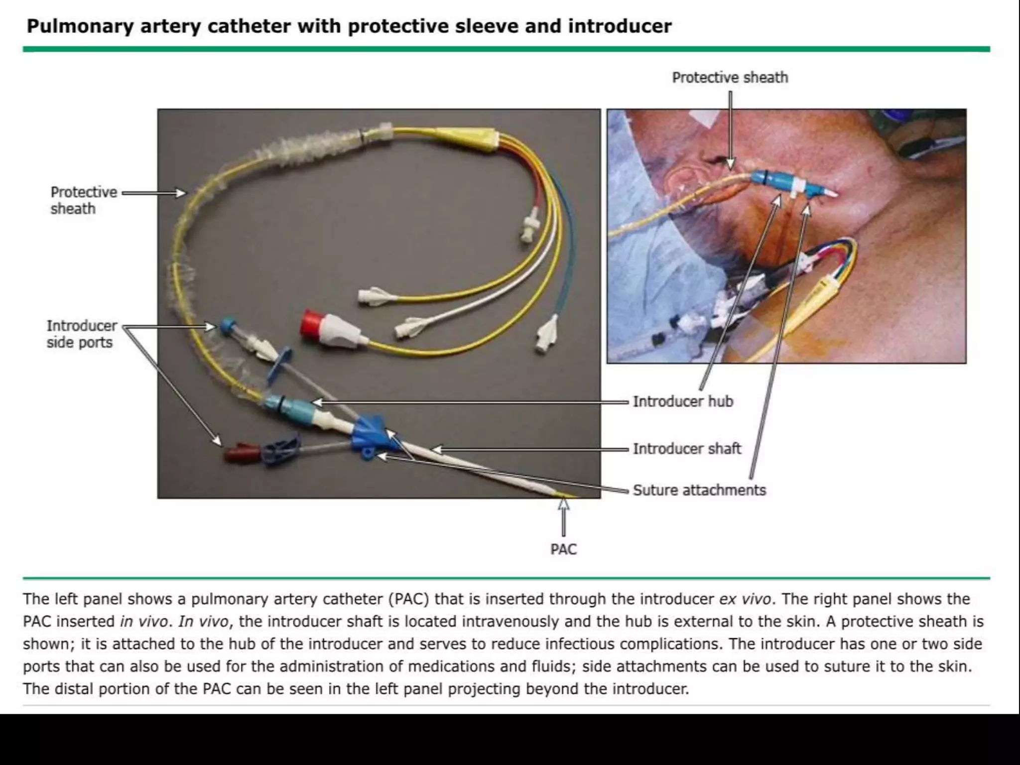
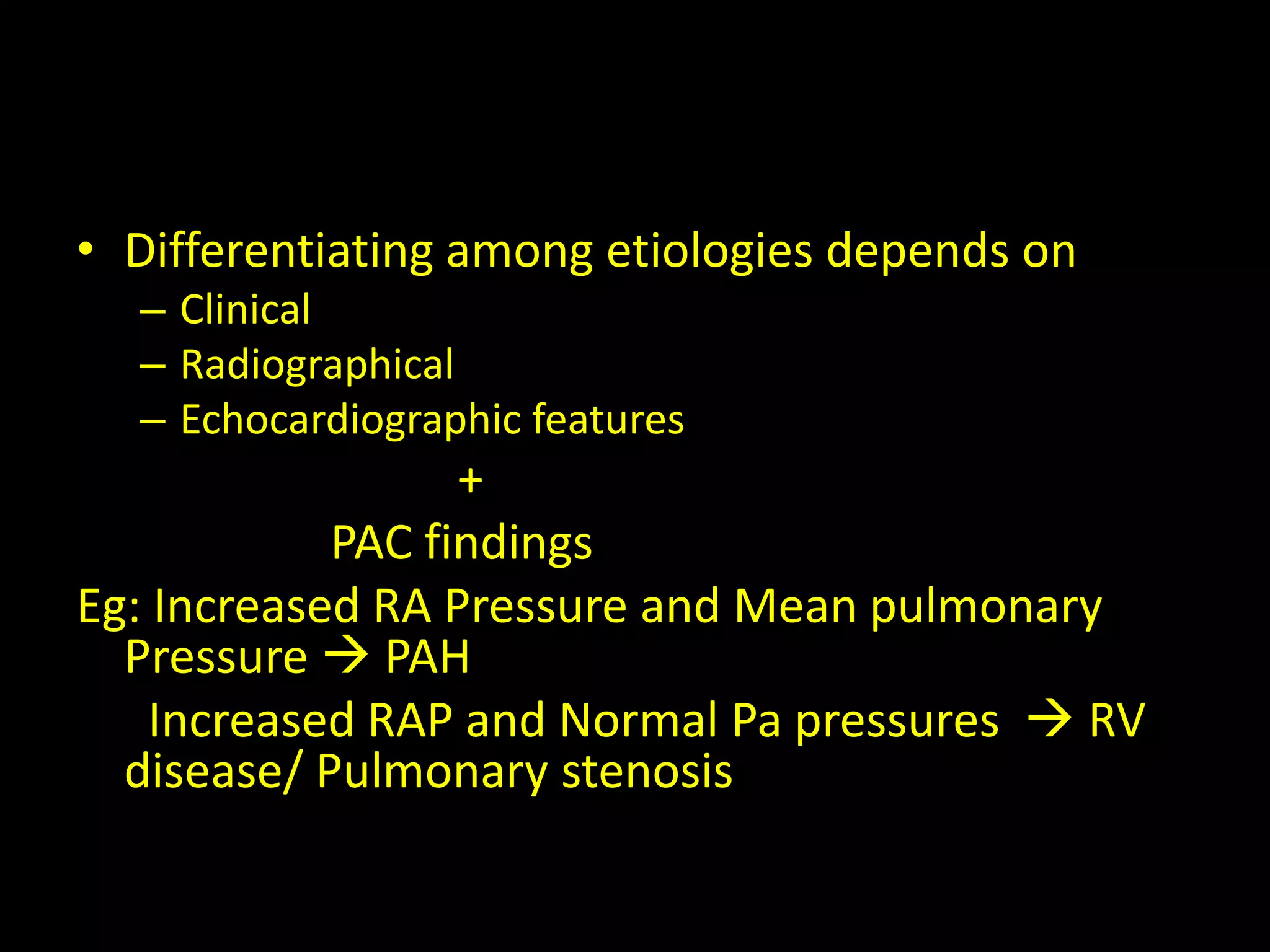
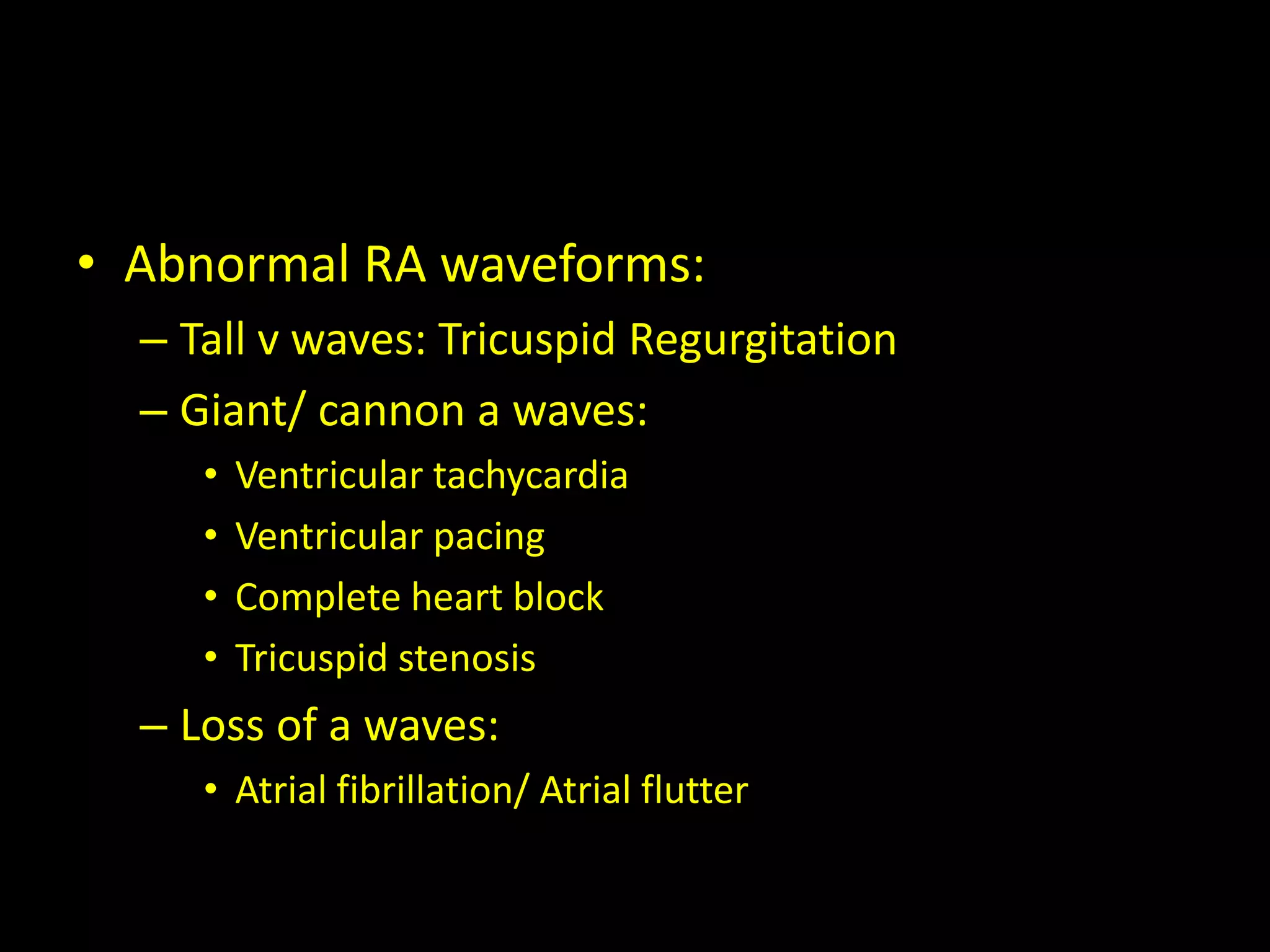
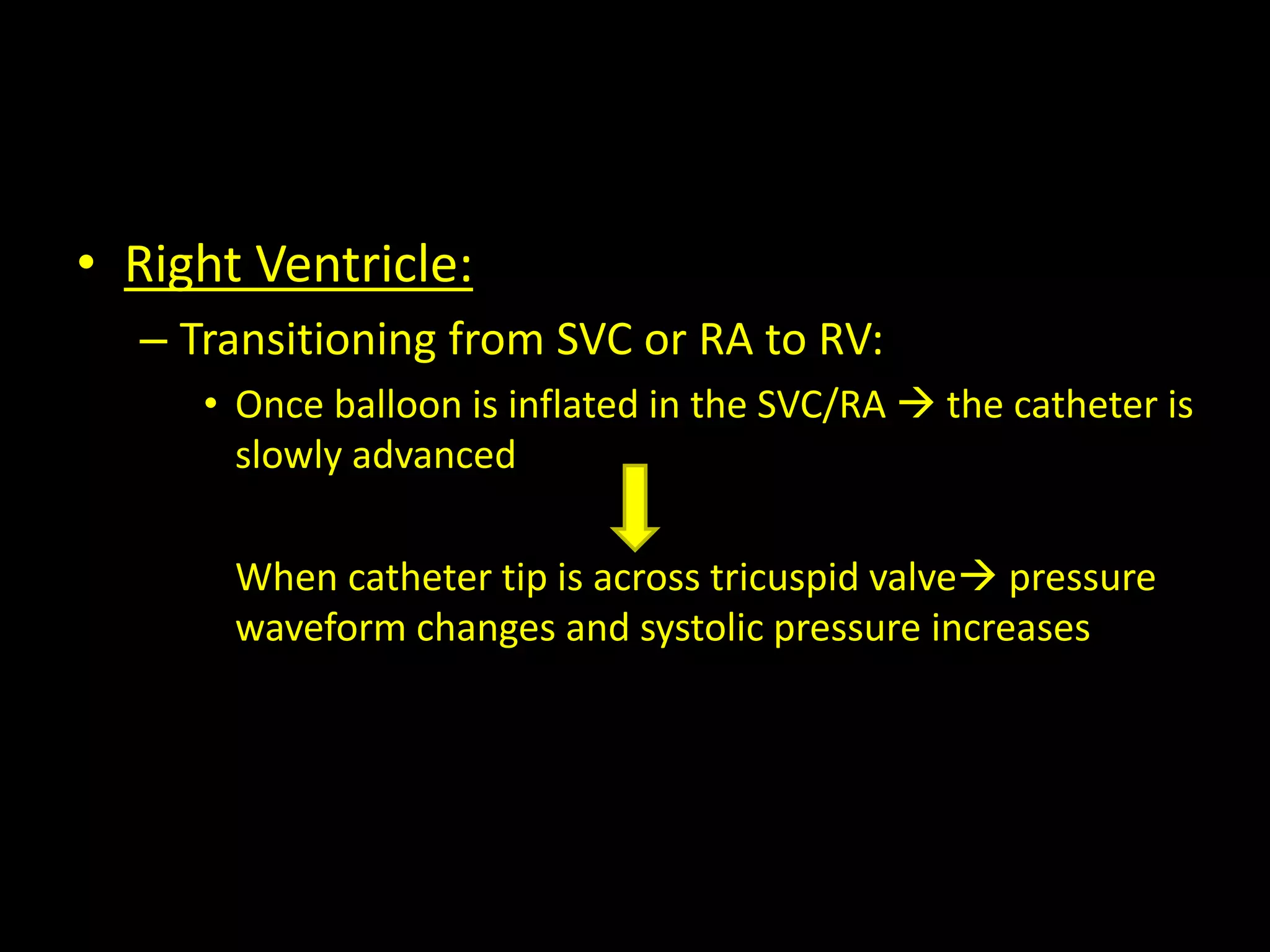
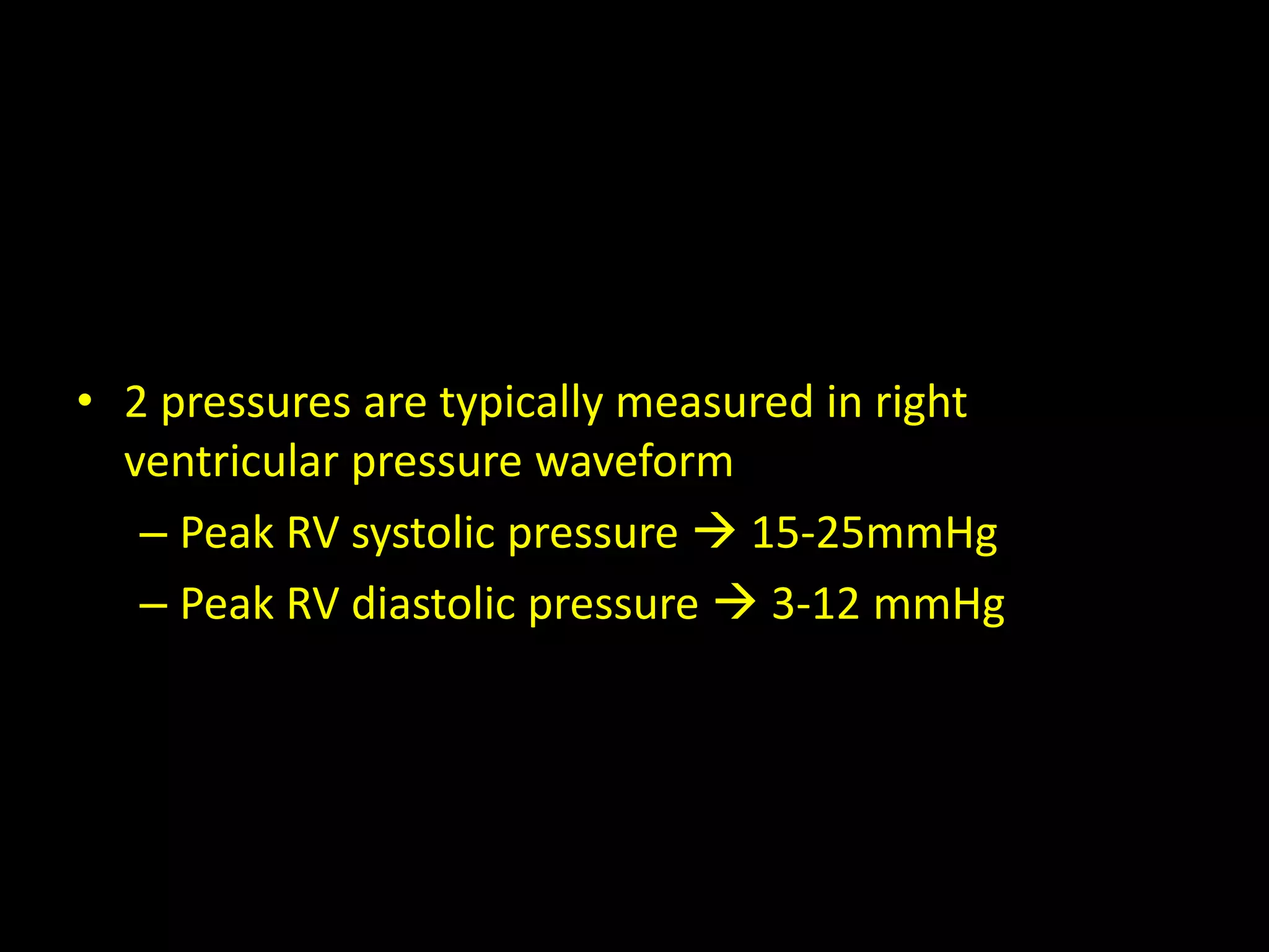
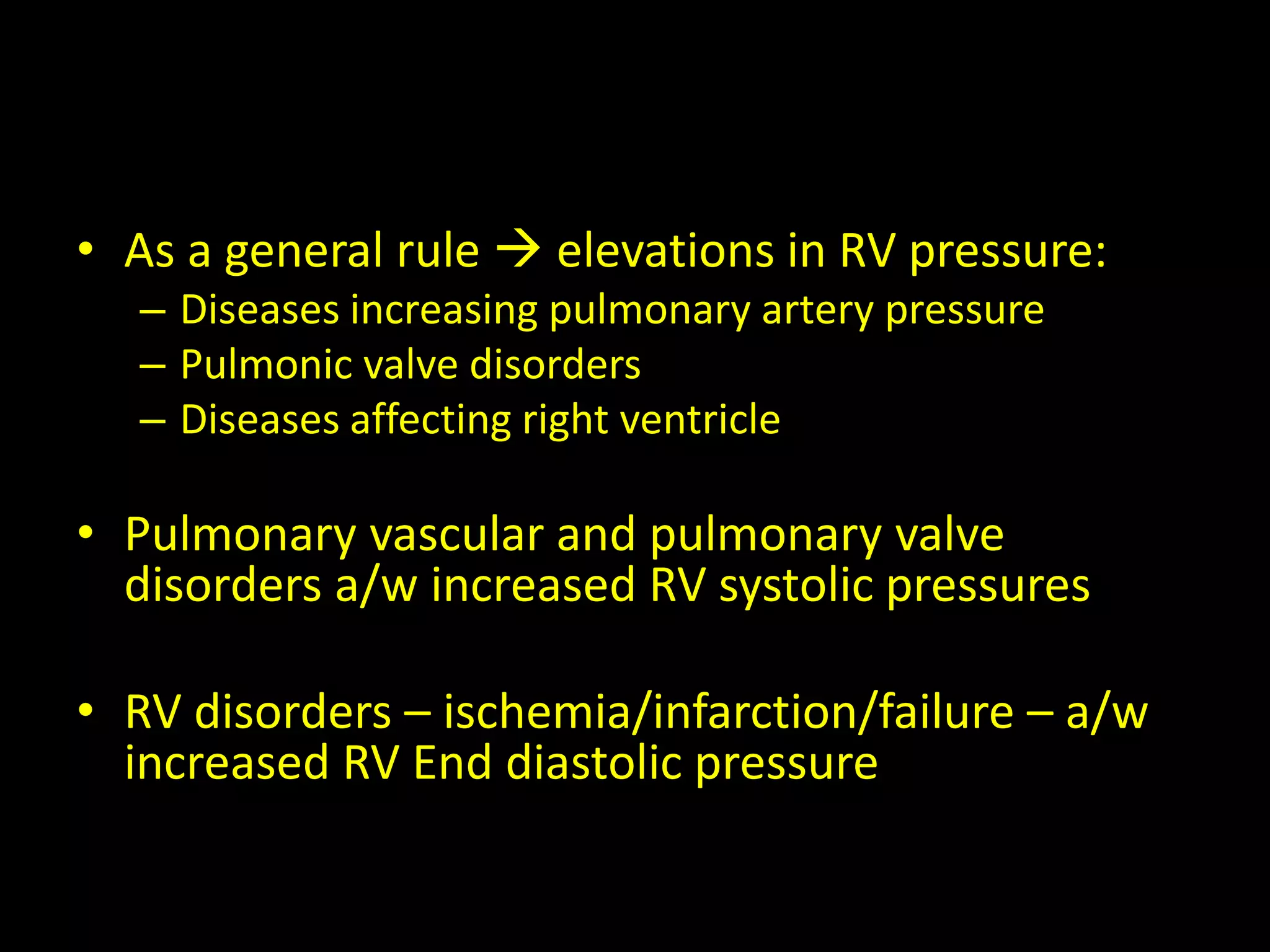
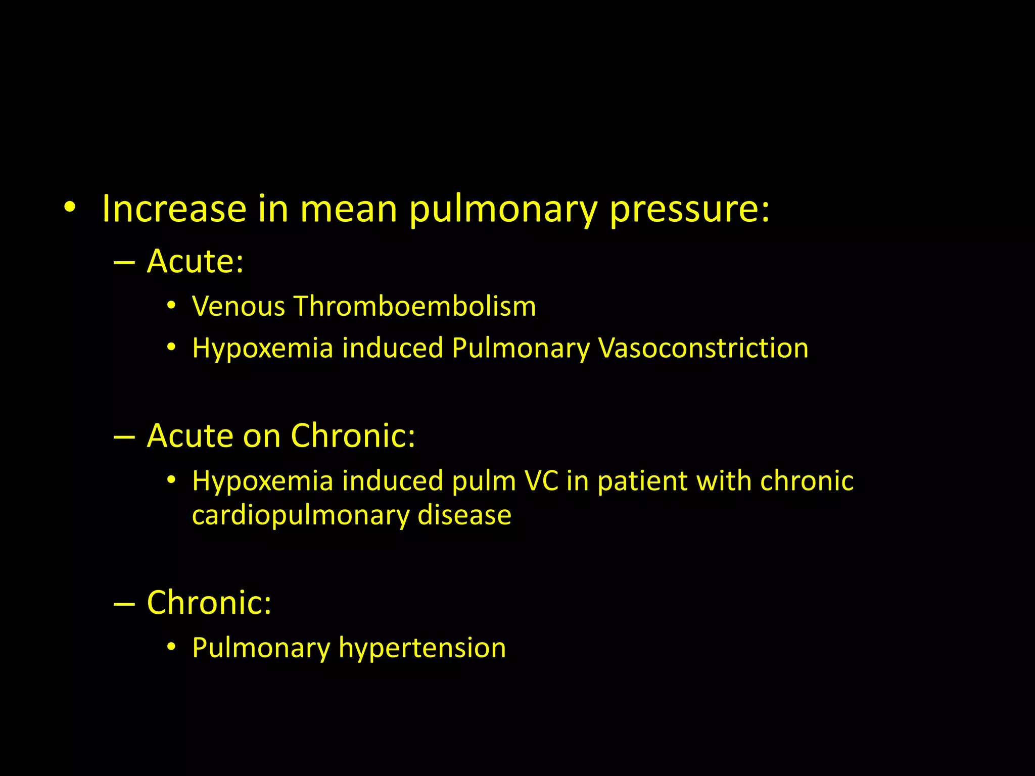
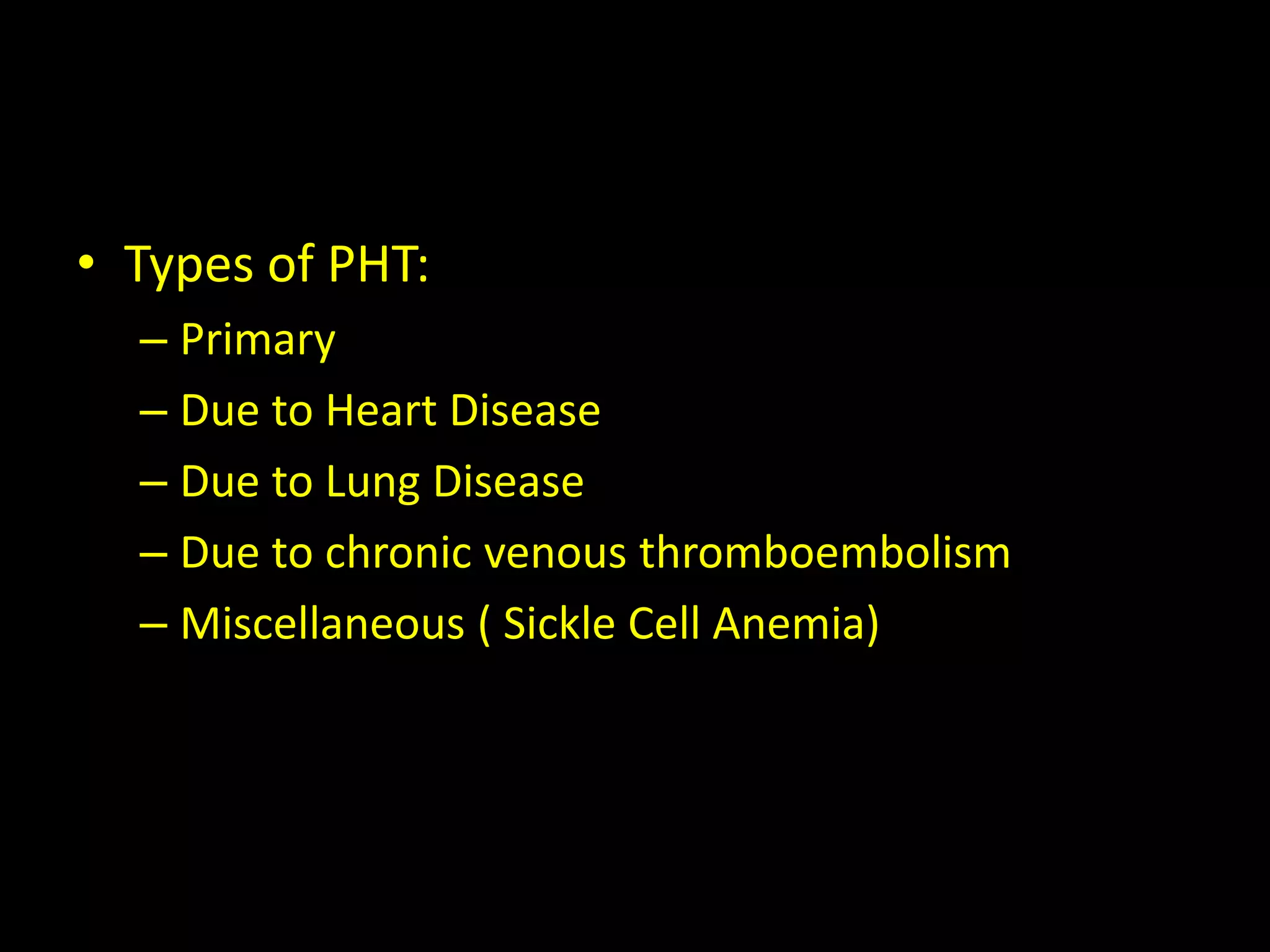
SlideShare a Scribd company logo
Pulmonary Artery Catheter
R.Srihari
• Introduction
• Physiologic Measurements
• Indications
• Contraindications
• Preparation
• Technique
• Interpretation of hemodynamic values and
waveforms
• Complications
Introduction
• Pulmonary artery catheters (also called as
Swan-Ganz catheter) are used for evaluation
of a range of condition
Although their routine use has fallen out of
favour, they are still occasionally placed for
management of critically ill patients
Physiological Measurements
• Direct measurements of the following can be
obtained from an accurately placed pulmonary
artery catheter(PAC)
– Central Venous Pressure(CVP)
– Right sided intracardiac pressures(RA/V)
– Pulmonary artery pressure(Pap)
– Pulmonary artery occlusion pressure (PAOP)
– Cardiac Output
– Mixed Venous Oxygen Saturation(SvO2)
• Indirect measurements that are possible:
– Systemic Vascular Resistance
– Pulmonary Vascular Resistance
– Cardiac Index
– Stroke volume index
– Oxygen delivery
– Oxygen uptake
Indications
• Diagnostic:
– Differentiation among causes of shock
– Differentiation between mechanisms of
pulmonary edema
– Evaluation of pulmonary hypertension
– Diagnosis of pericardial tamponade
– Diagnosis of right to left intracardiac shunts
– Unexplained dyspnea
• Therapeutic:
– Management of perioperative patients with unstable
cardiac status
– Management of complicated myocardial infarction
– Management of patients following cardiac
surgery/high risk surgery
– Management of severe preecclampsia
– Guide to pharmacologic therapy
– Burns/ Renal Failure/ Heart failure/Sepsis/
Decompensated cirrhosis
– Assess response to pulmonary hypertension specific
therapy
Contraindications
• Absolute:
• Infection at insertion
site
• Presence of RV assist
device
• Insertion during CPB
• Lack of consent
• Relative:
• Coagulopathy
• Thrombocytopenia
• Electrolyte disturbances
(K/Mg/Na/Ca)
• Severe Pulmonary HTN
Making decision to place pulmonary
artery catheter
• In critically ill or perioperative patients
decision to place a pulmonary artery catheter
should be based on patient’s hemodynamic
status or diagnosis
that cannot be answered satisfactory by
clinical or non-invasive assessment
Preparation
• Patient has to be monitored with continuous
ECG throughout the procedure, in supine
position regardless of the approach
• Aseptic precautions must be employed
• Cautions should be taken while cannulating
via IJV/ Subclavian vein
• Equipments:
– 2% chlorhexidine skin preparation solution
– Sterile gown, gloves, face shield and cap
– Sterile gauze pads
– 1% lidocaine -5 cc
– Seeker needle 23G
– Introducer needle  18G
– J-tip guidewire
– Transduction tubing
– Sterile catheter flush solution
– Sheath
– Pulonary catheter
– Sterile sleeve for catheter
– 2-0 silk suture
– Sterile dressing
Technique
1. Aseptic precautions undertaken
2. Local infiltration done
3. Check balloon integrity by inflating with 1.5ml of
air
4. Check lumens patency by flushing with saline
0.9%
5. Cover catheter with sterile sleeve provided
6. Cannulate vein with Seldinger technique
7. Place sheath
8. Pass catheter through sheath with tip curved
towards the heart
9. Once tip of catheter passed through
introducer sheath inflate balloon at level of
right ventricle
10. The progress of the catheter through right
atrium and ventricle into pulmonary artery
and wedge position can be monitored by
changes in pressure trace
11. After acquiring wedge pressure  deflate
balloon
• Important tip:
– When advancing catheter- always inflate tip
– When withdrawing catheter- always deflate
– Once in pulmonary artery - NEVER INFLATE
AGAINST RESISTANCE - RISK OF PULMONARY
ARTERY RUPTURE
Interpretation of hemodynamic values
and waveforms
• Ensuring accurate measurements:
– Zeroing and Referencing
– Correct placement
– Fast flush test
• Zeroing and Referencing:
– PAC must be appropriately zeroed and referenced
to obtain accurate readings  in supine
position/30 degrees semi-recumbent position
• Correct placement :
– By either pressure waveform/ fluoroscopic
guidance
• Rapid flush test:
Catheter waveforms and pressures
• Pressure waveforms can be obtained from
– Right atrium
– Right ventricle
– Pulmonary artery
• Right atrium:
– In presence of a a competent tricuspid valve, RA
pressure waveform reflect both
• Venous return to RA during ventricular systole
• RV End Diastolic Pressure
– Normal RA pressure: 0-7 mmHg
• Elevated RA pressure:
– Diseases of RV( infarction/ cardiomyopathy)
– Pulmonary hypertension
– Pulmonic stenosis
– Left to right shunts
– Pericardial diseases
– LV systolic failure
– Hypervolemia
• Differentiating among etiologies depends on
– Clinical
– Radiographical
– Echocardiographic features
+
PAC findings
Eg: Increased RA Pressure and Mean pulmonary
Pressure  PAH
Increased RAP and Normal Pa pressures  RV
disease/ Pulmonary stenosis
• Abnormal RA waveforms:
– Tall v waves: Tricuspid Regurgitation
– Giant/ cannon a waves:
• Ventricular tachycardia
• Ventricular pacing
• Complete heart block
• Tricuspid stenosis
– Loss of a waves:
• Atrial fibrillation/ Atrial flutter
• Right Ventricle:
– Transitioning from SVC or RA to RV:
• Once balloon is inflated in the SVC/RA  the catheter is
slowly advanced
When catheter tip is across tricuspid valve pressure
waveform changes and systolic pressure increases
• 2 pressures are typically measured in right
ventricular pressure waveform
– Peak RV systolic pressure  15-25mmHg
– Peak RV diastolic pressure  3-12 mmHg
• As a general rule  elevations in RV pressure:
– Diseases increasing pulmonary artery pressure
– Pulmonic valve disorders
– Diseases affecting right ventricle
• Pulmonary vascular and pulmonary valve
disorders a/w increased RV systolic pressures
• RV disorders – ischemia/infarction/failure – a/w
increased RV End diastolic pressure
• Pulmonary artery:
– The risk of arrhythmias is greatest while catheter
tip is in RV
Thus, catheter should be advanced from RV to PA
without delay
– When catheter tip passes pulmonary valve
Diastolic pressure increases and characteristic
dichrotic notch appears in waveform
• Normal pulmonary artery pressures:
– Systolic  15-25mmHg
– Diastolic  8-15 mmHg
– Mean  16 (10-22mmHg)
• Main components of PA tracing:
– Systolic and Diastolic pressure
– Dichrotic notch(due to closure of pulmonic valve)
• Increase in mean pulmonary pressure:
– Acute:
• Venous Thromboembolism
• Hypoxemia induced Pulmonary Vasoconstriction
– Acute on Chronic:
• Hypoxemia induced pulm VC in patient with chronic
cardiopulmonary disease
– Chronic:
• Pulmonary hypertension
• Types of PHT:
– Primary
– Due to Heart Disease
– Due to Lung Disease
– Due to chronic venous thromboembolism
– Miscellaneous ( Sickle Cell Anemia)
Pulmonary arterial occlusion pressure
• Once catheter tip has reached PA, it should be
advanced until PAOP is identified by decrease
in pressure and change in waveform
The balloon should then be deflated and PA
tracing should reappear
If PCOP tracing persists catheter should be
withdrawn with definitive PA tracing obtained
• Final position of the catheter within PA must
be such that PCOP tracing is obtained
whenever 75-100% of 1.5ml maximum
volume of balloon is insufflated
– If < 1ml of air is injected and PAOP is seen then it
is overwedged  needs to be withdrawn
– If after maximal inflation fails to result in PCOP
tracing or after 2-3 seconds delay  too proximal
– advanced with balloon inflated
• PCWP/PAOP  interprets Left atrial pressures
more importantly – LVEDP
– Best measured in
• Supine position
• At end of expiration
• Zone 3 (most dependent region)
– Normal PCWP- 6-15 mmHg ; Mean :9mmHg
• Abnormal PAOP:
– Increased LVEDP  Increased PAOP
• LV systolic HF
• LV Distolic HF
• Mitral and Aortic valve disease
• Hypertrophic cardiomyopathy
• Hypervolemia
• Large R-L shunts
• Pericardial disease
• Decreased PCWP:
– Hypovolemia
– Obstructive shock due to large pulmonary embolus
• Abnormal waveforms
– Large a waves:
• MS
• LV systolic /diastolic function
• LV volume overload
• MI
– Large v waves - MR
• Calculation of cardiac output:
– 2 methods
• Thermodilution method
• Fick’s Method
– Better measurement with Cardiac index
• Normal – 2.8- 4.2 l/min/m2
• Decreased CO:
– Systolic HF
– Diastolic HF
– MR
– Hypovolemia
– Pulmonary HT
– RVF
• Increased CO:
– Systemic A-V fistulas
– Anemia
– Beriberi
– Renal Disease
– Sepsis
• Other uses of pulmonary artery catheter:
– Detection of Left to right shunts
– Estimation of systemic and pulmonary vascular
resistance
Complications
• General:
– Immediate:
• Bleeding
• Arterial Puncture
• Air embolism
• Thoracic duct injury ( L side)
• Pneumothorax/hemothorax
– Delayed:
• Infections
• Thrombosis
• Related to insertion of PAC:
– Arrhythmias (most common- Ventricular/ RBBB)
– Misplacement
– Knotting
– Myocardial/valve/vessel rupture
• Related to maintenance and use of PAC:
– Pulmonary artery perforation
– Thromboembolism
– Infection
Thank You

More Related Content

What's hot

Pulmonary artery pressure monitoring
Pulmonary artery pressure monitoringPulmonary artery pressure monitoring
Pulmonary artery pressure monitoring
Princy Francis M
 
Cardiac output monitoring
Cardiac output monitoringCardiac output monitoring
Cardiac output monitoring
mauryaramgopal
 
Cardiac output monitoring
Cardiac output monitoringCardiac output monitoring
Cardiac output monitoringNIICS
 
Transesophageal echocardiography
Transesophageal echocardiographyTransesophageal echocardiography
Transesophageal echocardiography
Sheersha Pramanik 🇮🇳
 
CAPNOGRAPHY
CAPNOGRAPHYCAPNOGRAPHY
CAPNOGRAPHY
MAHESWARI JAIKUMAR
 
Central venous cannulation
Central venous cannulation Central venous cannulation
Central venous cannulation
ZIKRULLAH MALLICK
 
ECMO - extracorporeal membrane oxygenation
ECMO - extracorporeal membrane oxygenationECMO - extracorporeal membrane oxygenation
ECMO - extracorporeal membrane oxygenation
prapulla chandra
 
Central venous catheterization
Central venous catheterizationCentral venous catheterization
Central venous catheterization
suji kalai
 
cardiac output monitoring
cardiac output monitoringcardiac output monitoring
cardiac output monitoring
madhu chaitanya
 
Arterial line analysis
Arterial line analysisArterial line analysis
Arterial line analysis
samirelansary
 
Non invasive ventilation
Non invasive ventilationNon invasive ventilation
Non invasive ventilation
tbf413
 
Arterial lines by Dr.Tinku Joseph
Arterial lines by Dr.Tinku JosephArterial lines by Dr.Tinku Joseph
Arterial lines by Dr.Tinku Joseph
Dr.Tinku Joseph
 
Cardiac monitoring ppt
Cardiac monitoring pptCardiac monitoring ppt
Cardiac monitoring ppt
Manu Gupta
 
Hemodynamic monitoring in ICU
Hemodynamic monitoring in ICUHemodynamic monitoring in ICU
Hemodynamic monitoring in ICU
Manoj Prabhakar
 
Central venous pressure
Central venous pressureCentral venous pressure
Central venous pressure
Davis Kurian
 
PiCCO Monitor
PiCCO Monitor PiCCO Monitor
PiCCO Monitor
OsamaElazzouny
 
Double lumen tubes
Double lumen tubesDouble lumen tubes
Double lumen tubes
mauryaramgopal
 
Laryngeal mask-airway
Laryngeal mask-airwayLaryngeal mask-airway
Laryngeal mask-airway
Harith Daggupati
 
Non Invasive and Invasive Blood pressure monitoring RRT
Non Invasive and Invasive Blood pressure monitoring RRTNon Invasive and Invasive Blood pressure monitoring RRT
Non Invasive and Invasive Blood pressure monitoring RRT
Ranjith Thampi
 
Non invasive blood pressure monitoring
Non invasive blood pressure monitoringNon invasive blood pressure monitoring
Non invasive blood pressure monitoring
razishahid
 

What's hot (20)

Pulmonary artery pressure monitoring
Pulmonary artery pressure monitoringPulmonary artery pressure monitoring
Pulmonary artery pressure monitoring
 
Cardiac output monitoring
Cardiac output monitoringCardiac output monitoring
Cardiac output monitoring
 
Cardiac output monitoring
Cardiac output monitoringCardiac output monitoring
Cardiac output monitoring
 
Transesophageal echocardiography
Transesophageal echocardiographyTransesophageal echocardiography
Transesophageal echocardiography
 
CAPNOGRAPHY
CAPNOGRAPHYCAPNOGRAPHY
CAPNOGRAPHY
 
Central venous cannulation
Central venous cannulation Central venous cannulation
Central venous cannulation
 
ECMO - extracorporeal membrane oxygenation
ECMO - extracorporeal membrane oxygenationECMO - extracorporeal membrane oxygenation
ECMO - extracorporeal membrane oxygenation
 
Central venous catheterization
Central venous catheterizationCentral venous catheterization
Central venous catheterization
 
cardiac output monitoring
cardiac output monitoringcardiac output monitoring
cardiac output monitoring
 
Arterial line analysis
Arterial line analysisArterial line analysis
Arterial line analysis
 
Non invasive ventilation
Non invasive ventilationNon invasive ventilation
Non invasive ventilation
 
Arterial lines by Dr.Tinku Joseph
Arterial lines by Dr.Tinku JosephArterial lines by Dr.Tinku Joseph
Arterial lines by Dr.Tinku Joseph
 
Cardiac monitoring ppt
Cardiac monitoring pptCardiac monitoring ppt
Cardiac monitoring ppt
 
Hemodynamic monitoring in ICU
Hemodynamic monitoring in ICUHemodynamic monitoring in ICU
Hemodynamic monitoring in ICU
 
Central venous pressure
Central venous pressureCentral venous pressure
Central venous pressure
 
PiCCO Monitor
PiCCO Monitor PiCCO Monitor
PiCCO Monitor
 
Double lumen tubes
Double lumen tubesDouble lumen tubes
Double lumen tubes
 
Laryngeal mask-airway
Laryngeal mask-airwayLaryngeal mask-airway
Laryngeal mask-airway
 
Non Invasive and Invasive Blood pressure monitoring RRT
Non Invasive and Invasive Blood pressure monitoring RRTNon Invasive and Invasive Blood pressure monitoring RRT
Non Invasive and Invasive Blood pressure monitoring RRT
 
Non invasive blood pressure monitoring
Non invasive blood pressure monitoringNon invasive blood pressure monitoring
Non invasive blood pressure monitoring
 

Similar to Pulmonary artery catheter

pulmonaryarterycatheter-151008070656-lva1-app6892 (1) (1).pptx
pulmonaryarterycatheter-151008070656-lva1-app6892 (1) (1).pptxpulmonaryarterycatheter-151008070656-lva1-app6892 (1) (1).pptx
pulmonaryarterycatheter-151008070656-lva1-app6892 (1) (1).pptx
drashish05
 
PA cathrater ashish.pptx
PA cathrater ashish.pptxPA cathrater ashish.pptx
PA cathrater ashish.pptx
drashish05
 
Ccpa catheter basics07medicine
Ccpa catheter basics07medicineCcpa catheter basics07medicine
Ccpa catheter basics07medicineanjika
 
Basic hemodynamic monitoring for nurses
Basic hemodynamic monitoring for nurses Basic hemodynamic monitoring for nurses
Basic hemodynamic monitoring for nurses
Muhammad Asim Rana
 
Pulmonary artery-catheter2
Pulmonary artery-catheter2Pulmonary artery-catheter2
Pulmonary artery-catheter2Inba Aras
 
Cardiovascular monitoring Part II
Cardiovascular monitoring Part IICardiovascular monitoring Part II
Cardiovascular monitoring Part II
Siddhanta Choudhury
 
REVIEW OF HEMODYNAMIC MONITORING
REVIEW OF HEMODYNAMIC MONITORINGREVIEW OF HEMODYNAMIC MONITORING
REVIEW OF HEMODYNAMIC MONITORING
Ghaleb Almekhlafi
 
Monitoring of critically ill
Monitoring of critically illMonitoring of critically ill
Monitoring of critically ill
StudyTricksByIshu
 
Hemodynamic Monitoring .pptx
Hemodynamic Monitoring  .pptxHemodynamic Monitoring  .pptx
Hemodynamic Monitoring .pptx
anesthesia2023
 
Right heart cathterization AL-AMIN.pptx
Right heart cathterization AL-AMIN.pptxRight heart cathterization AL-AMIN.pptx
Right heart cathterization AL-AMIN.pptx
AlAmin837379
 
Hemodynamic monitoring
Hemodynamic monitoringHemodynamic monitoring
Hemodynamic monitoring
Manisha Shakya
 
Swan-Ganz-catheterisation_amit-panjwani.pdf
Swan-Ganz-catheterisation_amit-panjwani.pdfSwan-Ganz-catheterisation_amit-panjwani.pdf
Swan-Ganz-catheterisation_amit-panjwani.pdf
rambhoopal1
 
Monitoring in cardiac ananesthesia
Monitoring in cardiac ananesthesiaMonitoring in cardiac ananesthesia
Monitoring in cardiac ananesthesiaAbhishek Rathore
 
3. CVS monitoring.pptx
3. CVS monitoring.pptx3. CVS monitoring.pptx
3. CVS monitoring.pptx
FLOWERSOFPAKISTAN
 
12964620.ppt
12964620.ppt12964620.ppt
12964620.ppt
subhajit sahoo
 
ADVANCED HEMODYNAMIC MONITORING IN CRITICAL CARE MEDICINE
ADVANCED HEMODYNAMIC MONITORING IN CRITICAL CARE MEDICINEADVANCED HEMODYNAMIC MONITORING IN CRITICAL CARE MEDICINE
ADVANCED HEMODYNAMIC MONITORING IN CRITICAL CARE MEDICINE
DrAnzilAR
 
HAEMODYNAMIC MONITORING PART 1&2.pptx
HAEMODYNAMIC MONITORING PART 1&2.pptxHAEMODYNAMIC MONITORING PART 1&2.pptx
HAEMODYNAMIC MONITORING PART 1&2.pptx
ashishnair22
 
functional hemodynamic monitoring
functional hemodynamic monitoringfunctional hemodynamic monitoring
functional hemodynamic monitoring
anaesthesiaESICMCH
 
Cardiovascular monitoring final ppt.pptx
Cardiovascular monitoring final ppt.pptxCardiovascular monitoring final ppt.pptx
Cardiovascular monitoring final ppt.pptx
KLahari7
 
EVALUATION OF PULMONARY HEMODYNAMICS AND ALTERATION IN DISEASE STATES
EVALUATION OF PULMONARY HEMODYNAMICS AND ALTERATION IN DISEASE STATESEVALUATION OF PULMONARY HEMODYNAMICS AND ALTERATION IN DISEASE STATES
EVALUATION OF PULMONARY HEMODYNAMICS AND ALTERATION IN DISEASE STATES
soumenprasad
 

Similar to Pulmonary artery catheter (20)

pulmonaryarterycatheter-151008070656-lva1-app6892 (1) (1).pptx
pulmonaryarterycatheter-151008070656-lva1-app6892 (1) (1).pptxpulmonaryarterycatheter-151008070656-lva1-app6892 (1) (1).pptx
pulmonaryarterycatheter-151008070656-lva1-app6892 (1) (1).pptx
 
PA cathrater ashish.pptx
PA cathrater ashish.pptxPA cathrater ashish.pptx
PA cathrater ashish.pptx
 
Ccpa catheter basics07medicine
Ccpa catheter basics07medicineCcpa catheter basics07medicine
Ccpa catheter basics07medicine
 
Basic hemodynamic monitoring for nurses
Basic hemodynamic monitoring for nurses Basic hemodynamic monitoring for nurses
Basic hemodynamic monitoring for nurses
 
Pulmonary artery-catheter2
Pulmonary artery-catheter2Pulmonary artery-catheter2
Pulmonary artery-catheter2
 
Cardiovascular monitoring Part II
Cardiovascular monitoring Part IICardiovascular monitoring Part II
Cardiovascular monitoring Part II
 
REVIEW OF HEMODYNAMIC MONITORING
REVIEW OF HEMODYNAMIC MONITORINGREVIEW OF HEMODYNAMIC MONITORING
REVIEW OF HEMODYNAMIC MONITORING
 
Monitoring of critically ill
Monitoring of critically illMonitoring of critically ill
Monitoring of critically ill
 
Hemodynamic Monitoring .pptx
Hemodynamic Monitoring  .pptxHemodynamic Monitoring  .pptx
Hemodynamic Monitoring .pptx
 
Right heart cathterization AL-AMIN.pptx
Right heart cathterization AL-AMIN.pptxRight heart cathterization AL-AMIN.pptx
Right heart cathterization AL-AMIN.pptx
 
Hemodynamic monitoring
Hemodynamic monitoringHemodynamic monitoring
Hemodynamic monitoring
 
Swan-Ganz-catheterisation_amit-panjwani.pdf
Swan-Ganz-catheterisation_amit-panjwani.pdfSwan-Ganz-catheterisation_amit-panjwani.pdf
Swan-Ganz-catheterisation_amit-panjwani.pdf
 
Monitoring in cardiac ananesthesia
Monitoring in cardiac ananesthesiaMonitoring in cardiac ananesthesia
Monitoring in cardiac ananesthesia
 
3. CVS monitoring.pptx
3. CVS monitoring.pptx3. CVS monitoring.pptx
3. CVS monitoring.pptx
 
12964620.ppt
12964620.ppt12964620.ppt
12964620.ppt
 
ADVANCED HEMODYNAMIC MONITORING IN CRITICAL CARE MEDICINE
ADVANCED HEMODYNAMIC MONITORING IN CRITICAL CARE MEDICINEADVANCED HEMODYNAMIC MONITORING IN CRITICAL CARE MEDICINE
ADVANCED HEMODYNAMIC MONITORING IN CRITICAL CARE MEDICINE
 
HAEMODYNAMIC MONITORING PART 1&2.pptx
HAEMODYNAMIC MONITORING PART 1&2.pptxHAEMODYNAMIC MONITORING PART 1&2.pptx
HAEMODYNAMIC MONITORING PART 1&2.pptx
 
functional hemodynamic monitoring
functional hemodynamic monitoringfunctional hemodynamic monitoring
functional hemodynamic monitoring
 
Cardiovascular monitoring final ppt.pptx
Cardiovascular monitoring final ppt.pptxCardiovascular monitoring final ppt.pptx
Cardiovascular monitoring final ppt.pptx
 
EVALUATION OF PULMONARY HEMODYNAMICS AND ALTERATION IN DISEASE STATES
EVALUATION OF PULMONARY HEMODYNAMICS AND ALTERATION IN DISEASE STATESEVALUATION OF PULMONARY HEMODYNAMICS AND ALTERATION IN DISEASE STATES
EVALUATION OF PULMONARY HEMODYNAMICS AND ALTERATION IN DISEASE STATES
 

More from rajkumarsrihari

Anesthetic considerations for endocrine diseases – an overview
Anesthetic considerations for endocrine diseases – an overviewAnesthetic considerations for endocrine diseases – an overview
Anesthetic considerations for endocrine diseases – an overview
rajkumarsrihari
 
Perioperative management of a patient with diabetes mellitus
Perioperative management of a patient with diabetes mellitusPerioperative management of a patient with diabetes mellitus
Perioperative management of a patient with diabetes mellitus
rajkumarsrihari
 
Thyrotoxicosis and myxedema-Anesthetic implications
Thyrotoxicosis and myxedema-Anesthetic implicationsThyrotoxicosis and myxedema-Anesthetic implications
Thyrotoxicosis and myxedema-Anesthetic implications
rajkumarsrihari
 
Malignant hyperthermia and dantrolene sodium
Malignant hyperthermia and dantrolene sodiumMalignant hyperthermia and dantrolene sodium
Malignant hyperthermia and dantrolene sodium
rajkumarsrihari
 
Medical gas cylinders
Medical gas cylindersMedical gas cylinders
Medical gas cylinders
rajkumarsrihari
 
Rhabdomyolysis
RhabdomyolysisRhabdomyolysis
Rhabdomyolysis
rajkumarsrihari
 
Sensory and motor innervation of upper airway
Sensory and motor innervation of upper airwaySensory and motor innervation of upper airway
Sensory and motor innervation of upper airway
rajkumarsrihari
 
Blood components and preparation
Blood components and preparationBlood components and preparation
Blood components and preparation
rajkumarsrihari
 
Pain physiology
Pain physiologyPain physiology
Pain physiology
rajkumarsrihari
 
Meningomyelocoele
MeningomyelocoeleMeningomyelocoele
Meningomyelocoele
rajkumarsrihari
 
Oxygen dissociation curve
Oxygen dissociation curveOxygen dissociation curve
Oxygen dissociation curverajkumarsrihari
 

More from rajkumarsrihari (11)

Anesthetic considerations for endocrine diseases – an overview
Anesthetic considerations for endocrine diseases – an overviewAnesthetic considerations for endocrine diseases – an overview
Anesthetic considerations for endocrine diseases – an overview
 
Perioperative management of a patient with diabetes mellitus
Perioperative management of a patient with diabetes mellitusPerioperative management of a patient with diabetes mellitus
Perioperative management of a patient with diabetes mellitus
 
Thyrotoxicosis and myxedema-Anesthetic implications
Thyrotoxicosis and myxedema-Anesthetic implicationsThyrotoxicosis and myxedema-Anesthetic implications
Thyrotoxicosis and myxedema-Anesthetic implications
 
Malignant hyperthermia and dantrolene sodium
Malignant hyperthermia and dantrolene sodiumMalignant hyperthermia and dantrolene sodium
Malignant hyperthermia and dantrolene sodium
 
Medical gas cylinders
Medical gas cylindersMedical gas cylinders
Medical gas cylinders
 
Rhabdomyolysis
RhabdomyolysisRhabdomyolysis
Rhabdomyolysis
 
Sensory and motor innervation of upper airway
Sensory and motor innervation of upper airwaySensory and motor innervation of upper airway
Sensory and motor innervation of upper airway
 
Blood components and preparation
Blood components and preparationBlood components and preparation
Blood components and preparation
 
Pain physiology
Pain physiologyPain physiology
Pain physiology
 
Meningomyelocoele
MeningomyelocoeleMeningomyelocoele
Meningomyelocoele
 
Oxygen dissociation curve
Oxygen dissociation curveOxygen dissociation curve
Oxygen dissociation curve
 

Recently uploaded

Tom Selleck Health: A Comprehensive Look at the Iconic Actor’s Wellness Journey
Tom Selleck Health: A Comprehensive Look at the Iconic Actor’s Wellness JourneyTom Selleck Health: A Comprehensive Look at the Iconic Actor’s Wellness Journey
Tom Selleck Health: A Comprehensive Look at the Iconic Actor’s Wellness Journey
greendigital
 
ARTIFICIAL INTELLIGENCE IN HEALTHCARE.pdf
ARTIFICIAL INTELLIGENCE IN  HEALTHCARE.pdfARTIFICIAL INTELLIGENCE IN  HEALTHCARE.pdf
ARTIFICIAL INTELLIGENCE IN HEALTHCARE.pdf
Anujkumaranit
 
BRACHYTHERAPY OVERVIEW AND APPLICATORS
BRACHYTHERAPY OVERVIEW  AND  APPLICATORSBRACHYTHERAPY OVERVIEW  AND  APPLICATORS
BRACHYTHERAPY OVERVIEW AND APPLICATORS
Krishan Murari
 
Ophthalmology Clinical Tests for OSCE exam
Ophthalmology Clinical Tests for OSCE examOphthalmology Clinical Tests for OSCE exam
Ophthalmology Clinical Tests for OSCE exam
KafrELShiekh University
 
ACUTE SCROTUM.....pdf. ACUTE SCROTAL CONDITIOND
ACUTE SCROTUM.....pdf. ACUTE SCROTAL CONDITIONDACUTE SCROTUM.....pdf. ACUTE SCROTAL CONDITIOND
ACUTE SCROTUM.....pdf. ACUTE SCROTAL CONDITIOND
DR SETH JOTHAM
 
BENIGN PROSTATIC HYPERPLASIA.BPH. BPHpdf
BENIGN PROSTATIC HYPERPLASIA.BPH. BPHpdfBENIGN PROSTATIC HYPERPLASIA.BPH. BPHpdf
BENIGN PROSTATIC HYPERPLASIA.BPH. BPHpdf
DR SETH JOTHAM
 
basicmodesofventilation2022-220313203758.pdf
basicmodesofventilation2022-220313203758.pdfbasicmodesofventilation2022-220313203758.pdf
basicmodesofventilation2022-220313203758.pdf
aljamhori teaching hospital
 
ANATOMY AND PHYSIOLOGY OF URINARY SYSTEM.pptx
ANATOMY AND PHYSIOLOGY OF URINARY SYSTEM.pptxANATOMY AND PHYSIOLOGY OF URINARY SYSTEM.pptx
ANATOMY AND PHYSIOLOGY OF URINARY SYSTEM.pptx
Swetaba Besh
 
Evaluation of antidepressant activity of clitoris ternatea in animals
Evaluation of antidepressant activity of clitoris ternatea in animalsEvaluation of antidepressant activity of clitoris ternatea in animals
Evaluation of antidepressant activity of clitoris ternatea in animals
Shweta
 
Non-respiratory Functions of the Lungs.pdf
Non-respiratory Functions of the Lungs.pdfNon-respiratory Functions of the Lungs.pdf
Non-respiratory Functions of the Lungs.pdf
MedicoseAcademics
 
Pharynx and Clinical Correlations BY Dr.Rabia Inam Gandapore.pptx
Pharynx and Clinical Correlations BY Dr.Rabia Inam Gandapore.pptxPharynx and Clinical Correlations BY Dr.Rabia Inam Gandapore.pptx
Pharynx and Clinical Correlations BY Dr.Rabia Inam Gandapore.pptx
Dr. Rabia Inam Gandapore
 
Triangles of Neck and Clinical Correlation by Dr. RIG.pptx
Triangles of Neck and Clinical Correlation by Dr. RIG.pptxTriangles of Neck and Clinical Correlation by Dr. RIG.pptx
Triangles of Neck and Clinical Correlation by Dr. RIG.pptx
Dr. Rabia Inam Gandapore
 
Surat @ℂall @Girls ꧁❤8527049040❤꧂@ℂall @Girls Service Vip Top Model Safe
Surat @ℂall @Girls ꧁❤8527049040❤꧂@ℂall @Girls Service Vip Top Model SafeSurat @ℂall @Girls ꧁❤8527049040❤꧂@ℂall @Girls Service Vip Top Model Safe
Surat @ℂall @Girls ꧁❤8527049040❤꧂@ℂall @Girls Service Vip Top Model Safe
Savita Shen $i11
 
How to Give Better Lectures: Some Tips for Doctors
How to Give Better Lectures: Some Tips for DoctorsHow to Give Better Lectures: Some Tips for Doctors
How to Give Better Lectures: Some Tips for Doctors
LanceCatedral
 
Prix Galien International 2024 Forum Program
Prix Galien International 2024 Forum ProgramPrix Galien International 2024 Forum Program
Prix Galien International 2024 Forum Program
Levi Shapiro
 
Novas diretrizes da OMS para os cuidados perinatais de mais qualidade
Novas diretrizes da OMS para os cuidados perinatais de mais qualidadeNovas diretrizes da OMS para os cuidados perinatais de mais qualidade
Novas diretrizes da OMS para os cuidados perinatais de mais qualidade
Prof. Marcus Renato de Carvalho
 
The Normal Electrocardiogram - Part I of II
The Normal Electrocardiogram - Part I of IIThe Normal Electrocardiogram - Part I of II
The Normal Electrocardiogram - Part I of II
MedicoseAcademics
 
heat stroke and heat exhaustion in children
heat stroke and heat exhaustion in childrenheat stroke and heat exhaustion in children
heat stroke and heat exhaustion in children
SumeraAhmad5
 
Cervical & Brachial Plexus By Dr. RIG.pptx
Cervical & Brachial Plexus By Dr. RIG.pptxCervical & Brachial Plexus By Dr. RIG.pptx
Cervical & Brachial Plexus By Dr. RIG.pptx
Dr. Rabia Inam Gandapore
 
POST OPERATIVE OLIGURIA and its management
POST OPERATIVE OLIGURIA and its managementPOST OPERATIVE OLIGURIA and its management
POST OPERATIVE OLIGURIA and its management
touseefaziz1
 

Recently uploaded (20)

Tom Selleck Health: A Comprehensive Look at the Iconic Actor’s Wellness Journey
Tom Selleck Health: A Comprehensive Look at the Iconic Actor’s Wellness JourneyTom Selleck Health: A Comprehensive Look at the Iconic Actor’s Wellness Journey
Tom Selleck Health: A Comprehensive Look at the Iconic Actor’s Wellness Journey
 
ARTIFICIAL INTELLIGENCE IN HEALTHCARE.pdf
ARTIFICIAL INTELLIGENCE IN  HEALTHCARE.pdfARTIFICIAL INTELLIGENCE IN  HEALTHCARE.pdf
ARTIFICIAL INTELLIGENCE IN HEALTHCARE.pdf
 
BRACHYTHERAPY OVERVIEW AND APPLICATORS
BRACHYTHERAPY OVERVIEW  AND  APPLICATORSBRACHYTHERAPY OVERVIEW  AND  APPLICATORS
BRACHYTHERAPY OVERVIEW AND APPLICATORS
 
Ophthalmology Clinical Tests for OSCE exam
Ophthalmology Clinical Tests for OSCE examOphthalmology Clinical Tests for OSCE exam
Ophthalmology Clinical Tests for OSCE exam
 
ACUTE SCROTUM.....pdf. ACUTE SCROTAL CONDITIOND
ACUTE SCROTUM.....pdf. ACUTE SCROTAL CONDITIONDACUTE SCROTUM.....pdf. ACUTE SCROTAL CONDITIOND
ACUTE SCROTUM.....pdf. ACUTE SCROTAL CONDITIOND
 
BENIGN PROSTATIC HYPERPLASIA.BPH. BPHpdf
BENIGN PROSTATIC HYPERPLASIA.BPH. BPHpdfBENIGN PROSTATIC HYPERPLASIA.BPH. BPHpdf
BENIGN PROSTATIC HYPERPLASIA.BPH. BPHpdf
 
basicmodesofventilation2022-220313203758.pdf
basicmodesofventilation2022-220313203758.pdfbasicmodesofventilation2022-220313203758.pdf
basicmodesofventilation2022-220313203758.pdf
 
ANATOMY AND PHYSIOLOGY OF URINARY SYSTEM.pptx
ANATOMY AND PHYSIOLOGY OF URINARY SYSTEM.pptxANATOMY AND PHYSIOLOGY OF URINARY SYSTEM.pptx
ANATOMY AND PHYSIOLOGY OF URINARY SYSTEM.pptx
 
Evaluation of antidepressant activity of clitoris ternatea in animals
Evaluation of antidepressant activity of clitoris ternatea in animalsEvaluation of antidepressant activity of clitoris ternatea in animals
Evaluation of antidepressant activity of clitoris ternatea in animals
 
Non-respiratory Functions of the Lungs.pdf
Non-respiratory Functions of the Lungs.pdfNon-respiratory Functions of the Lungs.pdf
Non-respiratory Functions of the Lungs.pdf
 
Pharynx and Clinical Correlations BY Dr.Rabia Inam Gandapore.pptx
Pharynx and Clinical Correlations BY Dr.Rabia Inam Gandapore.pptxPharynx and Clinical Correlations BY Dr.Rabia Inam Gandapore.pptx
Pharynx and Clinical Correlations BY Dr.Rabia Inam Gandapore.pptx
 
Triangles of Neck and Clinical Correlation by Dr. RIG.pptx
Triangles of Neck and Clinical Correlation by Dr. RIG.pptxTriangles of Neck and Clinical Correlation by Dr. RIG.pptx
Triangles of Neck and Clinical Correlation by Dr. RIG.pptx
 
Surat @ℂall @Girls ꧁❤8527049040❤꧂@ℂall @Girls Service Vip Top Model Safe
Surat @ℂall @Girls ꧁❤8527049040❤꧂@ℂall @Girls Service Vip Top Model SafeSurat @ℂall @Girls ꧁❤8527049040❤꧂@ℂall @Girls Service Vip Top Model Safe
Surat @ℂall @Girls ꧁❤8527049040❤꧂@ℂall @Girls Service Vip Top Model Safe
 
How to Give Better Lectures: Some Tips for Doctors
How to Give Better Lectures: Some Tips for DoctorsHow to Give Better Lectures: Some Tips for Doctors
How to Give Better Lectures: Some Tips for Doctors
 
Prix Galien International 2024 Forum Program
Prix Galien International 2024 Forum ProgramPrix Galien International 2024 Forum Program
Prix Galien International 2024 Forum Program
 
Novas diretrizes da OMS para os cuidados perinatais de mais qualidade
Novas diretrizes da OMS para os cuidados perinatais de mais qualidadeNovas diretrizes da OMS para os cuidados perinatais de mais qualidade
Novas diretrizes da OMS para os cuidados perinatais de mais qualidade
 
The Normal Electrocardiogram - Part I of II
The Normal Electrocardiogram - Part I of IIThe Normal Electrocardiogram - Part I of II
The Normal Electrocardiogram - Part I of II
 
heat stroke and heat exhaustion in children
heat stroke and heat exhaustion in childrenheat stroke and heat exhaustion in children
heat stroke and heat exhaustion in children
 
Cervical & Brachial Plexus By Dr. RIG.pptx
Cervical & Brachial Plexus By Dr. RIG.pptxCervical & Brachial Plexus By Dr. RIG.pptx
Cervical & Brachial Plexus By Dr. RIG.pptx
 
POST OPERATIVE OLIGURIA and its management
POST OPERATIVE OLIGURIA and its managementPOST OPERATIVE OLIGURIA and its management
POST OPERATIVE OLIGURIA and its management
 

Pulmonary artery catheter

  • 2. • Introduction • Physiologic Measurements • Indications • Contraindications • Preparation • Technique • Interpretation of hemodynamic values and waveforms • Complications
  • 3. Introduction • Pulmonary artery catheters (also called as Swan-Ganz catheter) are used for evaluation of a range of condition Although their routine use has fallen out of favour, they are still occasionally placed for management of critically ill patients
  • 4. Physiological Measurements • Direct measurements of the following can be obtained from an accurately placed pulmonary artery catheter(PAC) – Central Venous Pressure(CVP) – Right sided intracardiac pressures(RA/V) – Pulmonary artery pressure(Pap) – Pulmonary artery occlusion pressure (PAOP) – Cardiac Output – Mixed Venous Oxygen Saturation(SvO2)
  • 5. • Indirect measurements that are possible: – Systemic Vascular Resistance – Pulmonary Vascular Resistance – Cardiac Index – Stroke volume index – Oxygen delivery – Oxygen uptake
  • 6. Indications • Diagnostic: – Differentiation among causes of shock – Differentiation between mechanisms of pulmonary edema – Evaluation of pulmonary hypertension – Diagnosis of pericardial tamponade – Diagnosis of right to left intracardiac shunts – Unexplained dyspnea
  • 7. • Therapeutic: – Management of perioperative patients with unstable cardiac status – Management of complicated myocardial infarction – Management of patients following cardiac surgery/high risk surgery – Management of severe preecclampsia – Guide to pharmacologic therapy – Burns/ Renal Failure/ Heart failure/Sepsis/ Decompensated cirrhosis – Assess response to pulmonary hypertension specific therapy
  • 8. Contraindications • Absolute: • Infection at insertion site • Presence of RV assist device • Insertion during CPB • Lack of consent • Relative: • Coagulopathy • Thrombocytopenia • Electrolyte disturbances (K/Mg/Na/Ca) • Severe Pulmonary HTN
  • 9. Making decision to place pulmonary artery catheter • In critically ill or perioperative patients decision to place a pulmonary artery catheter should be based on patient’s hemodynamic status or diagnosis that cannot be answered satisfactory by clinical or non-invasive assessment
  • 10. Preparation • Patient has to be monitored with continuous ECG throughout the procedure, in supine position regardless of the approach • Aseptic precautions must be employed • Cautions should be taken while cannulating via IJV/ Subclavian vein
  • 11. • Equipments: – 2% chlorhexidine skin preparation solution – Sterile gown, gloves, face shield and cap – Sterile gauze pads – 1% lidocaine -5 cc – Seeker needle 23G – Introducer needle  18G – J-tip guidewire – Transduction tubing – Sterile catheter flush solution – Sheath – Pulonary catheter – Sterile sleeve for catheter – 2-0 silk suture – Sterile dressing
  • 12.
  • 13.
  • 14. Technique 1. Aseptic precautions undertaken 2. Local infiltration done 3. Check balloon integrity by inflating with 1.5ml of air 4. Check lumens patency by flushing with saline 0.9% 5. Cover catheter with sterile sleeve provided 6. Cannulate vein with Seldinger technique 7. Place sheath 8. Pass catheter through sheath with tip curved towards the heart
  • 15. 9. Once tip of catheter passed through introducer sheath inflate balloon at level of right ventricle 10. The progress of the catheter through right atrium and ventricle into pulmonary artery and wedge position can be monitored by changes in pressure trace 11. After acquiring wedge pressure  deflate balloon
  • 16.
  • 17.
  • 18. • Important tip: – When advancing catheter- always inflate tip – When withdrawing catheter- always deflate – Once in pulmonary artery - NEVER INFLATE AGAINST RESISTANCE - RISK OF PULMONARY ARTERY RUPTURE
  • 19. Interpretation of hemodynamic values and waveforms • Ensuring accurate measurements: – Zeroing and Referencing – Correct placement – Fast flush test
  • 20. • Zeroing and Referencing: – PAC must be appropriately zeroed and referenced to obtain accurate readings  in supine position/30 degrees semi-recumbent position • Correct placement : – By either pressure waveform/ fluoroscopic guidance
  • 22. Catheter waveforms and pressures • Pressure waveforms can be obtained from – Right atrium – Right ventricle – Pulmonary artery
  • 23. • Right atrium: – In presence of a a competent tricuspid valve, RA pressure waveform reflect both • Venous return to RA during ventricular systole • RV End Diastolic Pressure – Normal RA pressure: 0-7 mmHg
  • 24.
  • 25. • Elevated RA pressure: – Diseases of RV( infarction/ cardiomyopathy) – Pulmonary hypertension – Pulmonic stenosis – Left to right shunts – Pericardial diseases – LV systolic failure – Hypervolemia
  • 26. • Differentiating among etiologies depends on – Clinical – Radiographical – Echocardiographic features + PAC findings Eg: Increased RA Pressure and Mean pulmonary Pressure  PAH Increased RAP and Normal Pa pressures  RV disease/ Pulmonary stenosis
  • 27. • Abnormal RA waveforms: – Tall v waves: Tricuspid Regurgitation – Giant/ cannon a waves: • Ventricular tachycardia • Ventricular pacing • Complete heart block • Tricuspid stenosis – Loss of a waves: • Atrial fibrillation/ Atrial flutter
  • 28.
  • 29.
  • 30. • Right Ventricle: – Transitioning from SVC or RA to RV: • Once balloon is inflated in the SVC/RA  the catheter is slowly advanced When catheter tip is across tricuspid valve pressure waveform changes and systolic pressure increases
  • 31. • 2 pressures are typically measured in right ventricular pressure waveform – Peak RV systolic pressure  15-25mmHg – Peak RV diastolic pressure  3-12 mmHg
  • 32.
  • 33. • As a general rule  elevations in RV pressure: – Diseases increasing pulmonary artery pressure – Pulmonic valve disorders – Diseases affecting right ventricle • Pulmonary vascular and pulmonary valve disorders a/w increased RV systolic pressures • RV disorders – ischemia/infarction/failure – a/w increased RV End diastolic pressure
  • 34. • Pulmonary artery: – The risk of arrhythmias is greatest while catheter tip is in RV Thus, catheter should be advanced from RV to PA without delay – When catheter tip passes pulmonary valve Diastolic pressure increases and characteristic dichrotic notch appears in waveform
  • 35. • Normal pulmonary artery pressures: – Systolic  15-25mmHg – Diastolic  8-15 mmHg – Mean  16 (10-22mmHg) • Main components of PA tracing: – Systolic and Diastolic pressure – Dichrotic notch(due to closure of pulmonic valve)
  • 36.
  • 37. • Increase in mean pulmonary pressure: – Acute: • Venous Thromboembolism • Hypoxemia induced Pulmonary Vasoconstriction – Acute on Chronic: • Hypoxemia induced pulm VC in patient with chronic cardiopulmonary disease – Chronic: • Pulmonary hypertension
  • 38. • Types of PHT: – Primary – Due to Heart Disease – Due to Lung Disease – Due to chronic venous thromboembolism – Miscellaneous ( Sickle Cell Anemia)
  • 39. Pulmonary arterial occlusion pressure • Once catheter tip has reached PA, it should be advanced until PAOP is identified by decrease in pressure and change in waveform The balloon should then be deflated and PA tracing should reappear If PCOP tracing persists catheter should be withdrawn with definitive PA tracing obtained
  • 40. • Final position of the catheter within PA must be such that PCOP tracing is obtained whenever 75-100% of 1.5ml maximum volume of balloon is insufflated – If < 1ml of air is injected and PAOP is seen then it is overwedged  needs to be withdrawn – If after maximal inflation fails to result in PCOP tracing or after 2-3 seconds delay  too proximal – advanced with balloon inflated
  • 41. • PCWP/PAOP  interprets Left atrial pressures more importantly – LVEDP – Best measured in • Supine position • At end of expiration • Zone 3 (most dependent region) – Normal PCWP- 6-15 mmHg ; Mean :9mmHg
  • 42.
  • 43.
  • 44. • Abnormal PAOP: – Increased LVEDP  Increased PAOP • LV systolic HF • LV Distolic HF • Mitral and Aortic valve disease • Hypertrophic cardiomyopathy • Hypervolemia • Large R-L shunts • Pericardial disease
  • 45. • Decreased PCWP: – Hypovolemia – Obstructive shock due to large pulmonary embolus • Abnormal waveforms – Large a waves: • MS • LV systolic /diastolic function • LV volume overload • MI – Large v waves - MR
  • 46. • Calculation of cardiac output: – 2 methods • Thermodilution method • Fick’s Method – Better measurement with Cardiac index • Normal – 2.8- 4.2 l/min/m2
  • 47. • Decreased CO: – Systolic HF – Diastolic HF – MR – Hypovolemia – Pulmonary HT – RVF • Increased CO: – Systemic A-V fistulas – Anemia – Beriberi – Renal Disease – Sepsis
  • 48. • Other uses of pulmonary artery catheter: – Detection of Left to right shunts – Estimation of systemic and pulmonary vascular resistance
  • 49. Complications • General: – Immediate: • Bleeding • Arterial Puncture • Air embolism • Thoracic duct injury ( L side) • Pneumothorax/hemothorax – Delayed: • Infections • Thrombosis
  • 50. • Related to insertion of PAC: – Arrhythmias (most common- Ventricular/ RBBB) – Misplacement – Knotting – Myocardial/valve/vessel rupture • Related to maintenance and use of PAC: – Pulmonary artery perforation – Thromboembolism – Infection