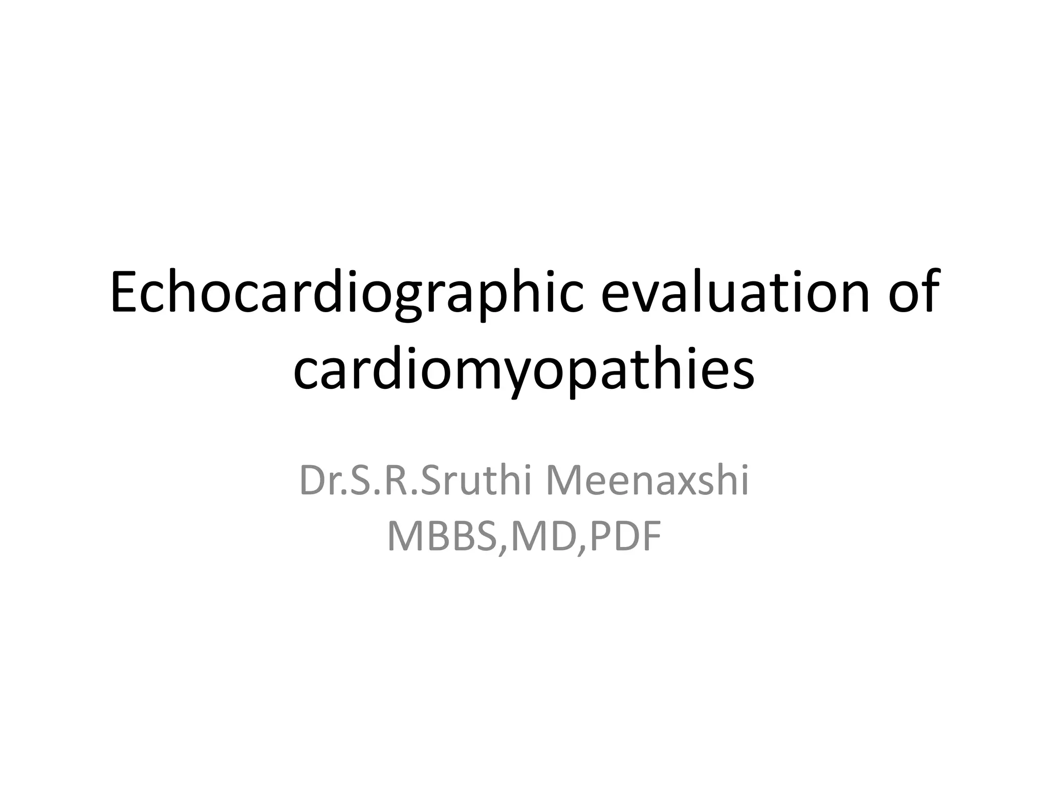
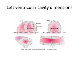
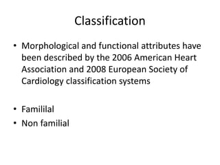
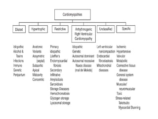
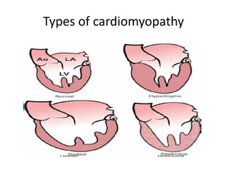
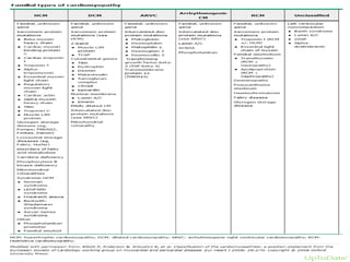
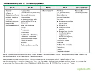
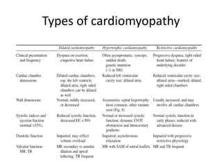
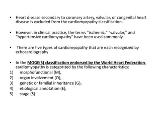
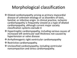
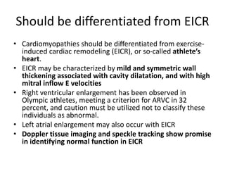
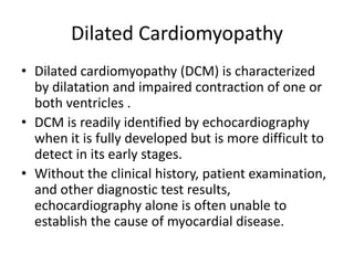
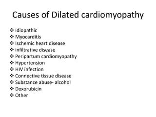
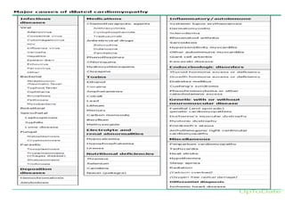
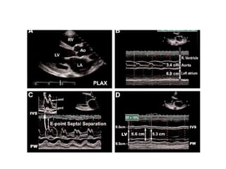
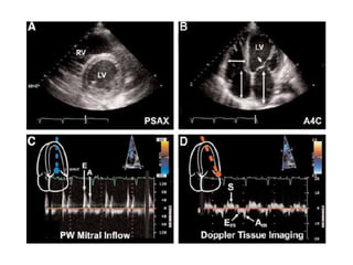
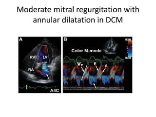
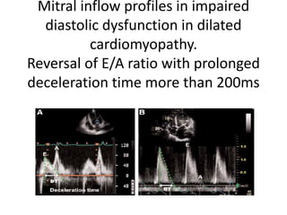
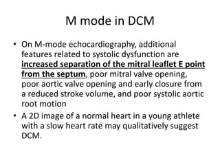
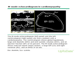
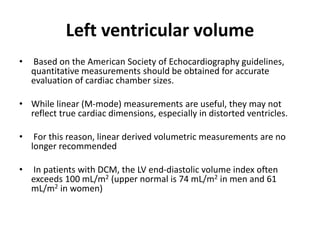
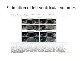
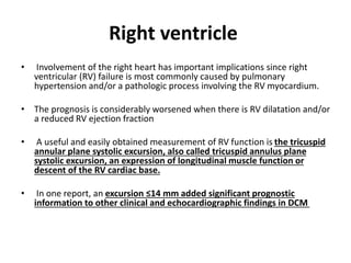
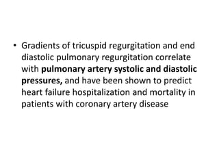
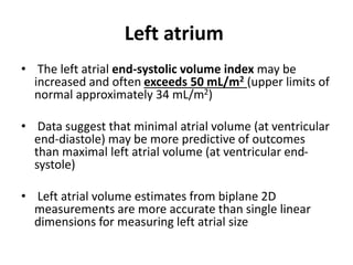
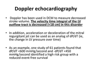
Echocardiographic evaluation of
cardiomyopathies
Dr.S.R.Sruthi Meenaxshi
MBBS,MD,PDF
Left ventricular cavity dimensions
• Cardiomyopathy is defined as a disease of
heart muscle.
• Cardiomyopathies include a variety of
myocardial disorders that manifest with
various structural and functional phenotypes
with familial and nonfamilial types.
• This topic will review the echocardiographic
features of the various types of
cardiomyopathy.
Classification
• Morphological and functional attributes have
been described by the 2006 American Heart
Association and 2008 European Society of
Cardiology classification systems
• Famililal
• Non familial
Types of cardiomyopathy
• Heart disease secondary to coronary artery, valvular, or congenital heart
disease is excluded from the cardiomyopathy classification.
• However, in clinical practice, the terms "ischemic," "valvular," and
"hypertensive cardiomyopathy" have been used commonly.
• There are five types of cardiomyopathy that are each recognized by
echocardiography
• In the MOGE(S) classification endorsed by the World Heart Federation,
cardiomyopathy is categorized by the following characteristics:
1) morphofunctional (M),
2) organ involvement (O),
3) genetic or familial inheritance (G),
4) etiological annotation (E),
5) stage (S)
Morphological classification
❖ Dilated cardiomyopathy arising as primary myocardial
disease of unknown etiology or as disorders of toxic,
familial, or infective origin. In clinical practice, ischemic
cardiomyopathy is frequently viewed as a type of dilated
cardiomyopathy, although current major society
classification systems exclude it
❖ Hypertrophic cardiomyopathy, including various causes of
increased left ventricular wall thickness not caused by
hypertension or valve disease
❖ Arrhythmogenic right ventricular cardiomyopathy
❖ Restrictive cardiomyopathy.
❖ Unclassified cardiomyopathy, including ventricular
noncompaction and stress cardiomyopathy.
Should be differentiated from EICR
• Cardiomyopathies should be differentiated from exercise-
induced cardiac remodeling (EICR), or so-called athlete’s
heart.
• EICR may be characterized by mild and symmetric wall
thickening associated with cavity dilatation, and with high
mitral inflow E velocities
• Right ventricular enlargement has been observed in
Olympic athletes, meeting a criterion for ARVC in 32
percent, and caution must be utilized not to classify these
individuals as abnormal.
• Left atrial enlargement may also occur with EICR
• Doppler tissue imaging and speckle tracking show promise
in identifying normal function in EICR
Dilated Cardiomyopathy
• Dilated cardiomyopathy (DCM) is characterized
by dilatation and impaired contraction of one or
both ventricles .
• DCM is readily identified by echocardiography
when it is fully developed but is more difficult to
detect in its early stages.
• Without the clinical history, patient examination,
and other diagnostic test results,
echocardiography alone is often unable to
establish the cause of myocardial disease.
Causes of Dilated cardiomyopathy
❖ Idiopathic
❖ Myocarditis
❖ Ischemic heart disease
❖ infiltrative disease
❖ Peripartum cardiomyopathy
❖ Hypertension
❖ HIV infection
❖ Connective tissue disease
❖ Substance abuse- alcohol
❖ Doxorubicin
❖ Other
Causes of dilated cardiomyopathy
Echocardiographic findings
• The most distinctive two-dimensional (2D)
echocardiographic findings in a DCM are left ventricular
(LV)
• spherical dilatation,
• normal or reduced wall thickness,
• poor systolic wall thickening
• reduced inward endocardial systolic motion
All of the systolic indices are reduced, including LV fractional
shortening, fractional area change, and ejection fraction.
Four chamber cardiac enlargement is often present .
Dilated cardiomyopathy
Moderate mitral regurgitation with
annular dilatation in DCM
Mitral inflow profiles in impaired
diastolic dysfunction in dilated
cardiomyopathy.
Reversal of E/A ratio with prolonged
deceleration time more than 200ms
M mode in DCM
• On M-mode echocardiography, additional
features related to systolic dysfunction are
increased separation of the mitral leaflet E point
from the septum, poor mitral valve opening,
poor aortic valve opening and early closure from
a reduced stroke volume, and poor systolic aortic
root motion
• A 2D image of a normal heart in a young athlete
with a slow heart rate may qualitatively suggest
DCM.
Left ventricular volume
• Based on the American Society of Echocardiography guidelines,
quantitative measurements should be obtained for accurate
evaluation of cardiac chamber sizes.
• While linear (M-mode) measurements are useful, they may not
reflect true cardiac dimensions, especially in distorted ventricles.
• For this reason, linear derived volumetric measurements are no
longer recommended
• In patients with DCM, the LV end-diastolic volume index often
exceeds 100 mL/m2 (upper normal is 74 mL/m2 in men and 61
mL/m2 in women)
Estimation of left ventricular volumes
Simpson method
End-systolic volume index
❖ We routinely measure the LV end-systolic volume index (ESVI)
because it provides important information in several clinical
settings.
❖ An ESVI that rises progressively from normal (<31 mL/m2 in men
and <24 mL/m2 in women is an important clinical indicator of
deteriorated global function .
❖ In patients with ischemic cardiomyopathy who have global
dysfunction with segmental evidence of infarction, an ESVI of 45
mL/m2 identifies patients with a poor outcome .
❖ In the later Heart and Soul study, the risk of an adverse outcome
began to increase sharply once the end systolic volume exceeded
25 mL/m
Strain echocardiography
• Global longitudinal strain (GLS) is a sensitive
measure of LV contractility and is an
independent predictor of all-cause mortality
in patients with systolic heart failure .
• In cardiomyopathy patients with recovered
LVEF, abnormal GLS predicts recurrent
reduced LVEF
Right ventricle
• Involvement of the right heart has important implications since right
ventricular (RV) failure is most commonly caused by pulmonary
hypertension and/or a pathologic process involving the RV myocardium.
• The prognosis is considerably worsened when there is RV dilatation and/or
a reduced RV ejection fraction
• A useful and easily obtained measurement of RV function is the tricuspid
annular plane systolic excursion, also called tricuspid annulus plane
systolic excursion, an expression of longitudinal muscle function or
descent of the RV cardiac base.
• In one report, an excursion ≤14 mm added significant prognostic
information to other clinical and echocardiographic findings in DCM
• Gradients of tricuspid regurgitation and end
diastolic pulmonary regurgitation correlate
with pulmonary artery systolic and diastolic
pressures, and have been shown to predict
heart failure hospitalization and mortality in
patients with coronary artery disease
Left atrium
• The left atrial end-systolic volume index may be
increased and often exceeds 50 mL/m2 (upper limits of
normal approximately 34 mL/m2)
• Data suggest that minimal atrial volume (at ventricular
end-diastole) may be more predictive of outcomes
than maximal left atrial volume (at ventricular end-
systole)
• Left atrial volume estimates from biplane 2D
measurements are more accurate than single linear
dimensions for measuring left atrial size
Doppler echocardiography
• Doppler has been used in DCM to measure decreased
stroke volume. The velocity time integral of the LV
outflow tract is decreased (<18 cm) in this disorder
• In addition, acceleration or deceleration of the mitral
regurgitant jet can be used as an analog of dP/dT (ie,
the change in LV pressure over time)
• As an example, one study of 61 patients found that
dP/dT <600 mmHg/second and -dP/dT <450
mmHg/second identified a high risk group with a
reduced event-free survival
• mitral regurgitation is a constant feature of DCM and is
usually mild to moderate in severity but, on occasion,
may be severe
• The pathophysiology of mitral regurgitation in DCM is
usually not due to organic leaflet disease but rather
due to abnormal leaflet tethering mandated by the
change in LV shape from ellipsoid to spherical.
• As cardiomyopathy progresses, the point of leaflet
coaptation migrates from its normal basal location to a
spot deeper in the LV cavity
DIASTOLIC DYSFUNCTION
• Evaluation of mitral inflow patterns can
identify patients with restrictive physiology
due to diastolic dysfunction.
• Restrictive or pseudonormal diastolic inflow
patterns connote a poor prognosis and aid in
guiding treatment
• Patients with DCM have abnormally low
diastolic suction and blunted capacity to
recruit suction
• The pulmonary vein flow signal should always be
sought as an adjunct to the mitral inflow pattern.
• Abnormal pulmonary venous systolic flow patterns
have been shown to correlate with future development
of pulmonary hypertension
• Loss of the systolic dominant flow pattern suggests
elevated filling pressure.
• Pulmonary venous flow reversal duration minus mitral
inflow duration at atrial contraction >30 ms predicts
mortality and hospitalization
• Similarly, Doppler tissue imaging has patterns
associated with elevated LV filling pressures.
• Cardiac synchrony — Echocardiographic
methods of evaluation for LV dyssynchrony
include:
• M-mode septal to posterior wall motion
delay,
• pre-ejection right and LV time intervals,
• Doppler tissue imaging delay,
• strain, strain rate, and tissue tracking
• features of specific types of dilated cardiomyopathy —
Although echocardiography cannot distinguish the specific
cause of DCM, certain types of disease processes have
characteristic echocardiographic features.
• Dilated LVs may occur after chemotherapy
• peri-partum; or
• may be tachycardia-mediated, premature ventricular
complex/contraction-mediated (premature ventricular
complex/contraction
• pacing-induced
• thyrotoxic, or
• muscular dystrophy-associated cardiomyopathy, among
other causes
Ischemic cardiomyopathy
• As noted above, ischemic heart disease is not considered a type of
cardiomyopathy in formal classification systems, although the term
"ischemic cardiomyopathy" is commonly used clinically to describe
significantly impaired LV function that results from coronary artery
disease.
• Wall motion abnormalities may be due to previous myocardial infarction
and/or associated with multivessel coronary artery disease.
• In both ischemic and idiopathic forms, LV wall motion abnormalities and
the intensity of scarring can be segmentally variable or heterogeneous.
• Ischemic cardiomyopathy is often associated with regional remodeling,
which is characterized by local segments that have their own radius of
curvature.
• Ischemic cardiomyopathy also tends to have areas of endocardial
brightening or scarring in infarcted areas.
chagas cardiomyopathy
• Chronic Chagas cardiomyopathy
• In relatively early stages of cardiac involvement, diastolic
dysfunction due to fibrotic myocardium and segmental wall motion
abnormalities may be seen, most commonly at the apex and
inferior to inferolateral walls.
• LV apical aneurysm has been described as the hallmark lesion in
Chagas heart disease, which helps differentiate it from other
cardiomyopathies.
• More advanced disease is characterized by global ventricular
dilation, diffuse hypokinesis, and longitudinal strain
abnormalities.
Echo in cardiomyopathies part 1

Echo in cardiomyopathies part 1

  • 1.
  • 2.
  • 4.
    • Cardiomyopathy isdefined as a disease of heart muscle. • Cardiomyopathies include a variety of myocardial disorders that manifest with various structural and functional phenotypes with familial and nonfamilial types. • This topic will review the echocardiographic features of the various types of cardiomyopathy.
  • 5.
    Classification • Morphological andfunctional attributes have been described by the 2006 American Heart Association and 2008 European Society of Cardiology classification systems • Famililal • Non familial
  • 7.
  • 10.
  • 11.
    • Heart diseasesecondary to coronary artery, valvular, or congenital heart disease is excluded from the cardiomyopathy classification. • However, in clinical practice, the terms "ischemic," "valvular," and "hypertensive cardiomyopathy" have been used commonly. • There are five types of cardiomyopathy that are each recognized by echocardiography • In the MOGE(S) classification endorsed by the World Heart Federation, cardiomyopathy is categorized by the following characteristics: 1) morphofunctional (M), 2) organ involvement (O), 3) genetic or familial inheritance (G), 4) etiological annotation (E), 5) stage (S)
  • 12.
    Morphological classification ❖ Dilatedcardiomyopathy arising as primary myocardial disease of unknown etiology or as disorders of toxic, familial, or infective origin. In clinical practice, ischemic cardiomyopathy is frequently viewed as a type of dilated cardiomyopathy, although current major society classification systems exclude it ❖ Hypertrophic cardiomyopathy, including various causes of increased left ventricular wall thickness not caused by hypertension or valve disease ❖ Arrhythmogenic right ventricular cardiomyopathy ❖ Restrictive cardiomyopathy. ❖ Unclassified cardiomyopathy, including ventricular noncompaction and stress cardiomyopathy.
  • 13.
    Should be differentiatedfrom EICR • Cardiomyopathies should be differentiated from exercise- induced cardiac remodeling (EICR), or so-called athlete’s heart. • EICR may be characterized by mild and symmetric wall thickening associated with cavity dilatation, and with high mitral inflow E velocities • Right ventricular enlargement has been observed in Olympic athletes, meeting a criterion for ARVC in 32 percent, and caution must be utilized not to classify these individuals as abnormal. • Left atrial enlargement may also occur with EICR • Doppler tissue imaging and speckle tracking show promise in identifying normal function in EICR
  • 14.
    Dilated Cardiomyopathy • Dilatedcardiomyopathy (DCM) is characterized by dilatation and impaired contraction of one or both ventricles . • DCM is readily identified by echocardiography when it is fully developed but is more difficult to detect in its early stages. • Without the clinical history, patient examination, and other diagnostic test results, echocardiography alone is often unable to establish the cause of myocardial disease.
  • 15.
    Causes of Dilatedcardiomyopathy ❖ Idiopathic ❖ Myocarditis ❖ Ischemic heart disease ❖ infiltrative disease ❖ Peripartum cardiomyopathy ❖ Hypertension ❖ HIV infection ❖ Connective tissue disease ❖ Substance abuse- alcohol ❖ Doxorubicin ❖ Other
  • 17.
    Causes of dilatedcardiomyopathy
  • 19.
    Echocardiographic findings • Themost distinctive two-dimensional (2D) echocardiographic findings in a DCM are left ventricular (LV) • spherical dilatation, • normal or reduced wall thickness, • poor systolic wall thickening • reduced inward endocardial systolic motion All of the systolic indices are reduced, including LV fractional shortening, fractional area change, and ejection fraction. Four chamber cardiac enlargement is often present .
  • 20.
  • 23.
    Moderate mitral regurgitationwith annular dilatation in DCM
  • 24.
    Mitral inflow profilesin impaired diastolic dysfunction in dilated cardiomyopathy. Reversal of E/A ratio with prolonged deceleration time more than 200ms
  • 29.
    M mode inDCM • On M-mode echocardiography, additional features related to systolic dysfunction are increased separation of the mitral leaflet E point from the septum, poor mitral valve opening, poor aortic valve opening and early closure from a reduced stroke volume, and poor systolic aortic root motion • A 2D image of a normal heart in a young athlete with a slow heart rate may qualitatively suggest DCM.
  • 31.
    Left ventricular volume •Based on the American Society of Echocardiography guidelines, quantitative measurements should be obtained for accurate evaluation of cardiac chamber sizes. • While linear (M-mode) measurements are useful, they may not reflect true cardiac dimensions, especially in distorted ventricles. • For this reason, linear derived volumetric measurements are no longer recommended • In patients with DCM, the LV end-diastolic volume index often exceeds 100 mL/m2 (upper normal is 74 mL/m2 in men and 61 mL/m2 in women)
  • 32.
    Estimation of leftventricular volumes
  • 34.
  • 35.
    End-systolic volume index ❖We routinely measure the LV end-systolic volume index (ESVI) because it provides important information in several clinical settings. ❖ An ESVI that rises progressively from normal (<31 mL/m2 in men and <24 mL/m2 in women is an important clinical indicator of deteriorated global function . ❖ In patients with ischemic cardiomyopathy who have global dysfunction with segmental evidence of infarction, an ESVI of 45 mL/m2 identifies patients with a poor outcome . ❖ In the later Heart and Soul study, the risk of an adverse outcome began to increase sharply once the end systolic volume exceeded 25 mL/m
  • 36.
    Strain echocardiography • Globallongitudinal strain (GLS) is a sensitive measure of LV contractility and is an independent predictor of all-cause mortality in patients with systolic heart failure . • In cardiomyopathy patients with recovered LVEF, abnormal GLS predicts recurrent reduced LVEF
  • 37.
    Right ventricle • Involvementof the right heart has important implications since right ventricular (RV) failure is most commonly caused by pulmonary hypertension and/or a pathologic process involving the RV myocardium. • The prognosis is considerably worsened when there is RV dilatation and/or a reduced RV ejection fraction • A useful and easily obtained measurement of RV function is the tricuspid annular plane systolic excursion, also called tricuspid annulus plane systolic excursion, an expression of longitudinal muscle function or descent of the RV cardiac base. • In one report, an excursion ≤14 mm added significant prognostic information to other clinical and echocardiographic findings in DCM
  • 38.
    • Gradients oftricuspid regurgitation and end diastolic pulmonary regurgitation correlate with pulmonary artery systolic and diastolic pressures, and have been shown to predict heart failure hospitalization and mortality in patients with coronary artery disease
  • 39.
    Left atrium • Theleft atrial end-systolic volume index may be increased and often exceeds 50 mL/m2 (upper limits of normal approximately 34 mL/m2) • Data suggest that minimal atrial volume (at ventricular end-diastole) may be more predictive of outcomes than maximal left atrial volume (at ventricular end- systole) • Left atrial volume estimates from biplane 2D measurements are more accurate than single linear dimensions for measuring left atrial size
  • 40.
    Doppler echocardiography • Dopplerhas been used in DCM to measure decreased stroke volume. The velocity time integral of the LV outflow tract is decreased (<18 cm) in this disorder • In addition, acceleration or deceleration of the mitral regurgitant jet can be used as an analog of dP/dT (ie, the change in LV pressure over time) • As an example, one study of 61 patients found that dP/dT <600 mmHg/second and -dP/dT <450 mmHg/second identified a high risk group with a reduced event-free survival
  • 42.
    • mitral regurgitationis a constant feature of DCM and is usually mild to moderate in severity but, on occasion, may be severe • The pathophysiology of mitral regurgitation in DCM is usually not due to organic leaflet disease but rather due to abnormal leaflet tethering mandated by the change in LV shape from ellipsoid to spherical. • As cardiomyopathy progresses, the point of leaflet coaptation migrates from its normal basal location to a spot deeper in the LV cavity
  • 45.
    DIASTOLIC DYSFUNCTION • Evaluationof mitral inflow patterns can identify patients with restrictive physiology due to diastolic dysfunction. • Restrictive or pseudonormal diastolic inflow patterns connote a poor prognosis and aid in guiding treatment • Patients with DCM have abnormally low diastolic suction and blunted capacity to recruit suction
  • 46.
    • The pulmonaryvein flow signal should always be sought as an adjunct to the mitral inflow pattern. • Abnormal pulmonary venous systolic flow patterns have been shown to correlate with future development of pulmonary hypertension • Loss of the systolic dominant flow pattern suggests elevated filling pressure. • Pulmonary venous flow reversal duration minus mitral inflow duration at atrial contraction >30 ms predicts mortality and hospitalization • Similarly, Doppler tissue imaging has patterns associated with elevated LV filling pressures.
  • 47.
    • Cardiac synchrony— Echocardiographic methods of evaluation for LV dyssynchrony include: • M-mode septal to posterior wall motion delay, • pre-ejection right and LV time intervals, • Doppler tissue imaging delay, • strain, strain rate, and tissue tracking
  • 48.
    • features ofspecific types of dilated cardiomyopathy — Although echocardiography cannot distinguish the specific cause of DCM, certain types of disease processes have characteristic echocardiographic features. • Dilated LVs may occur after chemotherapy • peri-partum; or • may be tachycardia-mediated, premature ventricular complex/contraction-mediated (premature ventricular complex/contraction • pacing-induced • thyrotoxic, or • muscular dystrophy-associated cardiomyopathy, among other causes
  • 49.
    Ischemic cardiomyopathy • Asnoted above, ischemic heart disease is not considered a type of cardiomyopathy in formal classification systems, although the term "ischemic cardiomyopathy" is commonly used clinically to describe significantly impaired LV function that results from coronary artery disease. • Wall motion abnormalities may be due to previous myocardial infarction and/or associated with multivessel coronary artery disease. • In both ischemic and idiopathic forms, LV wall motion abnormalities and the intensity of scarring can be segmentally variable or heterogeneous. • Ischemic cardiomyopathy is often associated with regional remodeling, which is characterized by local segments that have their own radius of curvature. • Ischemic cardiomyopathy also tends to have areas of endocardial brightening or scarring in infarcted areas.
  • 50.
    chagas cardiomyopathy • ChronicChagas cardiomyopathy • In relatively early stages of cardiac involvement, diastolic dysfunction due to fibrotic myocardium and segmental wall motion abnormalities may be seen, most commonly at the apex and inferior to inferolateral walls. • LV apical aneurysm has been described as the hallmark lesion in Chagas heart disease, which helps differentiate it from other cardiomyopathies. • More advanced disease is characterized by global ventricular dilation, diffuse hypokinesis, and longitudinal strain abnormalities.