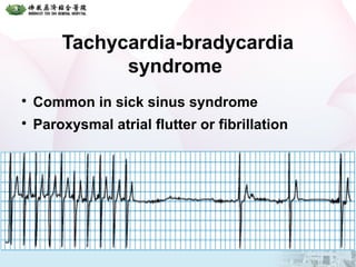
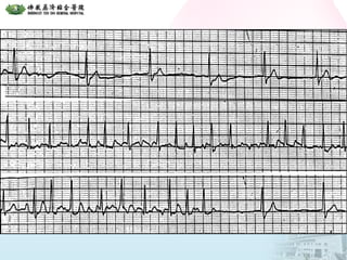
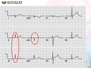
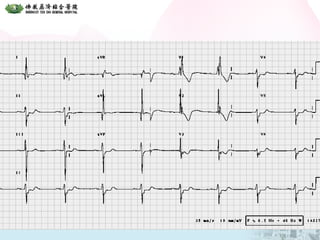
1. The document discusses various types of bradycardia and conduction blocks including sinus bradycardia, sick sinus syndrome, different degrees of AV block, bundle branch blocks, and fascicular blocks.
2. It provides details on the characteristics, causes, and electrocardiographic features of each type of block. First degree AV block results in a prolonged PR interval while second degree block causes intermittent failure of conduction.
3. Third degree or complete heart block results in independent atrial and ventricular rhythms. Bundle branch blocks involve the right or left bundle branches and fascicular blocks occur in the fascicles of the left bundle branch. Bifascicular and trifascicular blocks involve multiple levels of block
Single ventricle presentation for pediatricianLaxmi Ghimire
As the number of children who survive single ventricle physiology, it is very important for the pediatrician to understand about them to give them the best care.
ECG Lecture: Sinus arrest, sinoatrial exit block, AV block and escape rhythmsMichael-Joseph Agbayani
Simple ECG lecture about sinus arrest, sinoatrial exit block, AV block and escape rhythms. Slideshow was made with an audience of medical professionals in mind.
Printed aids combine words, pictures, and diagrams to convey accurate and clear information. It can be read at any available time and can be kept for record.
ABDOMINAL TRAUMA in pediatrics part one.drhasanrajab
Abdominal trauma in pediatrics refers to injuries or damage to the abdominal organs in children. It can occur due to various causes such as falls, motor vehicle accidents, sports-related injuries, and physical abuse. Children are more vulnerable to abdominal trauma due to their unique anatomical and physiological characteristics. Signs and symptoms include abdominal pain, tenderness, distension, vomiting, and signs of shock. Diagnosis involves physical examination, imaging studies, and laboratory tests. Management depends on the severity and may involve conservative treatment or surgical intervention. Prevention is crucial in reducing the incidence of abdominal trauma in children.
These lecture slides, by Dr Sidra Arshad, offer a quick overview of the physiological basis of a normal electrocardiogram.
Learning objectives:
1. Define an electrocardiogram (ECG) and electrocardiography
2. Describe how dipoles generated by the heart produce the waveforms of the ECG
3. Describe the components of a normal electrocardiogram of a typical bipolar lead (limb II)
4. Differentiate between intervals and segments
5. Enlist some common indications for obtaining an ECG
6. Describe the flow of current around the heart during the cardiac cycle
7. Discuss the placement and polarity of the leads of electrocardiograph
8. Describe the normal electrocardiograms recorded from the limb leads and explain the physiological basis of the different records that are obtained
9. Define mean electrical vector (axis) of the heart and give the normal range
10. Define the mean QRS vector
11. Describe the axes of leads (hexagonal reference system)
12. Comprehend the vectorial analysis of the normal ECG
13. Determine the mean electrical axis of the ventricular QRS and appreciate the mean axis deviation
14. Explain the concepts of current of injury, J point, and their significance
Study Resources:
1. Chapter 11, Guyton and Hall Textbook of Medical Physiology, 14th edition
2. Chapter 9, Human Physiology - From Cells to Systems, Lauralee Sherwood, 9th edition
3. Chapter 29, Ganong’s Review of Medical Physiology, 26th edition
4. Electrocardiogram, StatPearls - https://www.ncbi.nlm.nih.gov/books/NBK549803/
5. ECG in Medical Practice by ABM Abdullah, 4th edition
6. Chapter 3, Cardiology Explained, https://www.ncbi.nlm.nih.gov/books/NBK2214/
7. ECG Basics, http://www.nataliescasebook.com/tag/e-c-g-basics
Local Advanced Lung Cancer: Artificial Intelligence, Synergetics, Complex Sys...Oleg Kshivets
Overall life span (LS) was 1671.7±1721.6 days and cumulative 5YS reached 62.4%, 10 years – 50.4%, 20 years – 44.6%. 94 LCP lived more than 5 years without cancer (LS=2958.6±1723.6 days), 22 – more than 10 years (LS=5571±1841.8 days). 67 LCP died because of LC (LS=471.9±344 days). AT significantly improved 5YS (68% vs. 53.7%) (P=0.028 by log-rank test). Cox modeling displayed that 5YS of LCP significantly depended on: N0-N12, T3-4, blood cell circuit, cell ratio factors (ratio between cancer cells-CC and blood cells subpopulations), LC cell dynamics, recalcification time, heparin tolerance, prothrombin index, protein, AT, procedure type (P=0.000-0.031). Neural networks, genetic algorithm selection and bootstrap simulation revealed relationships between 5YS and N0-12 (rank=1), thrombocytes/CC (rank=2), segmented neutrophils/CC (3), eosinophils/CC (4), erythrocytes/CC (5), healthy cells/CC (6), lymphocytes/CC (7), stick neutrophils/CC (8), leucocytes/CC (9), monocytes/CC (10). Correct prediction of 5YS was 100% by neural networks computing (error=0.000; area under ROC curve=1.0).
Rasamanikya is a excellent preparation in the field of Rasashastra, it is used in various Kushtha Roga, Shwasa, Vicharchika, Bhagandara, Vatarakta, and Phiranga Roga. In this article Preparation& Comparative analytical profile for both Formulationon i.e Rasamanikya prepared by Kushmanda swarasa & Churnodhaka Shodita Haratala. The study aims to provide insights into the comparative efficacy and analytical aspects of these formulations for enhanced therapeutic outcomes.
Ozempic: Preoperative Management of Patients on GLP-1 Receptor Agonists Saeid Safari
Preoperative Management of Patients on GLP-1 Receptor Agonists like Ozempic and Semiglutide
ASA GUIDELINE
NYSORA Guideline
2 Case Reports of Gastric Ultrasound
NVBDCP.pptx Nation vector borne disease control programSapna Thakur
NVBDCP was launched in 2003-2004 . Vector-Borne Disease: Disease that results from an infection transmitted to humans and other animals by blood-feeding arthropods, such as mosquitoes, ticks, and fleas. Examples of vector-borne diseases include Dengue fever, West Nile Virus, Lyme disease, and malaria.
Basavarajeeyam is an important text for ayurvedic physician belonging to andhra pradehs. It is a popular compendium in various parts of our country as well as in andhra pradesh. The content of the text was presented in sanskrit and telugu language (Bilingual). One of the most famous book in ayurvedic pharmaceutics and therapeutics. This book contains 25 chapters called as prakaranas. Many rasaoushadis were explained, pioneer of dhatu druti, nadi pareeksha, mutra pareeksha etc. Belongs to the period of 15-16 century. New diseases like upadamsha, phiranga rogas are explained.
3. Bradycardia
A heart rate of <60 beats/min.
May be a normal physiological phenomenon
or result from a cardiac or non-cardiac
disorder.
Many patients tolerate heart rates of
40 beats/min
− Dizziness, near syncope, syncope, ischaemic
chest pain, and hypoxic seizures
8. Conditions associated with
sinoatrial node dysfunction
Age
Idiopathic fibrosis
Ischemia, including myocardial infarction
High vagal tone
Myocarditis
Drug toxicity, such as Digoxin
9. ECG feature of S.S.S.
Persistent sinus bradycardia
Periods of sinoatrial block
Sinus arrest
Junctional or ventricular escape rhythms
Tachycardia-bradycardia syndrome
Paroxysmal atrial flutter/fibrillation
12. Sinoatrial block
A transient failure of impulse of conduction
to the atrial myocardium
The pauses are the length of two or more
P-P intervals.
13. Sinus arrest
Sinus arrest with pause of 4.4 s before generation and
conduction of a junctional escape beat
Transient cessation of impulse formation at the
sinoatrial node.
A prolonged pause without P activity.
The pause is unrelated to the length of the P-P
cycle.
14. Escape rhythms
Escape rhythms are the result of
spontaneous activity from a subsidiary
pacemaker, located in the atria,
atrioventricular junction, or ventricles.
They take over when normal impulse
formation or conduction fails and may be
associated with any profound bradycardia.
15. Escape rhythms
A junctional escape
beat has a normal
QRS complex
shape with a rate of
40-60 beats/min.
A ventricular
escape rhythm has
broad complexes
and is slow (15-
40 beats/min)
17. Tachycardia-bradycardia
syndrome
Common in sick sinus syndrome.
Characterised by bursts of atrial tachycardia
interspersed with periods of bradycardia.
Paroxysmal atrial flutter or fibrillation may
also occur, and cardioversion may be
followed by a severe bradycardia.
22. Causes of atrioventricular
conduction block
Myocardial ischemia or infarction
Degeneration of the His-Purkinje system
Infection for example, Lyme disease,
diphtheria
Immunological disorders for example,
systemic lupus erythematosus
Surgery
Congenital disorders
23. First degree AV block
In first degree block there is a delay in
conduction of the atrial impulse to the
ventricles, usually at the level of the
atrioventricular node.
This results in prolongation of the PR
interval to >0.2 s.
A QRS complex follows each P wave, and
the PR interval remains constant.
27. Second degree AV block
In second degree block there is intermittent
failure of conduction between the atria and
ventricles.
Some P waves are not followed by a QRS
complex.
There are three types of second degree
block.
28. Mobitz type I block
(Wenckebach phenomenon)
Usually at the level of the atrioventricular
node, producing intermittent failure of
transmission of the atrial impulse to the
ventricles.
The initial PR interval is normal but
progressively lengthens until eventually
atrioventricular transmission is blocked
completely.
The PR interval then returns to normal, and
the cycle repeats.
31. Mobitz type II block
Less common but is more likely to produce
symptoms.
There is intermittent failure of conduction of
P waves.
The PR interval is constant.
The block is often at the level of the bundle
branches and is therefore associated with
wide QRS complexes.
33. Mobitz type II block
A complication of an inferior myocardial
infarction.
The PR interval is identical before and after the
P wave that is not conducted.
35. 2:1 atrioventricular block
2:1 atrioventricular block is difficult to
classify, but it is usually a Wenckebach
variant.
High degree atrioventricular block, which
occurs when a QRS complex is seen only
after every three, four, or more P waves,
may progress to complete third degree
atrioventricular block.
36.
37.
38. Third degree block
In third degree block there is complete
failure of conduction between the atria and
ventricles, with complete independence of
atrial and ventricular contractions.
The P waves bear no relation to the QRS
complexes and usually proceed at a faster
rate.
40. Third degree heart block
A pacemaker in the bundle of His produces a narrow
QRS complex (top)
More distal pacemakers tend to produce broader
complexes (bottom).
Arrows show P waves
43.
The bundle of His
divides into the right
and left bundle
branches.
The left bundle branch
then splits into
anterior and posterior
hemifascicles.
Conduction blocks in
any of these structures
produce characteristic
electrocardiographic
changes.
44.
45. Right Bundle Branch Block
In most cases right
bundle branch
block has a
pathological cause
though it is also
seen in healthy
individuals.
46. Conditions associated with right
bundle branch block
Rheumatic heart disease
Cor pulmonale/right ventricular
hypertrophy
Myocarditis or cardiomyopathy
Ischaemic heart disease
Degenerative disease of the conduction
system
Pulmonary embolus
Congenital heart disease for example, in
atrial septal defects
47. Diagnostic criteria for right
bundle branch block
QRS duration 0.12 s
A secondary R wave (R') in V1 or V2
Wide slurred S wave in leads I, V5, and V6
Associated feature
− ST segment depression and T wave inversion in
the right precordial leads
50. Left Bundle Branch Block
Most commonly caused by coronary artery
disease, hypertensive heart disease, or
dilated cardiomyopathy.
Unusual for left bundle branch block to exist
in the absence of organic disease.
The left bundle branch is supplied by both
the LAD & RCA.
Thus patients who develop left bundle
branch block generally have extensive
disease. This type of block is seen in 2-4%
of patients with AMI.
51.
QRS duration of 0.12 s
Broad monophasic R wave in leads 1, V5, and
V6
Absence of Q waves in leads V5 and V6
Associated features
− Displacement of ST segment and T wave in an
opposite direction to the dominant deflection of the
QRS complex (appropriate discordance)
− Poor R wave progression in the chest leads
− RS complex, rather than monophasic complex, in
leads V5 and V6
− Left axis deviation common but not invariable
finding
Diagnostic criteria for left
bundle branch block
54. Fascicular blocks
Block of the left anterior and posterior
hemifascicles gives rise to the hemiblocks.
Mainly affect the direction but not the duration of
the QRS complex, because the conduction
disturbance primarily involves the early phases of
activation.
55. Anterior fascicle of the LBB
The left anterior
fascicle crosses the
left ventricular outflow
tract and terminates in
the Purkinje system of
the anterolateral wall
of the left ventricle.
− Septal branches of the
LAD artery or by the
AV nodal artery
55
56. Left anterior hemiblock
Abnormal left axis deviation in the absence
of an inferior myocardial infarction or other
cause of left axis deviation.
rS morphology in leads II, III and aVF.
qR morphology in leads I and aVL.
56
57. Posterior fascicle of the LBB
Extension of the main bundle
and fans out extensively
posteriorly toward papillary
muscle and inferoposteriorly
to the free wall of the LV.
The proximal part
− AV nodal artery and septal
branches of the LAD artery.
The distal portion
− Dual blood supply from both
anterior and posterior septal
perforating arteries 57
58. Left posterior hemiblock
Frontal plane axis of >90° in the absence of
other causes of right axis deviation, such as
RVH or lung disease…
qR morphology in leads II, III and aVF.
rS morphology in leads I and aVL.
58
60. Bifascicular block
Right bundle branch block with left or right
axis deviation.
Right bundle branch block with left anterior
hemiblock is the commonest type of
bifascicular block.
The left posterior fascicle is fairly stout and
more resistant to damage, so right bundle
branch block with left posterior hemiblock
is rarely seen.
61.
62. Trifascicular block
Trifascicular block is present when
bifascicular block is associated with first
degree heart block.
If conduction in the dysfunctional fascicle
also fails completely, complete heart block
ensues.