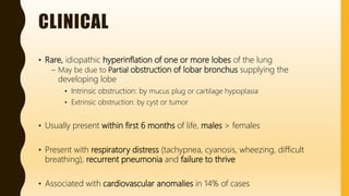
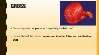
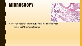
FLASHPATH
H A Z E M A L I
INFANTILE
(CONGENITAL)
LOBAR EMPHYSEMA
H A Z E M A L I
CLINICAL
• Rare, idiopathic hyperinflation of one or more lobes of the lung
– May be due to Partial obstruction of lobar bronchus supplying the
developing lobe
• Intrinsic obstruction: by mucus plug or cartilage hypoplasia
• Extrinsic obstruction: by cyst or tumor
• Usually present within first 6 months of life, males > females
• Present with respiratory distress (tachypnea, cyanosis, wheezing, difficult
breathing), recurrent pneumonia and failure to thrive
• Associated with cardiovascular anomalies in 14% of cases
GROSS
• Commonly affect upper lobes “ especially the left one”
• Hyperinflated lobe cause compression to other lobes and mediastinal
shift
MICROSCOPY
• Alveolar distension without actual wall destruction
– So it is not “true” emphysema
DIFFERENTIAL DIAGNOSIS
“ O t h e r c o n g e n i t a l / c y s t i c l u n g d i s e a s e s ”
• Congenital:
– Pulmonary sequestration
– Congenital pulmonary cysts
– Congenital pulmonary airway malformation
– Congenital lobar emphysema
• Acquired:
– Emphysema
– Healed abscess
– Honeycombing
• Mixed:
– Cystic fibrosis
DIFFERENTIAL DIAGNOSIS
Also can be misdiagnosed clinically as “pneumothorax”
• Pneumothorax lacks the linear bronchovascular and alveolar markings
seen radiologically in congenital lobar emphysema
• Treatments aimed at pneumothorax can worsen patient’s actual
congenital lobar emphysema
WWW.
DO NOT FORGET TO SEARCH FOR MORE PICS
AND VIRTUAL SLIDES
THANK YOU
H A Z E M A L I

FlashPath - Lung - Congenital Lobar Emphysema

  • 1.
    FLASHPATH H A ZE M A L I
  • 2.
  • 3.
    CLINICAL • Rare, idiopathichyperinflation of one or more lobes of the lung – May be due to Partial obstruction of lobar bronchus supplying the developing lobe • Intrinsic obstruction: by mucus plug or cartilage hypoplasia • Extrinsic obstruction: by cyst or tumor • Usually present within first 6 months of life, males > females • Present with respiratory distress (tachypnea, cyanosis, wheezing, difficult breathing), recurrent pneumonia and failure to thrive • Associated with cardiovascular anomalies in 14% of cases
  • 4.
    GROSS • Commonly affectupper lobes “ especially the left one” • Hyperinflated lobe cause compression to other lobes and mediastinal shift
  • 5.
    MICROSCOPY • Alveolar distensionwithout actual wall destruction – So it is not “true” emphysema
  • 6.
    DIFFERENTIAL DIAGNOSIS “ Ot h e r c o n g e n i t a l / c y s t i c l u n g d i s e a s e s ” • Congenital: – Pulmonary sequestration – Congenital pulmonary cysts – Congenital pulmonary airway malformation – Congenital lobar emphysema • Acquired: – Emphysema – Healed abscess – Honeycombing • Mixed: – Cystic fibrosis
  • 7.
    DIFFERENTIAL DIAGNOSIS Also canbe misdiagnosed clinically as “pneumothorax” • Pneumothorax lacks the linear bronchovascular and alveolar markings seen radiologically in congenital lobar emphysema • Treatments aimed at pneumothorax can worsen patient’s actual congenital lobar emphysema
  • 8.
    WWW. DO NOT FORGETTO SEARCH FOR MORE PICS AND VIRTUAL SLIDES
  • 9.
    THANK YOU H AZ E M A L I