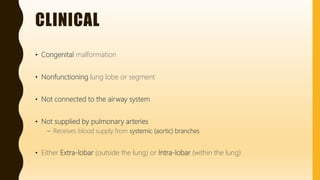
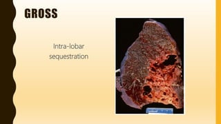
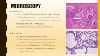
FLASHPATH
H A Z E M A L I
PULMONARY
SEQUESTRATION
H A Z E M A L I
CLINICAL
• Congenital malformation
• Nonfunctioning lung lobe or segment
• Not connected to the airway system
• Not supplied by pulmonary arteries
– Receives blood supply from systemic (aortic) branches
• Either Extra-lobar (outside the lung) or Intra-lobar (within the lung)
CLINICAL
Extra-lobar Intra-lobar
Incidence Less common More common
Age of presentation Infants
(below 1 year)
Older children and even
Adults
Gender Male > Females Equal
Foregut communication and
associated anomalies
Very Common
e.g. diaphragmatic hernia
Very rare
Chronic inflammation,
fibrosis and Honeycomb
changes
Very rare Very Common
GROSS
Extra-lobar Intra-lobar
Mass Solid, Spongy mass Solid, Fibrotic mass has
multiple, variable-sized
cystic areas
Location Outside the lung
anywhere above, within, or
below the diaphragm
Within the lung
Side Mainly left side Mainly left lung
(lower lobe)
Pleura Its own “separate”
pleural covering
The same pleural
covering of the lung
Venous drainage Systemic (azygous) Pulmonary
GROSS
Intra-lobar
sequestration
MICROSCOPY
• Intra-lobar:
– Marked chronic inflammation, fibrosis, cystic changes
• May be because it can be undiagnosed for long time,
it shows a higher risk for recurrent infections and fibrosis
– Not associated with CPAM type 2
(Congenital pulmonary airway malformation)
• Extra-lobar:
– No significant inflammation or fibrosis
• May be because it is often discovered early in life,
there is no time to develop infections or fibrosis
– Associated with (CPAM) type 2 in 50% of cases
• Areas of irregular, markedly dilated bronchioles
within parenchyma
DIFFERENTIAL DIAGNOSIS
O t h e r c y s t i c l u n g d i s e a s e s :
• Congenital:
– Bronchogenic cysts
– Congenital pulmonary cysts
– Congenital pulmonary airway malformation
– Congenital lobar emphysema
• Acquired:
– Emphysema
– Healed abscess
– Honeycombing
• Mixed:
– Cystic fibrosis
WWW.
DO NOT FORGET TO SEARCH FOR MORE PICS
AND VIRTUAL SLIDES
THANK YOU
H A Z E M A L I

Flashpath - lung - pulmonary sequestration

  • 1.
    FLASHPATH H A ZE M A L I
  • 2.
  • 3.
    CLINICAL • Congenital malformation •Nonfunctioning lung lobe or segment • Not connected to the airway system • Not supplied by pulmonary arteries – Receives blood supply from systemic (aortic) branches • Either Extra-lobar (outside the lung) or Intra-lobar (within the lung)
  • 4.
    CLINICAL Extra-lobar Intra-lobar Incidence Lesscommon More common Age of presentation Infants (below 1 year) Older children and even Adults Gender Male > Females Equal Foregut communication and associated anomalies Very Common e.g. diaphragmatic hernia Very rare Chronic inflammation, fibrosis and Honeycomb changes Very rare Very Common
  • 5.
    GROSS Extra-lobar Intra-lobar Mass Solid,Spongy mass Solid, Fibrotic mass has multiple, variable-sized cystic areas Location Outside the lung anywhere above, within, or below the diaphragm Within the lung Side Mainly left side Mainly left lung (lower lobe) Pleura Its own “separate” pleural covering The same pleural covering of the lung Venous drainage Systemic (azygous) Pulmonary
  • 6.
  • 7.
    MICROSCOPY • Intra-lobar: – Markedchronic inflammation, fibrosis, cystic changes • May be because it can be undiagnosed for long time, it shows a higher risk for recurrent infections and fibrosis – Not associated with CPAM type 2 (Congenital pulmonary airway malformation) • Extra-lobar: – No significant inflammation or fibrosis • May be because it is often discovered early in life, there is no time to develop infections or fibrosis – Associated with (CPAM) type 2 in 50% of cases • Areas of irregular, markedly dilated bronchioles within parenchyma
  • 8.
    DIFFERENTIAL DIAGNOSIS O th e r c y s t i c l u n g d i s e a s e s : • Congenital: – Bronchogenic cysts – Congenital pulmonary cysts – Congenital pulmonary airway malformation – Congenital lobar emphysema • Acquired: – Emphysema – Healed abscess – Honeycombing • Mixed: – Cystic fibrosis
  • 9.
    WWW. DO NOT FORGETTO SEARCH FOR MORE PICS AND VIRTUAL SLIDES
  • 10.
    THANK YOU H AZ E M A L I