21. OBSTRUCTION v. RESTRICTION
• OBSTRUCTION
• RESTRICTION
• Air or blood?
• Large or small?
• Inspiration or Expiration?
• “Compliance”
• “Infiltrative”
• Obstruction is
SMALL AIRWAY
EXPIRATION
• REDUCED lung VOLUME,
DYSPNEA, CYANOSIS
• REDUCED GAS
TRANSFER
obstruction, i.e., wheezing • “GROUND GLASS” on CXR
• HYPEREXPANSION on CXR
24. EMPHYSEMA
• COPD, or “END-STAGE” lung disease
• Centri-acinar, Pan-acinar, Paraseptal, Irregular
• (PROGRESSIVE) EXPIRATORY AIR TRAPPING,
i.e., WHEEZING
• Like cirrhosis, thought of as END-STAGE of
multiple chronic small airway obstructive
etiologies
• NON-specific
• IN-creased crepitance, BULLAE (BLEBS)
• Clinically likely to produce recurrent pneumonias,
and progressive failure
50. VASCULAR PULMONARY DISEASES
• PULMONARY EMBOLISM (with or usually
WITHOUT infarction)
• PULMONARY HYPERTENSION, leading to
cor pulmonale
• HEMORRHAGIC SYNDROMES
– GOODPASTURE SYNDROME
– HEMOSIDEROSIS, idiopathic
– WEGENER GRANULOMATOSIS
51. P.E.
• Usually secondary to debilitated states with
immobilization, or following surgery
• Usually deep leg and deep pelvic veins (DVT), NOT
superficial veins
• Follows Virchow’s triad, i.e., 1) flow problems, 2)
endothelial disruption, 3) hypercoagulabilty
• Usually do NOT infarct, usually ventilate
• When they DO infarct, the infarct is hemorrhagic
• Decreased PO2, acute chest pain, V/Q MIS-match
• DX: Chest CT, V/Q scan, angiogram
• RX: short term heparin, then long term coumadin
55. HEMORRHAGIC SYNDROMES
• GOODPASTURE Syndrome: Ab’s
to the alpha-3 chains of collagen
IV, GBM deposits too!
• IDIOPATHIC PULMONARY
HEMOSIDEROSIS, to be
differentiated from chronic CHF
• WEGENER GRANULOMATOSIS
58. PULMONARY INFECTIONS
COMMUNITY-ACQUIRED BACTERIAL ACUTE PNEUMONIAS (BACTERIAL)
Streptococcus Pneumoniae
Haemophilus Influenzae
Moraxella Catarrhalis
Staphylococcus Aureus
Klebsiella Pneumoniae
Pseudomonas Aeruginosa
Legionella Pneumophila
COMMUNITY-ACQUIRED ATYPICAL (VIRAL AND MYCOPLASMAL) PNEUMONIAS
(NON-BACTERIAL)
Influenza Infections
Severe Acute Respiratory Syndrome (SARS)
NOSOCOMIAL PNEUMONIA
ASPIRATION PNEUMONIA
LUNG ABSCESS
Etiology and Pathogenesis.
CHRONIC PNEUMONIA
Histoplasmosis, Morphology
Blastomycosis, Morphology
Coccidioidomycosis, Morphology
PNEUMONIA IN THE IMMUNOCOMPROMISED HOST
PULMONARY DISEASE IN HUMAN IMMUNODEFICIENCY VIRUS INFECTION
59. BASIC CONSIDERATIONS
• PNEUMONIA vs. PNEUMONITIS
• DIFFERENTIATION from INJURIES, OBSTRUCTIVE
DISEASES, RESTRICTIVE DISEASES, VASCULAR
DISEASES
• DIFFERENTIATION FROM NEOPLASMS
• CLASSICAL STAGES of INFLAMMATION
• LOBARvs. BRONCHO• INTERSTITIAL vs. ALVEOLAR
• COMMUNITY vs. NOSOCOMIAL
• ETIOLOGIC AGENTS vs. HOST IMMUNITY
• 2 PRESENTING SYMPTOMS: What are they?
• 2 DIAGNOSTIC METHODS: What are they?
• ANY ORGANISM CAN CAUSE PNEUMONIA!!!
60. PREDISPOSING FACTORS
• LOSS OF COUGH REFLEX
• DIMINISHED MUCIN or CILIA
FUNCTION
• ALVEOLAR MACROPHAGE
INTERFERENCE
• VASCULAR FLOW IMPAIRMENTS
• BRONCHIAL FLOW IMPAIRMENTS
61. Although pneumonia is
one of the most
common causes of
death, it usually does
NOT occur in healthy
people spontaneously
65. STREPTOCOCCUS
• The classic LOBAR pneumonia
• Normal flora in 20% of adults
• Only 20% of victims have + blood cultures
• “Penicillins” are often 100% curative
• Vaccines are often 100% preventive
66.
67. HAEMOPHILUS PNEUMONIA
• Commonest in CHILDREN <2, with
otitis, URI, meningitis, cellulitis,
osteomyelitis
• PNEUMONIAS in CHILDREN <2 are
often thought of as being H Flu until
proven otherwise, otitis, meningitis too
• Most common pneumonia from COPD
in adults
• BACTRIM (Trimethoprim-Sulfa) most
common treatment
68.
69. MORAXELLA CATARRHALIS
• 2nd most common COPD pneumonia,
after haemophilus
• Gram NEGATIVE coccobacillus, like H.
Flu
70. STAPH aureus
• Most common pneumonia
following viral pneumonias
• M.R.S.A., of course, is usually
NOT “community” acquired
72. PSEUDOMONAS Aeruginosa
• Usually NOT community acquired
but nosocomial
• CYSTIC FIBROSIS patients with
pneumonia are presumed to have
PSEUDOMONAS until proven
otherwise
79. INFLUENZA VIRUS
• A,B,C
• 1915, 1918, PAN-demics, type A
• Has MUTATED throughout history,
many STRAINS, avian swine, etc.
• B and C in children
• Exact strains can be ID’s by PCR
80.
81. SARS
(Severe Acute Respiratry Syndrome)
•
•
•
•
CORONA-VIRUS
2002 China outbreak
Spread CHIEFLY in Asia
Like most other NON-bacterial
pneumonias confirmed by PCR
• Like most viral pneumonias,
interstitium infiltrated, some giant
cells often present
83. Classifications of PNEUMONIAS
• COMMUNITY ACQUIRED
• COMMUNITY ACQUIRED, ATYPICAL
• NOSOCOMIAL
•
•
•
•
ASPIRATION
CHRONIC
NECROTIZING/ABSCESS FORMATION
PNEUMONIAS in
IMMUNOCOMPROMISED HOSTS
84. NOSOCOMIAL
• Acquired in HOSPITALS, also called “hospital acquired”,
versus “community acquired” pneumonias.
– DEBILITATION
– CATHETERS, VENTILATORS
– ENTEROBACTER, PSEUDOMONAS
– STAPH (MRSA)
– MRSA (MR=Methicillin Resistant)
• OTHER Common causes of Noso. Pneum.
P. aeruginosa
Klebsiella
E. coli
S. pneumoniae
H. influenzae
85. Classifications of PNEUMONIAS
• COMMUNITY ACQUIRED
• COMMUNITY ACQUIRED, ATYPICAL
• NOSOCOMIAL
• ASPIRATION
• CHRONIC (often granulomatous)
• NECROTIZING/ABSCESS FORMATION
• PNEUMONIAS in
IMMUNOCOMPROMISED HOSTS
86. ASPIRATION PNEUMONIAS
•
•
•
•
UNCONSCIOUS PATIENTS
PATIENTS IN PROLONGED BEDREST
LACK OF ABILITY TO SWALLOW OR GAG
USUALLY CAUSED BY ASPIRATION OF
GASTRIC CONTENTS
• POSTERIOR LOBES (gravity dependent)
MOST COMMONLY INVOLVED,
ESPECIALLY THE SUPERIOR SEGMENTS of
the LOWER LOBES
• Often lead to ABSCESSES
90. Classifications of PNEUMONIAS
•
•
•
•
COMMUNITY ACQUIRED
COMMUNITY ACQUIRED, ATYPICAL
NOSOCOMIAL
ASPIRATION
• CHRONIC
• NECROTIZING/ABSCESS FORMATION
• PNEUMONIAS in
IMMUNOCOMPROMISED HOSTS
91. CHRONIC Pneumonias
• USUALLY NOT persistences of the
community or nosocomial bacterial
infections, but CAN BE, at least
histologically
• Often SYNONYMOUS with the 4 classic
systemic fungal or granulomatous
pulmonary infections infections, i.e., TB,
Histo-, Blasto-, Coccidio• If you see pulmonary granulomas, think of a
CHRONIC process, often years
93. •
•
•
•
HISTOPLASMOSIS
Spores in bird or bat droppings
Mimics TB
Histoplasma CAPSULATUM
Pulmonary granulomas, often large and
calcified
• Tiny organisms live in macrophages
• Ohio, Mississippi valley
• MANY other organs
can be affected
94.
95. BLASTOMYCOSIS
• Spores in soil
• Mimics TB, like ALL the granulomatous lung
dideases do.
• Blastomyces DERMATIDIS
• Pulmonary granulomas, often large and
calcified
• Large distinct SPHERULES (larger than
coccidio)
• Ohio, Mississippi valley, Great Lakes,
WORLDWIDE
• MANY other organs can be affected,
especially SKIN
96.
97. •
•
•
•
•
•
•
•
COCCIDIOMYCOSIS
Spores in soil
Mimics TB
Coccidioides IMMITIS
Pulmonary granulomas, often large and
calcified
Smaller spherules than blasto.
Tiny organisms live in macrophages
American SOUTHWEST
MANY other organs can be affected
102. LUNG TRANSPLANTATION
Any end-stage lung disease in which the patient can tolerate
long term immunosuppression, and often just ONE lung is
enough, donors very SCARCE!
•
•
•
•
EMPHYSEMA
Pulmonary Fibrosis
Cystic Fibrosis
Pulmonary Hypertension
103. Lung Transplant Pathology
• Infections (patients are on
immunosuppressives )
– Bacterial
– Viral (CMV)
– Fungal
– PCP
• ACUTE rejection, pneumonias, usually
weeks to months
• CHRONIC rejection, HALF of all patients by
3-5 years, “bronchiolitis obliterans” (COP)
104.
105. LUNG TUMORS
• Benign, malignant, epithelial, mesenchymal,
but 90% are CARCINOMAS
• BIGGEST USA killer. Why? Ans: Prevalence
not as high as prostate or breast but
mortality higher. Only 15% 5 year survival.
• TOBACCO has polycyclic aromatic
hydrocarbons, such as benzopyrene,
anthracenes, radioactive isotopes
• Radiation, asbestos, radon
• C-MYC, K-RAS, EGFR, HER-2/neu
107. TWO TYPES
• NON-SMALL CELL
– SQUAMOUS CELL CARCINOMA
– ADENOCARCINOMA
– LARGE CELL CARCINOMA
• SMALL CELL CARCINOMA
108. •
•
•
•
•
•
•
•
•
•
•
The BIG list
Squamous cell carcinoma
Small cell carcinoma
Combined small cell carcinoma
Adenocarcinoma: Acinar, papillary,
bronchioloalveolar, solid, mixed subtypes
Large cell carcinoma
Large cell neuroendocrine carcinoma
Adenosquamous carcinoma
Carcinomas with pleomorphic, sarcomatoid, or
sarcomatous elements
Carcinoid tumor: Typical, atypical
Carcinomas of salivary gland type
Unclassified carcinoma
112. TNM, Lung
T1
Tumor <3 cm without pleural or main stem bronchus involvement
T2
Tumor >3 cm or involvement of main stem bronchus 2 cm from carina,
visceral pleural involvement, or lobar atelectasis
T3
Tumor with involvement of chest wall (including superior sulcus tumors),
diaphragm, mediastinal pleura, pericardium, main stem bronchus 2 cm from
carina, or entire lung atelectasis
T4
Tumor with invasion of mediastinum, heart, great vessels, trachea,
esophagus, vertebral body, or carina or with a malignant pleural
effusion
N0
No demonstrable metastasis to regional lymph nodes
N1
Ipsi-lateral hilar or peribronchial nodal involvement
Metastasis to ipsilateral mediastinal or subcarinal lymph nodes
Metastasis to contra-lateral mediastinal or hilar lymph nodes, ipsilateral
N2
N3
or contralateral scalene, or supraclavicular lymph nodes
M0
No (known) distant metastasis
M1
Distant metastasis present
113. LOCAL effects of LUNG CANCER
Clinical Feature
Pathologic Basis
Pneumonia, abscess, lobar
collapse
Tumor obstruction of airway
Lipid pneumonia
Tumor obstruction; accumulation of cellular
lipid in foamy macrophages
Pleural effusion
Tumor spread into pleura
Hoarseness
Recurrent laryngeal nerve invasion
Dysphagia
Esophageal invasion
Diaphragm paralysis
Phrenic nerve invasion
Rib destruction
Chest wall invasion
SVC syndrome
SVC compression by tumor
Horner syndrome
Sympathetic ganglia invasion
Pericarditis, tamponade
Pericardial involvement
114. SYSTEMIC effects of LUNG CANCER
(PARA-NEOPLASTIC SYNDROMES)~ 5%
ADH (hyponatremia)
ACTH (Cushing)
PTH (Hyper-CA)
CALCITONIN (Hypo-CA)
GONADOTROPINS
SEROTONIN/BRADYKININ
116. METASTATIC TUMORS
• LUNG is the MOST COMMON
site for all metastatic tumors,
regardless of site of origin
• It is the site of FIRST CHOICE
for metastatic sarcomas for
purely anatomic reasons!
121. MESOTHELIOMAS
• “Benign” vs. “Malignant”
differentiation does not matter, but a
self limited localized nodule can be
regarded as benign, and a spreading
tumor can be regarded as malignant
• Visceral or parietal pleura, pericardium,
or peritoneum
• Most are regarded as asbestos caused
or asbestos “related”
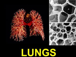
Typical normal 1000 gram lung (R550, L450), with lobes and bronchopulmonary segments, primary, secondary, tertiary bronchi, half billion alveoli, several hundred billion capillaries, etc. Pleura “smooth and glistening”, arteries traveling with bronchi, veins being rather independent of bronchopulmonary segments and lobes. Why is weight important? Why is “smooth and glistening” important? What is “crepitance”? Why is crepitance important? What is “compliance”? Why is compliance just as important as crepitance, in understanding lung diseases?
Classical classifications of diseases, degenerative, inflammatory, neoplastic. This classification still stands up today.
This is about a day’s (2-hour) job.
NON PNEUMONIAS (caused by pathogens)
NON TUMORS
This is also about a day’s (2-hour) job.
Typical normal 1000 gram lung (R550, L450), with lobes and bronchopulmonary segments, primary, secondary, tertiary bronchi, etc. Pleura “smooth and glistening”, arteries traveling with bronchi, veins being rather independent of bronchopulmonary segments and lobes. Why is weight important? Why is “smooth and glistening” important? What is “crepitance”? Why is crepitance important? Is crepitance the same as bubbliness? ANS: YES
Know the microscopic criteria for all the items delineated on the right, especially the kinds of simple epithelium which line them.
The “space” between the endothelium and the type-1 pneumocyte, is the blood air interface.
Type-I pneumocyte, Type-2 pneumocyte, Endothelial cell, Alveolar macrophages, Interstitial fibroblasts.
This simple embryology diagram may help explain most common congenital lung diseases. Why might this diagram be WRONG, especially the top figure? Ans: The RIGHT one is the main downward one, embryologically!
“NORMAL” chest X-Ray (CXR). What features of “normal” are the most important?
Infiltrates? Sharpness of costophrenic angles? Properly exposed? Hilar vasculature? Bronchi?
Why is a CXR to a radiologist like pathologists fingers?
By far, the most common scenario is for the baby to eat, and it comes back up WITHOUT food getting into the lungs.
ATALECTASIS is strictly an anatomic/physiologic/geometric CONCEPT, NOT a disease by itself, but seen in many disease states.
Reabsorption can be from a bronchial obstruction, such as a tumor.
Compression can be from, say, a pleural effusion, or pneomothorax.
Contraction can be from a diffuse lung fibrotic process.
FOUR main pathologic mechanisms of pulmonary edema.
In alveolar injury, the fluid is much more likely to be EXUDATE rather than TRANSUDATE, right?
*…..as opposed to NEONATAL Respiratory Distress Syndrome
ARDS can be thought of as NON-cardiac pulmonary edema, or, more correctly, edema related to alveolar INJURY. It is NON-specific!!! It is also sometimes called “shock lung” as we will see in the section on shock. Is the alveolar “edema” of ARDS more likely to have more protein than cardiac pulmonary edema?, i.e., exudate vs. transudate?
Think of ARDS morphologically as NON-cardiogenic pulmonary edema where much more leaks into the alveoli than just transudative fluid, i.e., fibrin, protein, cells, etc.
ARDS is generally SECONDARY to something else, when it ISN’T, we can call it ACUTE INTERSTITIAL PNEUMONIA. Histologically, they cannot be differentiated!
O vs. R constitute the majority of pulmonary diseases which are not infectious pneumonias.
Two EXTREMELY important concepts of pulmonary pathology.
OBSTRUCTION means SMALL AIRWAY EXPIRATORY obstruction, air trapping , wheezing, more lucency, less density.
RESTRICTION means REDUCED COMPLIACE, i.e., less sponginess, less gas transfer, more opacity, i.e., more density!
Are thesese all related? Generally, YES.
Sm,sm,sm,lg
Which one would feel more “hypercrepitant” at autopsy? Which one would be more severe? Answer: the MORE hypercrepitant one, which involvs the WHOLE acinus, rather than just the central portion of the acinus.
An acinus is NOT the same as an alveolus, the ACINUS is everything AFTER a terminal bronchiole.
Bullae, or “peripheral blebs” are hallmarks of chronic obstructive lung disease, COPD.
What would happen if a “bleb” was so paper-thin, that it ruptured? Ans: “Pneumo”-thorax
Calling something “hyperlucent” to a radiologist is like calling something “understained” to a pathologist, i.e., many technical factors have to be taken into consideration before you diagnose disease
Note the heavy inflammatory cell infiltrate around bronchioles and small bronchi.
Would the presence of a small amount of cartilage in the wall tend to make us call this a small bronchus rather than a bronchiole? Ans: Yes Do you think you might already see some overexpanded alveoli here?
What are the 4 classic histologic findings in bronchial asthma?
Answer: 1) Inflammation 2) Bronchial (luminal) narrowing 3) Increased Mucous 4) Smooth muscle hyperplasia
What is the 5th finding if the etiology is allergy? Ans: Increased eosinophils
Bronchiectasis is not a specific disease, but simply a condition in which LARGE bronchi are damaged and DILATED due to a variety of causes.
“-ectasis” is the root word for “dilatation”.
How do you know these are not bullae?
If you “squeezed” a lung with restrictive lung disease, you would note it wasn’t as “spongy” as a normal lung. This is the definition of reduced compliance. It simply will not “comply” when squeezed (or moved by respiratory motion either)! In contract to the “obstructive” lung diseases, the chest x-ray shows diffuse INCREASE in density, NOT DECREASED, usually. Compliance is NOT crepitance. Another common denominator of the “restrictive” lung diseases is that they have anatomic/functional barriers to the classic gas exchange between an endothelial cell and a type-1 pneumocyte.
“FIBROSING” is by far, the biggest category of restrictive lung diseases.
Fibrosis follows inflammation, but there is an absence of infectious organisms. What does that sound like? Ans: Autoimmune? Perhaps.
Would you see a lot of scar tissue here if you did a trichrome stain? Ans: yes
Could this have been preceded by an unknown infectious pathogen? Yes.
Often occurs in a transplanted lung.
Because these are also classified as “irritants” which may produce a bronchitic component as well, there may also be “chronic obstructive” components to these diseases.
Note that in this category, we do NOT include the systemic fungal diseases.
This image was “googled” from tumorboard.com, the internet’s FIRST diagnostic pathology image base which started even BEFORE there was a world wide web, when it was only a BBS.
The fact that this is a mesenteric lymph node will remind you that sarcoidosis is NOT limited to the lungs.
The classical difference between a “caseating” and a “non-caseating” granuloma, is often the difference between TB and sarcoid. Which one might culture out acid-fast bacteria? Ans: The one on the LEFT (i.e., caseating)
Why is it called “desquamative”? Ans: …because it looks like the alveolar wall “epithelium” is peeling off into the alveolus!
Pulmonary edema on left, PAP (Pulmonary Alveolar Proteinosis) on the right.
anti-GM-CSF autoantibodies in patients with PAP
Three types of “vascular” lung diseases.
Why would a PE usually NOT result in an infarct?
Routine chest x-rays are the very LEAST helpful in diagnosing PE. Why? Ans: there is no radiologic “infiltrate” unless there is infarction which is rare for PE. Also…..the lung still VENTILATES!
A general rule with COPD is: As the alveoli become wider, the arterioles become narrower! This is a totally non-scientific, but a TRUE observation.
A common finding in most cases of pulmonary hypertension, no matter what the cause is.
NORMAL thickness pulmonary arteriole on the LEFT.
Vicious cycle: Pulmonary hypertension is destructive on the pulmonary arterioles and causes, eventually, fibrotic narrowing. PLUS, narrowing from ANY reason causes pulmonary hypertension!
Wegener's granulomatosis is a form of vasculitis that affects the lungs, kidneys and other organs. Due to its end-organ damage, it can be a serious disease that requires long-term immunosuppression. It is named after Dr. Friedrich Wegener, who described the disease in 1936, it often has ANCAs, i.e., Anti-Neutrophil Cytoplasmic Antibodies.
Why do I LOVE “idiopathic” diseases? Because I don’t have to waste time talking about the etiology!
What is probably the most COMMON cause of a hemorrhagic part of the lung? Ans: INFARCT?
IPH has MUCH more hemosiderin in alveoli, usually, relative to chronic CHF. Acute CHF has NO hemosiderin. Why?
Biggest killer?
Biggest killer in hospitals?
Why is the term “chronic” pneumonia here, kind of a misnomer, classically?
PNEUMONIAS are also called LOWER respiratory infections, as opposed to UPPER.
Logically speaking, would impairments at ANY of these levels set the stage for pneumonias? Ans: OFF COURSE
I put this slide into my discussion of pneumonias 2-3 times. This is not enough times.
Of course these are NOT mutually exclusive classifications, e.g., ANY pneumonia may result in an abscess.
* Go back to the previous slide!
COMMUNITY vs. HOSPITAL
Know the gram staining properties of the common community acquired pneumonia organisms.
Do the upper two images demonstrate the “lobar-ness” of the pneumonia? Ans: Yes
H. Flu graphics
* Go to slides 61 or 74
This is the reason why after you feel so good about curing your patients pneumonia with antibiotics, you wonder if he will be back again, due to the underlying REAL reason he got the pneumonia!
Would a classical pneumonia produce more of a restrictive pattern or obstructive? Answer: Unfair question! (could be both). Functionally it might behave like a restrictive in the pulmonary blood gas lab, but it may be a complication of an obstructive.
Viral pneumonias, generally interstitial, bacterial pneumonias generally alveolar!!!
Can you see the RLL “subtle” infiltrate? Or do you want to call the radiologist?
Corona viruses are RNA, “enveloped”, i.e., “crowned” viruses
As soon as you step into a hospital, expect to be greeted by MRSA.
Two things live in hospitals: 1) bugs resistant to antibiotics 2) sick people
STREP, STAPH, H.FLU, PSEUDOMONAS are the most frequent secondary complicators.
This is not a TYPE of pneumonia, but a complication of ANY pneumonia!
An abscess can be thought of as a pneumonia in which all of the normal lung outline can no longer be seen, and there is 100% pus. Notice the increasing destruction of the alveolar framework as you progress closer to the center of the abscess.
The word “chronic” pneumonia classification may be a misnomer because most show a “granulomatous” pattern of inflammation, rather than pure “chronic”. i.e., monos. They are chronic CLINICALLY, however, if not strictly pathologically.
In this case CHRONIC means CLINICALLY CHRONIC, not PATHOLOGICALLY CHRONIC.
“Chronic” by classification, but “granulomatous” by histology.
Granulomatous reactions are commonly seen with mycobacteria, fungi, sarcoid, foreign bodies, and rarely with almost anything.
* Really jiroveci, not carinii any more.
PCP is the most common pneumonia in AIDS patients. It is so prevalent, many rationales consist in giving treatment for it prophylactically.
An interesting tidbit is that “cotton wool” or “wooly” exudates are described BOTH radiologically as well as histologically
*really “jiroveci”
Protozoan vs. Fungi?
Bronchiolitis obliterans, as seen with “COP”, occurs with chronic pulmonary transplant rejection
Different mutations may be occurring at different steps of this cascade!
The NON-small cell cancers behave and are treated similarly, the SMALL cell carcinomas are WORSE than the non-small cell carcinomas, but respond better to chemotherapy, often drastically!
Once again, the best way to classify tumors of ANY organ or tissue is to simply remember the histology. Tumors are clonal proliferations of native cells.
The classical squamous cell carcinoma starting in a large bronchus centrally, with bronchial obstruction.
Adenocarcinomas tend to be more peripheral. Note the features of malignant cells on sputum cytology.
Name the four most common histologic patterns of lung carcinoma and explain why!
Squamous, adeno, large, small, going clockwise.
TNM ALWAYS relates to BIOLOGIC BEHAVIOR!
Once again, the best way to classify tumors of ANY organ or tissue is to simply remember the histology. Tumors are clonal proliferations of native cells.
NB: OFTEN, the very first distant metastasis for a lung carcinoma is the adrenal!
LIVER:PORTAL DRAINED ORGANS::LUNG:SARCOMAS
Also recall that mesothelium does not only cover the lungs viscerally, as well as parietally, but also the pericardium and the peritoneum as well, so mesotheliomas and effusions of the pleura, and ALL diseases, are also have their corresponding counterparts in the pericardial space and peritoneal space as well. 12 possibilities ? Pneumo, hydro, hemo, chylo X pleura, pericardial, peritoneal
Pleuritis = Pleurisy
How would you differentiate a pleural transudate from an exudate? Ans: SPGR, cells, protein, LDH
The diagnosis of “MESOTHELIOMA” is RARELY preceded by the word benign or malignant on histology alone!
Typical growth appearance of a malignant mesothelioma, it compresses the lung from the OUTSIDE.
Mesothelial cells have MANY more microvilli than most epithelial cells and express a protein called CALRETININ, which epithelial cells do NOT.
The differentiation between mesothelioma and carcinoma may be crucially important!