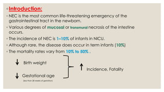
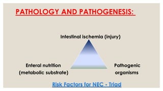
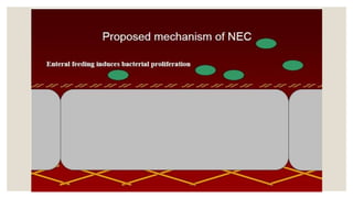
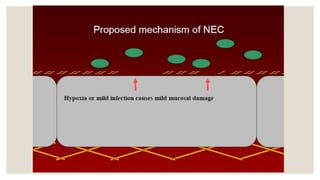
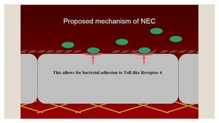
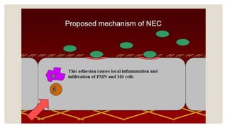
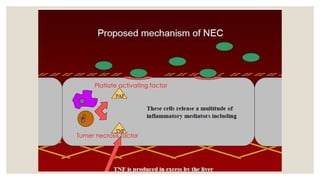
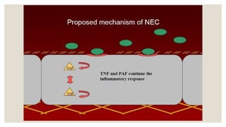
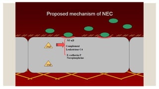
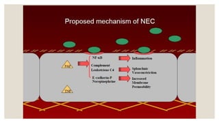
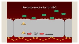
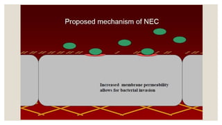
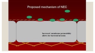
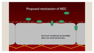
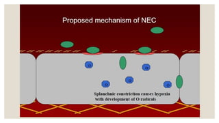
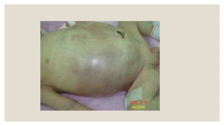
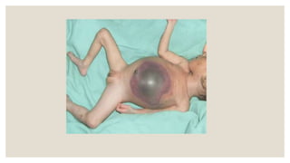
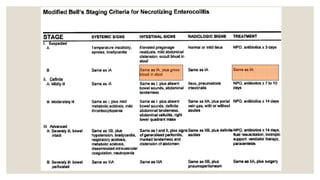
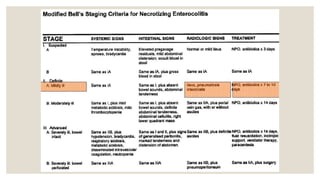
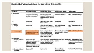
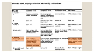
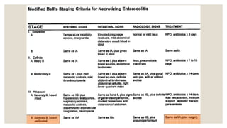
NEONATAL NECROTIZING
ENTEROCOLITIS
By :
Rasha Alqdi
Alwehda hospital
◦Introduction:
◦ NEC is the most common life-threatening emergency of the
gastrointestinal tract in the newborn.
◦ Various degrees of mucosal or transmural necrosis of the intestine
occurs.
◦ The incidence of NEC is 1–10% of infants in NICU.
◦ Although rare, the disease does occur in term infants (10%)
◦ The mortality rates vary from 10% to 50% .
Birth weight
Incidence, Fatality
Gestational age
(less than 28 weeks of gestation)
◦Necrotizing Enterocolitis In The Beginning
◦ NEC was unknown as a disease before the 1950’s
◦ First described in 1950’s by Schmid and Quaiser
◦ – Case reports describing neonates who died from necrotizing lesions
of their GI tracts.
NEC became recognized as a clinical entity in 1960’s and 1970’s
◦ – At this time NEC’s mortality exceeded 70%
◦ – NEC was initially described as idiopathic gastrointestinal perforations
So what is NEC ?
Def. of NEC
Is an acquired neonatal disorder representing an
end expression of serious intestinal injury after a
combination of vascular, mucosal, and metabolic
(and other unidentified) insults to a relatively
immature gut.
Risk factors
◦ Prematurity. The lower the gestational age the greater the risk of the NEC, because of the
immaturity of the circulatory, gastrointestinal, and immune systems.
◦ Asphyxia and acute cardiopulmonary collapse, as they lead to low cardiac output and
diminished intestinal perfusion.
◦ Enteral feeding: NEC is rare in unfed infants. About 90-95% of infants with NEC received at least
one enteral feeding.
◦ Enteral feeding provides a substrate for proliferation of enteric pathogens.
◦ Hyperosmolar formula may cause direct damage to intestinal mucosa.
◦ Lack of the immuno-protective factors in commercially prepared formula.
◦ Breast-feeding significantly lowers the risk of NEC ( suggesting that breast milk factors, including growth
factors, antibodies, and cellular immune factors, might be protective).
◦ Polycythymia and hyperviscosity syndrome.
◦ Exchange transfusion.
◦ Large feeding volumes and rapid advancement of enteral feedings.
◦ Enteric pathogenic organisms comprising bacterial and viral pathogens. These include E.coli,
Klebsiella, salmonella, rotaviruses, and enteroviruses.
Why Exchange transfusion is a risk
factor for NEC ?
Answer :
◦By using Umbilical Catheter for
Exchange transfusion , increase in
portal venous pressure that can result
in decrease in ilial and colonic blood
flow
ETIOLOGY:
◦ Etiology of NEC is unclear; May be multifactorial.
◦ Prematurity is the single greatest risk factor.
◦ Infants exposed to cocaine have a 2.5 times increased risk
of developing NEC.
The mean gestational age of infants with NEC is 30 to 32
weeks, and the infants generally are weight appropriate
for gestational age.
PATHOLOGY AND PATHOGENESIS:
Intestinal ischemia (injury)
Enteral nutrition Pathogenic
(metabolic substrate) organisms
What is the most common site of NEC?
◦A. Stomach
◦B. Jejunum
◦C. Ileum
◦D. Colon
Terminal ileum/
Proximal Colon
In fatal cases, gangrene may extend from the stomach to the rectum.
PATHOLOGY AND PATHOGENESIS: contd..
◦ NEC probably results from an interaction between loss of mucosal integrity due to factors like
ischemia, infection, inflammation,
and the host's response to that injury like circulatory, immunologic, inflammatory responses
resulting in necrosis of the affected area.
◦ Various bacterial and viral agents, including Escherichia coli, Klebsiella, Clostridium perfringens,
Staphylococcus epidermidis, and rotavirus, have been recovered from cultures.
◦ However, in most situations, no pathogen is identified.
◦ NEC rarely occurs before the initiation of enteral feeding and is much less common in infants
fed human milk.
◦ Aggressive enteral feeding may predispose to the development of NEC.
◦ Coagulation necrosis is the characteristic histologic finding of intestinal specimens.
Question :
◦Why drugs like theophylline , sodium
bicarbonate and calcium supplements ,
increase the risk of NEC ?
Answer :
◦All the previouse medications increase
the osmolarityof the stomach and
intestine .
Why the Preterm Gut is Different
◦Decreased IgA
◦Decreased Intestinal T lymphocytes
◦Poor Antibody Response
◦Higher Membrane Permeability of GI Epithelial
Lining
◦Lower Gastric Motility
◦More Scant and More Permeable Mucin Blanket
Intestinal Archetecture
Keep in mind we will be
looking at a small slice of an
enormous organ that has
multiple layers of complexity…
Platlate activating factor
Tumer necrosis factor
CLINICAL MANIFESTATIONS:
◦ Onset can be insidious or rapid.
◦ Onset time: The onset of NEC varies; in very low birth weight (VLBW)
infants, NEC is usually diagnosed between 14-20 days of life. In full-term
infants, the age of onset is usually during the first week of life.
◦ The postnatal age at onset is inversely related to birth weight and
gestational age.
◦ It is unusual for the disease to progress from mild to severe after 72 hr.
CLINICAL MANIFESTATIONS:
◦ The 1st signs of impending disease may be
-Nonspecific including lethargy and temperature instability
or
-Related to gastrointestinal pathology such as abdominal
distention and gastric retention.
◦ Obvious bloody stools are seen in 25% of patients.
◦ The spectrum of illness is broad and ranges from
-Mild disease with only guaiac-positive stools to
-Severe illness with bowel perforation, peritonitis, systemic
inflammatory response syndrome, shock, and death.
What do we use to determine severity
and medical management in NEC?
◦Modified Bell’s Staging
True or False:
Gastroschisis carries
an
increased risk of
developing NEC…
Diagnosis of NEC
◦ Very high index of suspicion.
◦ 1. Plain abdominal x-rays :
◦ Pneumatosis intestinalis (air in the bowel wall) is diagnostic;
◦ Portal venous gas is a sign of severe disease, and
◦ Pneumoperitoneum indicates a perforation.
◦ 2. Hepatic ultrasonography:
◦ May detect portal venous gas despite normal abdominal Xray.
◦ 3. Analysis of stool for blood and carbohydrate
◦ Carbohydrate malabsorption - positive stool Clinitest result, can be a frequent and
early indicator of NEC.
4. Blood studies:
Thrombocytopenia
COMMON TRIAD
OF SIGNS
Persistent Severe Refractory
Metabolic Acidosis Hyponatremia
Pneumoperitoneum
“football” sign
◦ The median umbilical ligament and falciform ligament
are sometimes included in the description of this sign
Portal vein gas
Pneumoperitoneum/scrotum
◦ Intestinal perforation.
◦ Abdominal Xray in NEC demonstrates marked distention and massive
pneumoperitoneum
Free air below the anterior abdominal wall.
◦ Differential diagnosis of NEC :
◦ Specific infections (systemic or intestinal)- Pneumonia,
Sepsis.
◦ Gastrointestinal obstruction, volvulus, malrotation,
◦ Isolated intestinal perforation.
◦ Severe Inherited Metabolic disorders. (e.g., galactosemia
with Escherichia coli sepsis)
◦ Feeding intolerance
◦ Severe allergic colitis
◦ Idiopathic focal intestinal perforation can occur
spontaneously or after the early use of postnatal steroids
and indomethacin.
◦ TREATMENT:
◦ Rapid initiation of therapy is required for suspected as
well as proven NEC cases.
◦ There is no definitive treatment for established NEC
and, therapy is directed at supportive care and
preventing further injury with
-Cessation of feeding,
-Nasogastric decompression, and
-Administration of intravenous fluids.
◦ Once blood has been drawn for culture, systemic
antibiotics (with broad coverage for gram-positive,
gram-negative, and anaerobic organisms) should be
started immediately.
◦ TREATMENT: Contd..
◦ Umbilical catheters if present should be removed.
◦ Ventilation should be assisted as required.
◦ Intravascular volume replacement with crystalloid or blood
products.
◦ Cardiovascular support with volume and/or inotropes.
◦ Correction of hematologic, metabolic, and electrolyte
abnormalities.
◦ Careful attention to respiratory status, coagulation profile, and
acid-base and electrolyte balance are important.
◦MONITORING:
◦ Sequential anteroposterior and cross-table lateral or lateral
decubitus abdominal x-rays to detect intestinal perforation;
◦ Serial determination of hematologic status,
◦ Serial determination of electrolyte status, and
◦ Serial determination of acid-base status.
◦ Indications for surgery :
◦ Absolute indications:
◦ Evidence of perforation on abdominal roentgenograms
(pneumoperitoneum) or
◦ Positive abdominal paracentesis (stool or organism on Gram stain
from peritoneal fluid).
◦ Relative indications:
◦ Failure of medical management,
◦ Single fixed bowel loop on roentgenograms,
◦ Abdominal wall erythema, or
◦ A palpable mass.
◦ Ideally, surgery should be performed after intestinal necrosis
develops, but before perforation and peritonitis occurs.
◦ Peritoneal drainage may be helpful for patients with peritonitis
who are too unstable to undergo surgery. Peritoneal drainage is
more successful in patients with isolated intestinal perforation.
PROGNOSIS.:
◦ Medical management fails in about 20–40% of patients with
pneumatosis intestinalis at diagnosis; of these, 10–30% die.
◦ Early postoperative complications : Wound infection,
dehiscence, and stomal problems (prolapse, necrosis).
◦ Later complications : Intestinal strictures develop at the site of the
necrotizing lesion in about 10% of surgically or medically
managed patients.
◦ PROGNOSIS….
◦ After massive intestinal resection,
-Complications from postoperative NEC include short-bowel
syndrome (malabsorption, growth failure, malnutrition),
◦ Premature infants with NEC who require surgical intervention or who
have concomitant bacteremia are at increased risk for adverse
growth and neurodevelopmental outcome.
◦ The overall mortality is 9% to 28% regardless of surgical or medical
intervention.
◦ PREVENTION:
◦ Always better than cure!
◦ Newborns exclusively breast-fed have a reduced risk of NEC.
◦ Early initiation of aggressive feeding may increase the risk of NEC in VLBW
infants.
◦ Gut stimulation protocol of minimal enteral feeds followed by judicious
volume advancement may decrease the risk.
◦ Probiotic preparations have also decreased the incidence of NEC. .
Induction of GI maturation.
◦ Incidence of NEC is significantly reduced after prenatal steroid therapy.
◦ Alteration of the immunologic status of the intestine using immunoglobulin
A (IgA) and immunoglobulin G (IgG) supplementation.

Neonatal necrotizing enterocolitis

  • 1.
  • 2.
    ◦Introduction: ◦ NEC isthe most common life-threatening emergency of the gastrointestinal tract in the newborn. ◦ Various degrees of mucosal or transmural necrosis of the intestine occurs. ◦ The incidence of NEC is 1–10% of infants in NICU. ◦ Although rare, the disease does occur in term infants (10%) ◦ The mortality rates vary from 10% to 50% . Birth weight Incidence, Fatality Gestational age (less than 28 weeks of gestation)
  • 3.
    ◦Necrotizing Enterocolitis InThe Beginning ◦ NEC was unknown as a disease before the 1950’s ◦ First described in 1950’s by Schmid and Quaiser ◦ – Case reports describing neonates who died from necrotizing lesions of their GI tracts. NEC became recognized as a clinical entity in 1960’s and 1970’s ◦ – At this time NEC’s mortality exceeded 70% ◦ – NEC was initially described as idiopathic gastrointestinal perforations
  • 4.
  • 5.
    Def. of NEC Isan acquired neonatal disorder representing an end expression of serious intestinal injury after a combination of vascular, mucosal, and metabolic (and other unidentified) insults to a relatively immature gut.
  • 6.
    Risk factors ◦ Prematurity.The lower the gestational age the greater the risk of the NEC, because of the immaturity of the circulatory, gastrointestinal, and immune systems. ◦ Asphyxia and acute cardiopulmonary collapse, as they lead to low cardiac output and diminished intestinal perfusion. ◦ Enteral feeding: NEC is rare in unfed infants. About 90-95% of infants with NEC received at least one enteral feeding. ◦ Enteral feeding provides a substrate for proliferation of enteric pathogens. ◦ Hyperosmolar formula may cause direct damage to intestinal mucosa. ◦ Lack of the immuno-protective factors in commercially prepared formula. ◦ Breast-feeding significantly lowers the risk of NEC ( suggesting that breast milk factors, including growth factors, antibodies, and cellular immune factors, might be protective). ◦ Polycythymia and hyperviscosity syndrome. ◦ Exchange transfusion. ◦ Large feeding volumes and rapid advancement of enteral feedings. ◦ Enteric pathogenic organisms comprising bacterial and viral pathogens. These include E.coli, Klebsiella, salmonella, rotaviruses, and enteroviruses.
  • 7.
    Why Exchange transfusionis a risk factor for NEC ?
  • 8.
    Answer : ◦By usingUmbilical Catheter for Exchange transfusion , increase in portal venous pressure that can result in decrease in ilial and colonic blood flow
  • 9.
    ETIOLOGY: ◦ Etiology ofNEC is unclear; May be multifactorial. ◦ Prematurity is the single greatest risk factor. ◦ Infants exposed to cocaine have a 2.5 times increased risk of developing NEC. The mean gestational age of infants with NEC is 30 to 32 weeks, and the infants generally are weight appropriate for gestational age.
  • 10.
    PATHOLOGY AND PATHOGENESIS: Intestinalischemia (injury) Enteral nutrition Pathogenic (metabolic substrate) organisms
  • 11.
    What is themost common site of NEC? ◦A. Stomach ◦B. Jejunum ◦C. Ileum ◦D. Colon
  • 12.
    Terminal ileum/ Proximal Colon Infatal cases, gangrene may extend from the stomach to the rectum.
  • 13.
    PATHOLOGY AND PATHOGENESIS:contd.. ◦ NEC probably results from an interaction between loss of mucosal integrity due to factors like ischemia, infection, inflammation, and the host's response to that injury like circulatory, immunologic, inflammatory responses resulting in necrosis of the affected area. ◦ Various bacterial and viral agents, including Escherichia coli, Klebsiella, Clostridium perfringens, Staphylococcus epidermidis, and rotavirus, have been recovered from cultures. ◦ However, in most situations, no pathogen is identified. ◦ NEC rarely occurs before the initiation of enteral feeding and is much less common in infants fed human milk. ◦ Aggressive enteral feeding may predispose to the development of NEC. ◦ Coagulation necrosis is the characteristic histologic finding of intestinal specimens.
  • 14.
    Question : ◦Why drugslike theophylline , sodium bicarbonate and calcium supplements , increase the risk of NEC ?
  • 15.
    Answer : ◦All thepreviouse medications increase the osmolarityof the stomach and intestine .
  • 16.
    Why the PretermGut is Different ◦Decreased IgA ◦Decreased Intestinal T lymphocytes ◦Poor Antibody Response ◦Higher Membrane Permeability of GI Epithelial Lining ◦Lower Gastric Motility ◦More Scant and More Permeable Mucin Blanket
  • 18.
    Intestinal Archetecture Keep inmind we will be looking at a small slice of an enormous organ that has multiple layers of complexity…
  • 35.
  • 44.
    CLINICAL MANIFESTATIONS: ◦ Onsetcan be insidious or rapid. ◦ Onset time: The onset of NEC varies; in very low birth weight (VLBW) infants, NEC is usually diagnosed between 14-20 days of life. In full-term infants, the age of onset is usually during the first week of life. ◦ The postnatal age at onset is inversely related to birth weight and gestational age. ◦ It is unusual for the disease to progress from mild to severe after 72 hr.
  • 45.
    CLINICAL MANIFESTATIONS: ◦ The1st signs of impending disease may be -Nonspecific including lethargy and temperature instability or -Related to gastrointestinal pathology such as abdominal distention and gastric retention. ◦ Obvious bloody stools are seen in 25% of patients. ◦ The spectrum of illness is broad and ranges from -Mild disease with only guaiac-positive stools to -Severe illness with bowel perforation, peritonitis, systemic inflammatory response syndrome, shock, and death.
  • 49.
    What do weuse to determine severity and medical management in NEC?
  • 50.
  • 58.
    True or False: Gastroschisiscarries an increased risk of developing NEC…
  • 60.
    Diagnosis of NEC ◦Very high index of suspicion. ◦ 1. Plain abdominal x-rays : ◦ Pneumatosis intestinalis (air in the bowel wall) is diagnostic; ◦ Portal venous gas is a sign of severe disease, and ◦ Pneumoperitoneum indicates a perforation. ◦ 2. Hepatic ultrasonography: ◦ May detect portal venous gas despite normal abdominal Xray. ◦ 3. Analysis of stool for blood and carbohydrate ◦ Carbohydrate malabsorption - positive stool Clinitest result, can be a frequent and early indicator of NEC.
  • 61.
    4. Blood studies: Thrombocytopenia COMMONTRIAD OF SIGNS Persistent Severe Refractory Metabolic Acidosis Hyponatremia
  • 63.
    Pneumoperitoneum “football” sign ◦ Themedian umbilical ligament and falciform ligament are sometimes included in the description of this sign
  • 64.
  • 65.
  • 66.
    ◦ Intestinal perforation. ◦Abdominal Xray in NEC demonstrates marked distention and massive pneumoperitoneum Free air below the anterior abdominal wall.
  • 67.
    ◦ Differential diagnosisof NEC : ◦ Specific infections (systemic or intestinal)- Pneumonia, Sepsis. ◦ Gastrointestinal obstruction, volvulus, malrotation, ◦ Isolated intestinal perforation. ◦ Severe Inherited Metabolic disorders. (e.g., galactosemia with Escherichia coli sepsis) ◦ Feeding intolerance ◦ Severe allergic colitis ◦ Idiopathic focal intestinal perforation can occur spontaneously or after the early use of postnatal steroids and indomethacin.
  • 68.
    ◦ TREATMENT: ◦ Rapidinitiation of therapy is required for suspected as well as proven NEC cases. ◦ There is no definitive treatment for established NEC and, therapy is directed at supportive care and preventing further injury with -Cessation of feeding, -Nasogastric decompression, and -Administration of intravenous fluids. ◦ Once blood has been drawn for culture, systemic antibiotics (with broad coverage for gram-positive, gram-negative, and anaerobic organisms) should be started immediately.
  • 69.
    ◦ TREATMENT: Contd.. ◦Umbilical catheters if present should be removed. ◦ Ventilation should be assisted as required. ◦ Intravascular volume replacement with crystalloid or blood products. ◦ Cardiovascular support with volume and/or inotropes. ◦ Correction of hematologic, metabolic, and electrolyte abnormalities. ◦ Careful attention to respiratory status, coagulation profile, and acid-base and electrolyte balance are important.
  • 70.
    ◦MONITORING: ◦ Sequential anteroposteriorand cross-table lateral or lateral decubitus abdominal x-rays to detect intestinal perforation; ◦ Serial determination of hematologic status, ◦ Serial determination of electrolyte status, and ◦ Serial determination of acid-base status.
  • 71.
    ◦ Indications forsurgery : ◦ Absolute indications: ◦ Evidence of perforation on abdominal roentgenograms (pneumoperitoneum) or ◦ Positive abdominal paracentesis (stool or organism on Gram stain from peritoneal fluid). ◦ Relative indications: ◦ Failure of medical management, ◦ Single fixed bowel loop on roentgenograms, ◦ Abdominal wall erythema, or ◦ A palpable mass.
  • 72.
    ◦ Ideally, surgeryshould be performed after intestinal necrosis develops, but before perforation and peritonitis occurs. ◦ Peritoneal drainage may be helpful for patients with peritonitis who are too unstable to undergo surgery. Peritoneal drainage is more successful in patients with isolated intestinal perforation.
  • 73.
    PROGNOSIS.: ◦ Medical managementfails in about 20–40% of patients with pneumatosis intestinalis at diagnosis; of these, 10–30% die. ◦ Early postoperative complications : Wound infection, dehiscence, and stomal problems (prolapse, necrosis). ◦ Later complications : Intestinal strictures develop at the site of the necrotizing lesion in about 10% of surgically or medically managed patients.
  • 74.
    ◦ PROGNOSIS…. ◦ Aftermassive intestinal resection, -Complications from postoperative NEC include short-bowel syndrome (malabsorption, growth failure, malnutrition), ◦ Premature infants with NEC who require surgical intervention or who have concomitant bacteremia are at increased risk for adverse growth and neurodevelopmental outcome. ◦ The overall mortality is 9% to 28% regardless of surgical or medical intervention.
  • 75.
    ◦ PREVENTION: ◦ Alwaysbetter than cure! ◦ Newborns exclusively breast-fed have a reduced risk of NEC. ◦ Early initiation of aggressive feeding may increase the risk of NEC in VLBW infants. ◦ Gut stimulation protocol of minimal enteral feeds followed by judicious volume advancement may decrease the risk. ◦ Probiotic preparations have also decreased the incidence of NEC. . Induction of GI maturation. ◦ Incidence of NEC is significantly reduced after prenatal steroid therapy. ◦ Alteration of the immunologic status of the intestine using immunoglobulin A (IgA) and immunoglobulin G (IgG) supplementation.

Editor's Notes

  • #3 NEC is predominantly a disorder of preterm infants, with an incidence of 6-10% in infants weighing <1.5 kg. The incidence increases with decreasing gestational age. Seventy to 90% of cases occur in high-risk low birth weight infants, whereas 10-25% occur in full-term newborns. Infants with NEC represent 2-5% of neonatal intensive care unit (NICU) admissions. (clinical manual ) It has been estimated that 90% of cases occur in premature infants and that NEC may develop in 1% to 10% of infants hospitalized in neonatal intensive care units (84). Significant intercenter differences in the prevalence of NEC have been reported (79). The mortality rates vary from 10% to 50%. The age of onset of NEC is related to birth weight and gestational age. Smaller, more immature infants (less than 28 weeks of gestation) tend to have NEC at an older age than larger, more mature (greater than 31 weeks of age) infants . ( avery )
  • #6 Spontaneous intestinal perforation is a clinical syndrome of undetermined cause resembling NEC with less systemic involvement and a less severe clinical course. It may represent a variant of classic NEC.
  • #16 Delay gastic emptying