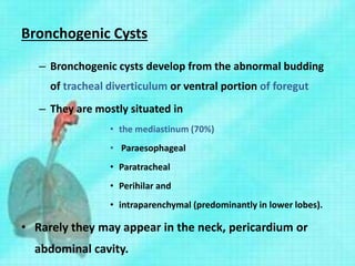
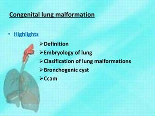
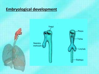
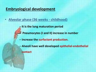
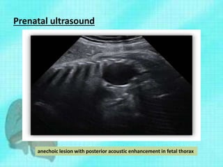
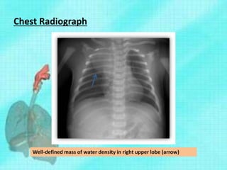
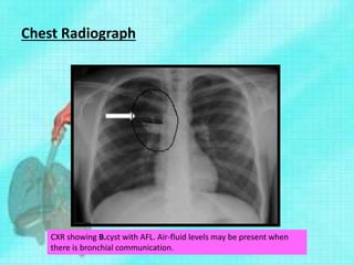
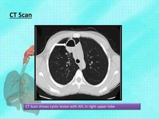
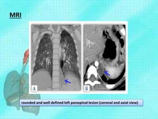
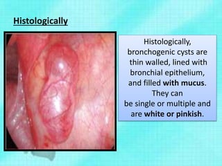
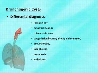
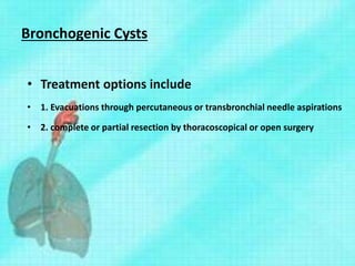
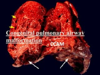
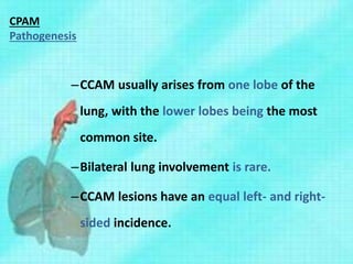
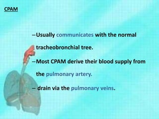
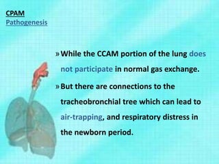
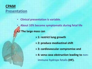
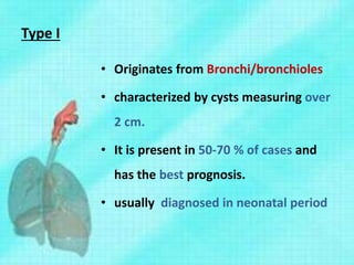
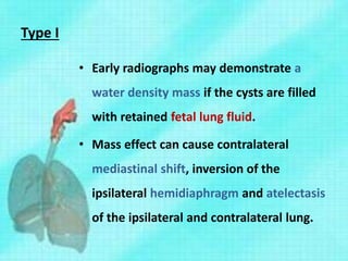
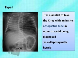
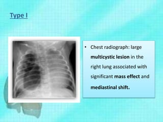
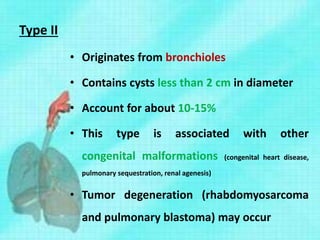
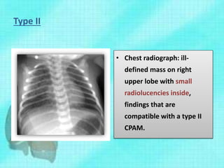
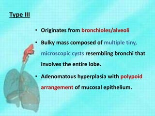
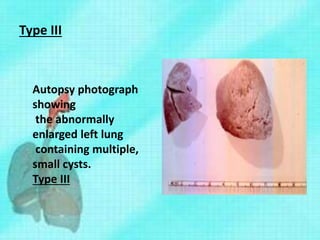
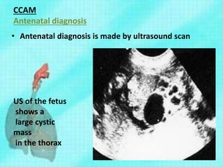
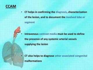
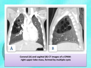
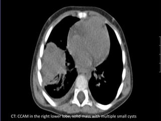
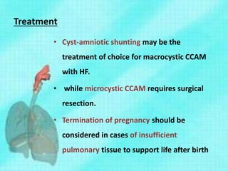
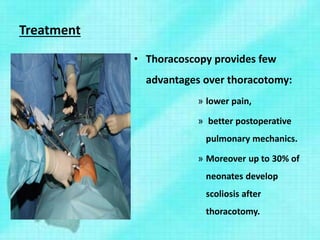
Presenter
Dr. Faheemul Hassan Andrabi
M Ch resident
Moderator
Dr. Aejaz A. Baba
Associateprofessor
PediatricSurgerySKIMS
Congenital lung malformation
• Highlights
Definition
Embryology of lung
Clasification of lung malformations
Bronchogenic cyst
Ccam
Congenital lung malformation
• Pulmonary malformations are also called congenital
anomalies of the primitive foregut
• Represent broad spectrum abnormalities with a
common origin at some point of the embryological
development of the foregut / lung.
Congenital lung malformation
• These anomalies are detected in most of cases in
infants and children
• Asymptomatic cases can be diagnosed in adults.
• Use of prenatal ultrasound has seen a large increase
in the diagnosis of these anomalies
Classification
• 1. Abnormal lung with normal vasculature
–congenital lobar hyperinflation
–bronchial atresia,
–bronchial cyst
• 2. Abnormal vasculature and normal lung
–arteriovenus malformation
Classification
• 3. Both parenchyma and vascular abnormalities
–pulmonary sequestration
–hypogenetic lung syndrome
Embryological development
• Development of lung has 5 stages
–Embryonic stage
–Pseudoglandular stage
–Canalicular stage
–Terminal sac period
–Alveolar period
Embryological development
• Embryonic phase (3-7 weeks)
– Begins with the formation of the respiratory
diverticulum from the ventral wall of the primitive
foregut.
– Around 33 days of gestation, the distal end of the
respiratory diverticulum bifurcates into two buds
(left and right primary bronchi)
Embryological development
• Pseudoglandular phase (5-17 weeks):
–Segmental and sub-segmental bronchi are
formed through a series of divisions
–Repeated branching forms upto terminal
bronchioles
–No respiratory bronchioles and alveoli are
present
Embryological development
• Canalicular phase (17-24 weeks)
–The diameter of the tube increases
–Respiratory bronchioles and alveolar ducts
develop.
–airspaces are being canalized and
approximated by a network of capillaries.
Embryological development
• Saccular phase (25-36 weeks)
–Primitive alveoli are formed
–By 26 weeks the alveoli have become
vascularised
Embryological development
• Alveolar phase (36 weeks - childhood)
– It is the lung maturation period
– Pneumocytes (I and II) increase in number
– increase the surfactant production.
– Alveoli have well developed epithelial-endothelial
contact
Embryological development
Pathogenesis
• It's difficult and in most of cases confusing to
understand the pathogenesis of lung malformations.
• The proposed theories are
– defective foregut budding and differentiation.
– airway obstruction with secondary pulmonary
dysplastic changes.
Types of broncopulmonary malformations
–Bronchogenic Cysts
–Congenital pulmonary airway malformation
–Pulmonary sequestration
–Bronchial atresia
–Congenital lobar hyperinflation
Bronchogenic Cysts
– Bronchogenic cysts develop from the abnormal budding
of tracheal diverticulum or ventral portion of foregut
– They are mostly situated in
• the mediastinum (70%)
• Paraesophageal
• Paratracheal
• Perihilar and
• intraparenchymal (predominantly in lower lobes).
• Rarely they may appear in the neck, pericardium or
abdominal cavity.
Bronchogenic Cysts
– Bronchogenic cysts are classified primarily
into intrapulmonary and mediastinal types
– Mediastinal is further classified by Maier into
5 types
• Type I (paratracheal),
• Type II (bronchial bifurcational),
• Type III (hilar),
• Type IV (paraesophageal) and
• Type V others
Bronchogenic Cysts
– It is usually
– unilocular,
– Has usually no communication with the bronchial
tree and
– Is filled with clear mucous secretion.
– It is almost always closely attached to major airways or
the esophagus by dense fibrous tissue
Bronchogenic Cysts
• Most bronchogenic cysts are asymptomatic and are
found incidentally.
• The symptoms are generally caused by compression
of the trachea, bronchi or esophagus, leading to
– wheezing, coughing
– stridor,
– dyspnea, and dysphagia.
– Atelectasis
Bronchogenic Cysts
• The clinical course of the cyst is strongly influenced
by the presence or absence of communication with
the parent bronchia.
• Non-communicating cysts cause symptoms by local
compression but in most of the cases are
asymptomatic.
Bronchogenic Cysts
• On the other hand a cyst-bronchial communication
can cause two complications
–tension cyst and
–infection
• A tension cyst is a rapidly expanding cyst which
causes acute distress and needs immediate
intervention
Bronchogenic Cysts
• Cyst infection is more frequent for the
intrapulmonary cysts
• They may present with
– Fever
– Cough
– sputum or even hemoptysis.
• Infections are recurrent and have poor response to
treatment
Bronchogenic Cysts
• Spontaneous pneumothorax is rare.
• Malignancy although rare but has been reported in
longstanding Bronchogenic cysts
Prenatal ultrasound
anechoic lesion with posterior acoustic enhancement in fetal thorax
Chest Radiograph
Well-defined mass of water density in right upper lobe (arrow)
Chest Radiograph
CXR showing B.cyst with AFL. Air-fluid levels may be present when
there is bronchial communication.
CT Scan
CT Scan shows cystic lesion with AFL in right upper lobe
MRI
rounded and well defined left paraspinal lesion (coronal and axial view)
Histologically
Histologically,
bronchogenic cysts are
thin walled, lined with
bronchial epithelium,
and filled with mucus.
They can
be single or multiple and
are white or pinkish.
Bronchogenic Cysts
• Differential diagnoses
• Foreign body
• Bronchial stenosis
• Lobar emphysema
• congenital pulmonary airway malformation,
• pneumatocele,
• lung abscess,
• pneumonia
• Hydatic cyst
Bronchogenic Cysts
• Treatment options include
• 1. Evacuations through percutaneous or transbronchial needle aspirations
• 2. complete or partial resection by thoracoscopical or open surgery
Bronchogenic Cysts
Treatment
• Needle aspiration is required
– in case of a tension cyst that expands rapidly
and can lead to death by acute
cardiorespiratory embarrassment.
• Otherwise the cyst should be surgically
removed.
Bronchogenic Cysts
Treatment
• For intrapulmonary cysts the excision
options are segmentectomy, lobectomy
or simple cyst removal.
• Extrapulmonary cysts can be removed by
thoracoscopic surgery.
Bronchogenic Cysts
Treatment
• The precise vascular supply to these
lesions is difficult to determine.
• Both extra- and intrapulmonary
bronchogenic cysts are supplied by
numerous small branches.
• Therefore special attention should be paid
during the dissection of the cyst.
Bronchogenic Cysts
Treatment
• When cyst is adherent to a vital structure (trachea,
main vessels, heart).
– can be partially resected and cyst epithelium
destroyed with electrocautery to prevent
recurrence or malignant degeneration.
•
Congenital pulmonary airway
malformation
CCAM
Congenital pulmonary airway malformation CPAM
• CCAM is characterized by an
“adenomatoid” increase of terminal
bronchioles that form cysts of various sizes.
• It is the most commonly diagnosed lung
malformation in prenatal period
• Accounts for about 25% of all congenital
bronchopulmonary malformations.
CPAM
• Incidence between 1/25000 and 1/35000.
• These malformations are characterized
– 1: by an abnormal branching of immature
bronchioles, with a lack of normal alveolar
development.
– 2: hamartomatous proliferation of cysts which
resemble bronchioles (airways without cartilage)
•
CPAM
Pathogenesis
• The cause of CCAM is unknown but the error occurs around sixth
week of gestation in the pseudoglandular stage of lung
development. The proposed theories of pathogenesis are
• increased cell proliferation and markedly decreased
apoptosis
• increased platelet-derived growth factor (PDGF) gene
expression
• FGF-10 (fibroblast growth factor) over expression in the proximal
tracheobronchial tree during the Pseudoglandular stage of
lung development.
CPAM
Pathogenesis
–CCAM usually arises from one lobe of the
lung, with the lower lobes being the most
common site.
–Bilateral lung involvement is rare.
–CCAM lesions have an equal left- and right-
sided incidence.
CPAM
–Usually communicates with the normal
tracheobronchial tree.
–Most CPAM derive their blood supply from
the pulmonary artery.
– drain via the pulmonary veins.
CPAM
Pathogenesis
• CCAM is distinguished from other lesions and normal
lung by following histological features
» (1) polypoid projections of the mucosa,
» (2) an increase in smooth muscle within cyst walls,
» (3) an absence of cartilage
» (4) the presence of mucus-secreting cells, and
» (5) the absence of inflammation.
CPAM
Pathogenesis
»While the CCAM portion of the lung does
not participate in normal gas exchange.
»But there are connections to the
tracheobronchial tree which can lead to
air-trapping, and respiratory distress in
the newborn period.
CPAM
Presentation
• Clinical presentation is variable.
• About 10% become symptomatic during fetal life
• The large mass can
» 1: restrict lung growth
» 2: produce mediastinal shift
» 3: cardiovascular compromise and
» 4: vena cava obstruction leading to non-
immune hydrops fetalis (HF).
CPAM
Presentation
• 60% of patients become symptomatic
within one month of life.
• another 10% between one and six
months
• and 15 % by adolescence
CCAM
Presentation
• The key symptom in neonatal period is
the respiratory distress.
• Outside of the neonatal period
recurrent pulmonary infections are the
presenting features of CCAM
CCAM
Stocker’s classification( according to the cysts size)
• Stocker defined three types of CCAM based on cyst
size,
–1: Single or multiple cysts, > 2 cm, lined by
pseudostratified columnar epithelium
–2: Single or multiple cysts <2 cm. Cuboidal
or columnar epithelial lining
–3: Predominantly solid lesions, with small
(under 0.5 cm) cysts, lined by cuboidal
epithelium
However, this categorization has little clinical relevance
Graphic representation of the congenital pulmonary
airway malformation (CPAM)
Type I
• Originates from Bronchi/bronchioles
• characterized by cysts measuring over
2 cm.
• It is present in 50-70 % of cases and
has the best prognosis.
• usually diagnosed in neonatal period
Type I
• Early radiographs may demonstrate a
water density mass if the cysts are filled
with retained fetal lung fluid.
• Mass effect can cause contralateral
mediastinal shift, inversion of the
ipsilateral hemidiaphragm and atelectasis
of the ipsilateral and contralateral lung.
Type I
It is essential to take
the X-ray with an in situ
nasogastric tube in
order to avoid being
diagnosed
as a diaphragmatic
hernia
Type I
• Chest radiograph: large
multicystic lesion in the
right lung associated with
significant mass effect and
mediastinal shift.
Type II
• Originates from bronchioles
• Contains cysts less than 2 cm in diameter
• Account for about 10-15%
• This type is associated with other
congenital malformations (congenital heart disease,
pulmonary sequestration, renal agenesis)
• Tumor degeneration (rhabdomyosarcoma
and pulmonary blastoma) may occur
Type II
• Chest radiograph: ill-
defined mass on right
upper lobe with small
radiolucencies inside,
findings that are
compatible with a type II
CPAM.
Type III
• Originates from bronchioles/alveoli
• Bulky mass composed of multiple tiny,
microscopic cysts resembling bronchi that
involves the entire lobe.
• Adenomatous hyperplasia with polypoid
arrangement of mucosal epithelium.
Type III
Autopsy photograph
showing
the abnormally
enlarged left lung
containing multiple,
small cysts.
Type III
CCAM
Antenatal diagnosis
• Antenatal diagnosis is made by ultrasound scan
US of the fetus
shows a
large cystic
mass
in the thorax
CCAM
Antenatal diagnosis
• Antenatal USG may show
– Polyhydramnios- Due to esophageal
compression
– Fetal hydrops -From venocaval
obstruction and cardiac compression
– Ascites
CCAM
• CT helps in confirming the diagnosis, characterization
of the lesion, and to document the involved lobe or
segment
• Intravenous contrast media must be used to define
the presence of any systemic arterial vessels
supplying the lesion
• CT also helps to diagnose other associated congenital
malformations
Coronal (A) and sagittal (B) CT images of a CPAM:
right upper lobe mass, formed by multiple cysts
CT: CCAM in the right lower lobe, solid mass with multiple small cysts
CCAM
• Other diagnostic means include (MRI) and
Bronchoscopy.
• One of the most recent developments in the
area is virtual bronchoscopy (VB).
• VB is a non-invasive three-dimensional (3D)
technique that uses multidetector CT-generated
image.
• It can evaluate the airways down to the sixth- to
seventh-generation bronchial subdivisions
CCAM
Appearance of CCAM on virtual bronchoscopy
Differential diagnoses
• Congenital lobar emphysema
• Bronchogenic cyst
• Bronchopulmonary sequestration
• Prior infection with pneumatocele
formation
• Diaphragmatic hernia
Treatment
• The natural history of antenatal
diagnosed CCAM is difficult to
determine accurately
• spontaneous resolution in utero of
an antenatally diagnosed CCAM is
reported to occur in up to 15% of
patients.
Treatment
• Although largely silent during fetal life
there are certain cases when fetal
intervention is required like:
• mediastinal shift
• lung hypoplasia.
• Cardiovascular compression
• presence of HF
Treatment
• In order to help predict whether a fetus
may be at risk of hydrops, Crumbleholme
et al. proposed sonographic CPAM
volume ratio (CVR)
• CVR= (CPAM Length × Height × Width ×
0.52)/head circumference.
Treatment
• A CVR >1.6 predicts fetal hydrops in 75%
of patients.
• postnatal intubation was required in 88%
of patients with CVR of >1.6, and survival
was only 53%.
Treatment
• The optimal type of fetal intervention is
still a debated subject. Treatment
options include:
»Fetal surgical resection
»Cyst -amniotic shunting
»Thoracentesis
»And steroid therapy.
Treatment
• Cyst-amniotic shunting may be the
treatment of choice for macrocystic CCAM
with HF.
• while microcystic CCAM requires surgical
resection.
• Termination of pregnancy should be
considered in cases of insufficient
pulmonary tissue to support life after birth
Treatment
• After birth management is dictated by the
clinical status of the new-born.
• Prompt surgical resection should be performed
in symptomatic newborn with respiratory
distress.
• In asymptomatic patients delaying resection
until infancy is reasonable and allows somatic
growth that may facilitate the ease of
pulmonary resection.
Treatment
• Surgical resection of CCAM lesions is
necessary even for asymptomatic
patients in order to:
» prevent infection
»and avoid malignant
transformation of the lesion
Treatment
• Excision of the CCAM is accomplished by
» Lobectomy
» segmentectomy and
» Pneumonectomy in certain cases
• The usual intervention is lobectomy via
thoracotomy or thoracoscopical approach.
• Lobectomy is preferred because of potential
early air-leak after segmentectomy and long-
term complications after pneumonectomy
Treatment
• Thoracoscopy provides few
advantages over thoracotomy:
» lower pain,
» better postoperative
pulmonary mechanics.
» Moreover up to 30% of
neonates develop
scoliosis after
thoracotomy.
Complications of surgery
• Early complications include:
» Sepsis
» Air leaks with pneumothorax,
» Bronchopleural fistula,
» Wound infections.
• Later complications and sequelae are
» Incomplete excision,
» asthma,
» pneumonia
Prognosis
• Prognosis depends of several factors such as:
» cyst volume
» the presence of complications during fetal life
» the presence and severity of early respiratory
distress.
» Histological type (type I has the best prognosis)
» microcystic lesions involve a relatively large
amount of lung tissue
Thank you.
drfaheemandrabi@gmail.com

Lung malformation part 1

  • 2.
    Presenter Dr. Faheemul HassanAndrabi M Ch resident Moderator Dr. Aejaz A. Baba Associateprofessor PediatricSurgerySKIMS
  • 3.
    Congenital lung malformation •Highlights Definition Embryology of lung Clasification of lung malformations Bronchogenic cyst Ccam
  • 4.
    Congenital lung malformation •Pulmonary malformations are also called congenital anomalies of the primitive foregut • Represent broad spectrum abnormalities with a common origin at some point of the embryological development of the foregut / lung.
  • 5.
    Congenital lung malformation •These anomalies are detected in most of cases in infants and children • Asymptomatic cases can be diagnosed in adults. • Use of prenatal ultrasound has seen a large increase in the diagnosis of these anomalies
  • 6.
    Classification • 1. Abnormallung with normal vasculature –congenital lobar hyperinflation –bronchial atresia, –bronchial cyst • 2. Abnormal vasculature and normal lung –arteriovenus malformation
  • 7.
    Classification • 3. Bothparenchyma and vascular abnormalities –pulmonary sequestration –hypogenetic lung syndrome
  • 8.
    Embryological development • Developmentof lung has 5 stages –Embryonic stage –Pseudoglandular stage –Canalicular stage –Terminal sac period –Alveolar period
  • 9.
    Embryological development • Embryonicphase (3-7 weeks) – Begins with the formation of the respiratory diverticulum from the ventral wall of the primitive foregut. – Around 33 days of gestation, the distal end of the respiratory diverticulum bifurcates into two buds (left and right primary bronchi)
  • 10.
  • 11.
    Embryological development • Pseudoglandularphase (5-17 weeks): –Segmental and sub-segmental bronchi are formed through a series of divisions –Repeated branching forms upto terminal bronchioles –No respiratory bronchioles and alveoli are present
  • 12.
    Embryological development • Canalicularphase (17-24 weeks) –The diameter of the tube increases –Respiratory bronchioles and alveolar ducts develop. –airspaces are being canalized and approximated by a network of capillaries.
  • 13.
    Embryological development • Saccularphase (25-36 weeks) –Primitive alveoli are formed –By 26 weeks the alveoli have become vascularised
  • 14.
    Embryological development • Alveolarphase (36 weeks - childhood) – It is the lung maturation period – Pneumocytes (I and II) increase in number – increase the surfactant production. – Alveoli have well developed epithelial-endothelial contact
  • 15.
  • 16.
    Pathogenesis • It's difficultand in most of cases confusing to understand the pathogenesis of lung malformations. • The proposed theories are – defective foregut budding and differentiation. – airway obstruction with secondary pulmonary dysplastic changes.
  • 17.
    Types of broncopulmonarymalformations –Bronchogenic Cysts –Congenital pulmonary airway malformation –Pulmonary sequestration –Bronchial atresia –Congenital lobar hyperinflation
  • 18.
    Bronchogenic Cysts – Bronchogeniccysts develop from the abnormal budding of tracheal diverticulum or ventral portion of foregut – They are mostly situated in • the mediastinum (70%) • Paraesophageal • Paratracheal • Perihilar and • intraparenchymal (predominantly in lower lobes). • Rarely they may appear in the neck, pericardium or abdominal cavity.
  • 19.
    Bronchogenic Cysts – Bronchogeniccysts are classified primarily into intrapulmonary and mediastinal types – Mediastinal is further classified by Maier into 5 types • Type I (paratracheal), • Type II (bronchial bifurcational), • Type III (hilar), • Type IV (paraesophageal) and • Type V others
  • 20.
    Bronchogenic Cysts – Itis usually – unilocular, – Has usually no communication with the bronchial tree and – Is filled with clear mucous secretion. – It is almost always closely attached to major airways or the esophagus by dense fibrous tissue
  • 21.
    Bronchogenic Cysts • Mostbronchogenic cysts are asymptomatic and are found incidentally. • The symptoms are generally caused by compression of the trachea, bronchi or esophagus, leading to – wheezing, coughing – stridor, – dyspnea, and dysphagia. – Atelectasis
  • 22.
    Bronchogenic Cysts • Theclinical course of the cyst is strongly influenced by the presence or absence of communication with the parent bronchia. • Non-communicating cysts cause symptoms by local compression but in most of the cases are asymptomatic.
  • 23.
    Bronchogenic Cysts • Onthe other hand a cyst-bronchial communication can cause two complications –tension cyst and –infection • A tension cyst is a rapidly expanding cyst which causes acute distress and needs immediate intervention
  • 24.
    Bronchogenic Cysts • Cystinfection is more frequent for the intrapulmonary cysts • They may present with – Fever – Cough – sputum or even hemoptysis. • Infections are recurrent and have poor response to treatment
  • 25.
    Bronchogenic Cysts • Spontaneouspneumothorax is rare. • Malignancy although rare but has been reported in longstanding Bronchogenic cysts
  • 26.
    Prenatal ultrasound anechoic lesionwith posterior acoustic enhancement in fetal thorax
  • 27.
    Chest Radiograph Well-defined massof water density in right upper lobe (arrow)
  • 28.
    Chest Radiograph CXR showingB.cyst with AFL. Air-fluid levels may be present when there is bronchial communication.
  • 29.
    CT Scan CT Scanshows cystic lesion with AFL in right upper lobe
  • 30.
    MRI rounded and welldefined left paraspinal lesion (coronal and axial view)
  • 31.
    Histologically Histologically, bronchogenic cysts are thinwalled, lined with bronchial epithelium, and filled with mucus. They can be single or multiple and are white or pinkish.
  • 32.
    Bronchogenic Cysts • Differentialdiagnoses • Foreign body • Bronchial stenosis • Lobar emphysema • congenital pulmonary airway malformation, • pneumatocele, • lung abscess, • pneumonia • Hydatic cyst
  • 33.
    Bronchogenic Cysts • Treatmentoptions include • 1. Evacuations through percutaneous or transbronchial needle aspirations • 2. complete or partial resection by thoracoscopical or open surgery
  • 34.
    Bronchogenic Cysts Treatment • Needleaspiration is required – in case of a tension cyst that expands rapidly and can lead to death by acute cardiorespiratory embarrassment. • Otherwise the cyst should be surgically removed.
  • 35.
    Bronchogenic Cysts Treatment • Forintrapulmonary cysts the excision options are segmentectomy, lobectomy or simple cyst removal. • Extrapulmonary cysts can be removed by thoracoscopic surgery.
  • 36.
    Bronchogenic Cysts Treatment • Theprecise vascular supply to these lesions is difficult to determine. • Both extra- and intrapulmonary bronchogenic cysts are supplied by numerous small branches. • Therefore special attention should be paid during the dissection of the cyst.
  • 37.
    Bronchogenic Cysts Treatment • Whencyst is adherent to a vital structure (trachea, main vessels, heart). – can be partially resected and cyst epithelium destroyed with electrocautery to prevent recurrence or malignant degeneration. •
  • 38.
  • 39.
    Congenital pulmonary airwaymalformation CPAM • CCAM is characterized by an “adenomatoid” increase of terminal bronchioles that form cysts of various sizes. • It is the most commonly diagnosed lung malformation in prenatal period • Accounts for about 25% of all congenital bronchopulmonary malformations.
  • 40.
    CPAM • Incidence between1/25000 and 1/35000. • These malformations are characterized – 1: by an abnormal branching of immature bronchioles, with a lack of normal alveolar development. – 2: hamartomatous proliferation of cysts which resemble bronchioles (airways without cartilage) •
  • 41.
    CPAM Pathogenesis • The causeof CCAM is unknown but the error occurs around sixth week of gestation in the pseudoglandular stage of lung development. The proposed theories of pathogenesis are • increased cell proliferation and markedly decreased apoptosis • increased platelet-derived growth factor (PDGF) gene expression • FGF-10 (fibroblast growth factor) over expression in the proximal tracheobronchial tree during the Pseudoglandular stage of lung development.
  • 42.
    CPAM Pathogenesis –CCAM usually arisesfrom one lobe of the lung, with the lower lobes being the most common site. –Bilateral lung involvement is rare. –CCAM lesions have an equal left- and right- sided incidence.
  • 43.
    CPAM –Usually communicates withthe normal tracheobronchial tree. –Most CPAM derive their blood supply from the pulmonary artery. – drain via the pulmonary veins.
  • 44.
    CPAM Pathogenesis • CCAM isdistinguished from other lesions and normal lung by following histological features » (1) polypoid projections of the mucosa, » (2) an increase in smooth muscle within cyst walls, » (3) an absence of cartilage » (4) the presence of mucus-secreting cells, and » (5) the absence of inflammation.
  • 45.
    CPAM Pathogenesis »While the CCAMportion of the lung does not participate in normal gas exchange. »But there are connections to the tracheobronchial tree which can lead to air-trapping, and respiratory distress in the newborn period.
  • 46.
    CPAM Presentation • Clinical presentationis variable. • About 10% become symptomatic during fetal life • The large mass can » 1: restrict lung growth » 2: produce mediastinal shift » 3: cardiovascular compromise and » 4: vena cava obstruction leading to non- immune hydrops fetalis (HF).
  • 47.
    CPAM Presentation • 60% ofpatients become symptomatic within one month of life. • another 10% between one and six months • and 15 % by adolescence
  • 48.
    CCAM Presentation • The keysymptom in neonatal period is the respiratory distress. • Outside of the neonatal period recurrent pulmonary infections are the presenting features of CCAM
  • 49.
    CCAM Stocker’s classification( accordingto the cysts size) • Stocker defined three types of CCAM based on cyst size, –1: Single or multiple cysts, > 2 cm, lined by pseudostratified columnar epithelium –2: Single or multiple cysts <2 cm. Cuboidal or columnar epithelial lining –3: Predominantly solid lesions, with small (under 0.5 cm) cysts, lined by cuboidal epithelium However, this categorization has little clinical relevance
  • 50.
    Graphic representation ofthe congenital pulmonary airway malformation (CPAM)
  • 51.
    Type I • Originatesfrom Bronchi/bronchioles • characterized by cysts measuring over 2 cm. • It is present in 50-70 % of cases and has the best prognosis. • usually diagnosed in neonatal period
  • 52.
    Type I • Earlyradiographs may demonstrate a water density mass if the cysts are filled with retained fetal lung fluid. • Mass effect can cause contralateral mediastinal shift, inversion of the ipsilateral hemidiaphragm and atelectasis of the ipsilateral and contralateral lung.
  • 53.
    Type I It isessential to take the X-ray with an in situ nasogastric tube in order to avoid being diagnosed as a diaphragmatic hernia
  • 54.
    Type I • Chestradiograph: large multicystic lesion in the right lung associated with significant mass effect and mediastinal shift.
  • 55.
    Type II • Originatesfrom bronchioles • Contains cysts less than 2 cm in diameter • Account for about 10-15% • This type is associated with other congenital malformations (congenital heart disease, pulmonary sequestration, renal agenesis) • Tumor degeneration (rhabdomyosarcoma and pulmonary blastoma) may occur
  • 56.
    Type II • Chestradiograph: ill- defined mass on right upper lobe with small radiolucencies inside, findings that are compatible with a type II CPAM.
  • 57.
    Type III • Originatesfrom bronchioles/alveoli • Bulky mass composed of multiple tiny, microscopic cysts resembling bronchi that involves the entire lobe. • Adenomatous hyperplasia with polypoid arrangement of mucosal epithelium.
  • 58.
    Type III Autopsy photograph showing theabnormally enlarged left lung containing multiple, small cysts. Type III
  • 59.
    CCAM Antenatal diagnosis • Antenataldiagnosis is made by ultrasound scan US of the fetus shows a large cystic mass in the thorax
  • 60.
    CCAM Antenatal diagnosis • AntenatalUSG may show – Polyhydramnios- Due to esophageal compression – Fetal hydrops -From venocaval obstruction and cardiac compression – Ascites
  • 61.
    CCAM • CT helpsin confirming the diagnosis, characterization of the lesion, and to document the involved lobe or segment • Intravenous contrast media must be used to define the presence of any systemic arterial vessels supplying the lesion • CT also helps to diagnose other associated congenital malformations
  • 62.
    Coronal (A) andsagittal (B) CT images of a CPAM: right upper lobe mass, formed by multiple cysts
  • 63.
    CT: CCAM inthe right lower lobe, solid mass with multiple small cysts
  • 64.
    CCAM • Other diagnosticmeans include (MRI) and Bronchoscopy. • One of the most recent developments in the area is virtual bronchoscopy (VB). • VB is a non-invasive three-dimensional (3D) technique that uses multidetector CT-generated image. • It can evaluate the airways down to the sixth- to seventh-generation bronchial subdivisions
  • 65.
    CCAM Appearance of CCAMon virtual bronchoscopy
  • 66.
    Differential diagnoses • Congenitallobar emphysema • Bronchogenic cyst • Bronchopulmonary sequestration • Prior infection with pneumatocele formation • Diaphragmatic hernia
  • 67.
    Treatment • The naturalhistory of antenatal diagnosed CCAM is difficult to determine accurately • spontaneous resolution in utero of an antenatally diagnosed CCAM is reported to occur in up to 15% of patients.
  • 68.
    Treatment • Although largelysilent during fetal life there are certain cases when fetal intervention is required like: • mediastinal shift • lung hypoplasia. • Cardiovascular compression • presence of HF
  • 69.
    Treatment • In orderto help predict whether a fetus may be at risk of hydrops, Crumbleholme et al. proposed sonographic CPAM volume ratio (CVR) • CVR= (CPAM Length × Height × Width × 0.52)/head circumference.
  • 70.
    Treatment • A CVR>1.6 predicts fetal hydrops in 75% of patients. • postnatal intubation was required in 88% of patients with CVR of >1.6, and survival was only 53%.
  • 71.
    Treatment • The optimaltype of fetal intervention is still a debated subject. Treatment options include: »Fetal surgical resection »Cyst -amniotic shunting »Thoracentesis »And steroid therapy.
  • 72.
    Treatment • Cyst-amniotic shuntingmay be the treatment of choice for macrocystic CCAM with HF. • while microcystic CCAM requires surgical resection. • Termination of pregnancy should be considered in cases of insufficient pulmonary tissue to support life after birth
  • 73.
    Treatment • After birthmanagement is dictated by the clinical status of the new-born. • Prompt surgical resection should be performed in symptomatic newborn with respiratory distress. • In asymptomatic patients delaying resection until infancy is reasonable and allows somatic growth that may facilitate the ease of pulmonary resection.
  • 74.
    Treatment • Surgical resectionof CCAM lesions is necessary even for asymptomatic patients in order to: » prevent infection »and avoid malignant transformation of the lesion
  • 75.
    Treatment • Excision ofthe CCAM is accomplished by » Lobectomy » segmentectomy and » Pneumonectomy in certain cases • The usual intervention is lobectomy via thoracotomy or thoracoscopical approach. • Lobectomy is preferred because of potential early air-leak after segmentectomy and long- term complications after pneumonectomy
  • 76.
    Treatment • Thoracoscopy providesfew advantages over thoracotomy: » lower pain, » better postoperative pulmonary mechanics. » Moreover up to 30% of neonates develop scoliosis after thoracotomy.
  • 77.
    Complications of surgery •Early complications include: » Sepsis » Air leaks with pneumothorax, » Bronchopleural fistula, » Wound infections. • Later complications and sequelae are » Incomplete excision, » asthma, » pneumonia
  • 78.
    Prognosis • Prognosis dependsof several factors such as: » cyst volume » the presence of complications during fetal life » the presence and severity of early respiratory distress. » Histological type (type I has the best prognosis) » microcystic lesions involve a relatively large amount of lung tissue
  • 79.