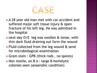
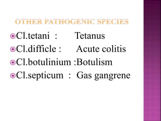
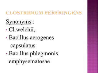
Clostridium
perfringens
Dr.V.S.Vatkar
Asso Pro
D Y Patil Medical College
Kolhapur
 A 28 year old man met with car accident and
suffered major soft tissue injury & open
fracture of his left leg. He was admitted in
the hospital
 next day O/E leg was swollen & tense, with
thin dark fluid draining out form the wound
 Fluid collected from the leg wound & send
for microbiological examination
 Gram stain : GPB (thick rods , no spores)
 Non motile, on B A : large β-hemolytic
colonies seen (anaerobic condition)
Anaerobic
Spore forming
Gram positive bacilli
“Kloster” meaning a
spindle
Kingdom: Bacteria
Division: Firmicutes
Class: Clostridia
Order: Clostridiales
Family: Clostridiaceae
Genus: Clostridium
Species: perfringens
Cl.tetani : Tetanus
Cl.difficle : Acute colitis
Cl.botulinium :Botulism
Cl.septicum : Gas gangrene
Synonyms :
• Cl.welchii,
• Bacillus aerogenes
capsulatus
• Bacillus phlegmonis
emphysematosae
Originally cultivated by Achalme (1891)
Welch & Nuttall (1892): isolated it form
blood & organs of a cadaver.
Normal inhabitant of the large intestine of
man and the most imp species, causing
diseases like Gas gangrene, food
poisoning and necrotic enteritis.
 Gram positive
bacillus, 4-6µ x 1µ
 Straight parallel sides
with rounded ends
 Occurs singly, in
chains or in small
bundles
 spores: central/
subterminal: rarely
seen in artificial
cultures or sample
collected from lesion
• Pleomorphic
• Capsulated, non-motile
Anaerobe, can grow under
microaerophilic conditions.
Culture do not die on exposure to air
Ph 5.0 to 8.0
Temp. 20°C to 50°C,opt temp=45 C,
generation time at this temp may be as
short as 10 min
Grows on Blood agar, RCM and on
Thioglycollate broth within 24-48 hrs.
In litmus milk : lactose fermentation
leads to acid formation, color of
litmus from blue to red. Acid
coagulates casein(acid clot) &
clotted milk disrupted due to
vigorosus gas production
Stormy fermentation: paraffin plug
pushed up
On blood agar
containing rabbit,
sheep or human :
overnight incubation
shows
 ‘Target haemolysis’:
narrow zone of
hemolysis due to θ
toxin
 Zone of hemolysis
fade on prolong
incubation
 Ferments glucose,maltose,lactose and sucrose
with production of acid and gas
 Indole : - ve
 M.R. : + ve
 V.P. : - ve
 H2S is formed abundantly
 Most strains reduce nitrates
 Spores usually destroyed within 5 minutes by
boiling but those of food poisoning strains of
Type A & Type C ,resist boiling for 1 -3 hours
 Spores are resistant to antiseptics &
disinfectants commonly used
 Autoclaving at 121°C for 15 minutes is lethal
Cl.perfringens produces at least
12 distinct toxins
Based on the four major one’s
(alpha,beta,epsilon and iota) , the
strains are classified into 5 types,
from A to E
Type A strains : produce α toxin
Type B strains : produce α,β and
epsilon
Type C strains : produce α and β
Type D strains : produce α and
epsilon
Type E strains : produce α and iota
toxins
Produced by all strains of Cl.perfringens
but predominantly by type A strains
Most imp. toxin biologically & is
responsible for toxemia of gas gangrene
Chemically it is a phospholipidase (lecithinase C),
which, in the presence of Ca++ and Mg++ ions,
splits
Lecithin
Phosphoryl choline Diglyceride
Alpha toxin is lethal, heat stable and
dermonecrotic
It is haemolytic for the red cells of most
species
The haemolytic action is due to its action
on phospholipids, situated on RBC
memb
Beta(β), epsilon(ε) and iota(ι)
toxins :lethal and necrotising
properties
Gamma(γ) : minor lethal actions
Delta(δ) toxin :lethal effect and is
haemolytic for RBC’s of
sheep,goat,pig and cattle
Theta (θ) toxin : an O2 labile
haemolysin and antigenically related to
streptolysin O, shows a cytolytic activity
Kappa(κ) toxin: a collagenase
Lambda(λ) toxin : a protienase &
gelatinase
Mu(μ) toxin : a hyaluronidase
Nu (v) toxin : a deoxyribonuclease
1) Enzymes which destroy the blood group
subs. A and H
2) A neuraminidase which destroys myxovirus
receptors on the RBC’s
3) A substance which renders the RBC’s
panagglutinable
4) A haemagglutinin active against human
RBC’s
5) Fibrinolysin
6) Haemolysin – distinct from α, ϴ and ɗ toxins
7) Histamine
8) A bursting factor with specific action
on muscle tissues
9) A circulatory factor which can cause an
increase in adrenaline sensitivity of the
capillary bed & can also inhibit
phagocytosis
 produced by some strains of type A causing
diarrhoea and other symptoms of food
poisoning
 It interacts with epithelial cell tight junction
proteins, leading to diarrhea and intestinal
cramping caused by leakage of water and ions
 Production of enterotoxin is co-regulated with
sporulation, the toxin is released when
vegetative cells undergo lysis.
The diseases caused ,range from simple
wound contamination to disease like Gas
gangrene
3 imp. pathogenic lesions caused are:
a. Gas gangrene
b. Food poisoning
c. Necrotising enteritis
As defined by Oakley in 1954 :
A rapidly spreading ,oedematous
myonecrosis,occuring in association
with severe wounds of extensive muscle
masses that have been contaminated
with pathogenic
Clostridia,esp.Cl.perfringens
Came into prominence in World war I
(1914), Disease of the war
In civilian life, the disease follows road
accidents and crush injuries
Rarely it may follow surgical operations
Clostridia enter the wounds along with
foreign particles like soil, dust, bits of
clothing and shrapnel
Cl.novy & Cl.septicum, Cl.histolyticum,
Cl.bifermentans also causes inf.
 MacLennan – 3 types of wound
infections
a. Simple wound contamination : no
invasion of underlying tissues
b. Anaerobic cellulitis : Cl. invade the
tissues but with min. toxin production
and without invasion of the muscles
c. Gas gangrene : Cl. invade the healthy
muscle tissues with abundant
formation of exotoxins
Crushing or tearing of an artery
Muscle anoxia
Capillary pressure increases
Blood supply decreases
Ph of damaged tissue falls
Breakdown of carbohydrates & liberation of amino acids
Ideal platform for proliferation of anaerobes
The Clostridia multiply and elaborate
toxins which further cause tissue damage
Muscle anoxia is due to increased
capillary permeability which is caused by
Lecithinases ,by damaging the cell
membranes
Haemolytic anaemia and haemoglobinuria is
due to the lysis of RBC’s by the alpha toxin
- High grade fever
- I P: 7 hrs to 6 wks after wounding
- brownish pus
- discolouration
- foul odour
- watery discharge from the wound
- Underlying tissue becomes crepitant
- death occurs due to circulatory failure
 Caused by some strains of type A which are
called as food poisoning strains (cooked
meat/poultry)
Ingestion of contaminated food
liberation of heat labile enterotoxin in
small intestine
After an IP of 8-24 hours, abdo.pain, diarrhoea
and vomiting set in
Necrotising enteritis
A severe and fatal necrotising jejunitis is caused by type C
strains of Cl.perfringens
Gangrenous appendicitis
Caused by Cl.perfringens type A and sometimes by
type D strains
Necrotising colitis
A rare but fatal disease in which large segments of
the colon become gangrenous
Biliary tract infections
Cl.perfringens has been reported to produce 2
rare but serious infections of the biliary tract :
1) Acute emphysematous
cholecystitis
2) Post- cholecystectomy
septicaemia
Panophthalmitis
occasionally following penetrating eye injuries
Thoracic infections
seen commonly as battle wound injuries than in
civilian ones
Urogenital infections
*infection of the urinary tract may sometimes
follow a surgical procedure like nephrectomy
*Cl.Infection of the uterus is a serious condition
asso.with septic abortion . Septicaemia is common
in this condition
Lab. diagnosis of gas gangrene :
Made primararily on clinical grounds.
The lab. provides only the confirmation
of clinical diagnosis, identification and
enumeration of the infecting organisms.
 Specimens to be collected :
1.Films from the muscles from the edge of the
affected area & from the tissues from the
necrotic area
2. A swab from the exudate in the deeper parts of
the wound
3. Necrotic tissue & muscle fragments
a) Direct microscopy :
Gram stained smears give presumptive
diagnosis about Clostridial species & their relative
numbers
Accordingly -
* Presence of large no. of regularly shaped gram + bacilli,
WITHOUT SPORES ,is strongly suggestive of
Cl.perfringens infection
* Citron bodies and boat or leaf shaped pleomorphic bacilli
with irregular staining – Cl.septicum
* Large bacilli with oval or sub terminal spores –
Cl.oedematiens
* Slender bacilli with round terminal spores – Cl.tetani or
Cl.tetanomorphum
 Microscopy also helps to distinguish gas
gangrene from anaerobic streptococcal
myositis – both are almost similar clinically in
the early stages of the disease.
In Strept.myositis ,large no. of pus cells &
streptococci are seen but not the bacilli while in
gas gangrene scanty pus cells & diverse
bacterial flora is seen
b) Culture
The specimens are inoculated on fresh and
heated blood agar and on RCM
. On blood agar – target haemolysis is seen
. after 24-48 hours .Most strains produce beta
haemolysis and few are nonhaemolytic
Nagler’s reaction :
A useful test for the rapid detection of Cl.perfringens
from clinical specimens
A plate of serum or egg yolk is used
Cl.perfringens is grown on a medium with
6% agar
5% Filde’s peptic digest of sheep blood
20 % Human serum
 On one half of the plate,
antitoxin is spread on the
surface & is kept for
incubation for 24 hours
 The colonies, on the side
with the antitoxin, show no
opacity, due to specific
neutralization of the alpha
toxin
 The colonies, on the side without
the antitoxin , will be surrounded
by an opacity
To differentiate from group B streptococci
- A blood agar plate is inoculated with single streak of
suspected Cl.perfringens
- Inoculate a gr.B strep. with a single streak at an angle
of 90°, within few mm from Cl.perfringens
Incubate the plate anaerobically for 24-48 hours
Positive : Arrow head of synergistic haemolysis
is seen
 Toxigenicity of the strain can be analyzed by animal
inoculation
0.1 ml of a 24 hr.growth in RCMM ,is injected IM into
a healthy guinea pig. The animal dies within 24 hours.
A control animal, protected with the antiserum, is also
included.
On autopsy, bacteria can be recovered from the heart
and spleen of the test animal.
Made by isolating heat resistant Cl.perfringens type A
from faeces & from food
RCM is inoculated and heated at 100°C for 30 mins.
After cooling it is incubated at 37°C for 18 hours.
Subcultured on selective medium and incubated
anaerobically
The isolates are identified by morphology ,c/c,
biochem.reactions and by Nagler’s reaction
1) Surgery :
Most imp. prophylactic & therapeutic method in
gas gangrene.
All damaged tissues should be removed & the
wound is flushed with antiseptic solution to remove
blood clots, necrotic tissues and foreign materials
2) Antibiotics :
Penicillin's are known to be particularly active
against C. perfringens. Resistance to penicillin
is very rare and β- lactamase has not been
demonstrated in C. perfringens.
A low degree of resistance to ampicillin has been
reported in several studies
Tetracycline resistance is the most common antibiotic
resistance trait found in C. perfringens . It has been
reported that C. perfringens can carry the tetracycline-
resistance genes tetA(P), tetB(P), tetM and tetQ
Resistance to MLS antibiotics is also a common trait. It
is caused by methylation of the 23S rRNA gene
Chloramphenicol resistance in C. perfringens is not
common, though several resistant isolates have been
identified
3) Antitoxins :
Passive immunization with polyvalent
antitoxin (anti gas gangrene serum) is given as
a prophylactic measure in gas gangrene.
The dose given –
10,000 U of Cl.per
10,000 U of Cl.oede I.M.or I.V
5,000 U of Cl.sep
4) Hyperbaric oxygen :
Introduced in the depth of the wound
to reduce anaerobiosis
5) Active immunization :
with toxoids, to induce antitoxic response
HEPTAVAC
Contains 7:1 Clostridial vaccine
It is an opaque fluid vaccine containing purified toxoids
of Cl.perfringens,types B,C and D , together with
formalin killed cells and a purified formal toxoid
The antigen is absorbed into aluminum hydroxide & is
preserved with 0.013 % thiomersal BP
A multiplex PCR which can detect all the
major toxin genes of Cl.perfringens is
developed
Also PCR system for the detection of 2
other imp. toxin genes ,cpe and cpb2 is
also available
 For more detailed characterization of
Cl.perfringens isolates, diff.methods
have been used .
1. Plasmid profiling
2. Multilocus enzyme electrophoresis
(MLEE)
3. Ribotyping
4. Amplified fragment length polymorphism
(AFLP)
THANK YOU

5.cl.perfringens

  • 1.
  • 2.
     A 28year old man met with car accident and suffered major soft tissue injury & open fracture of his left leg. He was admitted in the hospital  next day O/E leg was swollen & tense, with thin dark fluid draining out form the wound  Fluid collected from the leg wound & send for microbiological examination  Gram stain : GPB (thick rods , no spores)  Non motile, on B A : large β-hemolytic colonies seen (anaerobic condition)
  • 3.
    Anaerobic Spore forming Gram positivebacilli “Kloster” meaning a spindle
  • 4.
    Kingdom: Bacteria Division: Firmicutes Class:Clostridia Order: Clostridiales Family: Clostridiaceae Genus: Clostridium Species: perfringens
  • 5.
    Cl.tetani : Tetanus Cl.difficle: Acute colitis Cl.botulinium :Botulism Cl.septicum : Gas gangrene
  • 6.
    Synonyms : • Cl.welchii, •Bacillus aerogenes capsulatus • Bacillus phlegmonis emphysematosae
  • 7.
    Originally cultivated byAchalme (1891) Welch & Nuttall (1892): isolated it form blood & organs of a cadaver. Normal inhabitant of the large intestine of man and the most imp species, causing diseases like Gas gangrene, food poisoning and necrotic enteritis.
  • 8.
     Gram positive bacillus,4-6µ x 1µ  Straight parallel sides with rounded ends  Occurs singly, in chains or in small bundles  spores: central/ subterminal: rarely seen in artificial cultures or sample collected from lesion • Pleomorphic • Capsulated, non-motile
  • 9.
    Anaerobe, can growunder microaerophilic conditions. Culture do not die on exposure to air Ph 5.0 to 8.0 Temp. 20°C to 50°C,opt temp=45 C, generation time at this temp may be as short as 10 min Grows on Blood agar, RCM and on Thioglycollate broth within 24-48 hrs.
  • 10.
    In litmus milk: lactose fermentation leads to acid formation, color of litmus from blue to red. Acid coagulates casein(acid clot) & clotted milk disrupted due to vigorosus gas production Stormy fermentation: paraffin plug pushed up
  • 11.
    On blood agar containingrabbit, sheep or human : overnight incubation shows  ‘Target haemolysis’: narrow zone of hemolysis due to θ toxin  Zone of hemolysis fade on prolong incubation
  • 12.
     Ferments glucose,maltose,lactoseand sucrose with production of acid and gas  Indole : - ve  M.R. : + ve  V.P. : - ve  H2S is formed abundantly  Most strains reduce nitrates
  • 13.
     Spores usuallydestroyed within 5 minutes by boiling but those of food poisoning strains of Type A & Type C ,resist boiling for 1 -3 hours  Spores are resistant to antiseptics & disinfectants commonly used  Autoclaving at 121°C for 15 minutes is lethal
  • 14.
    Cl.perfringens produces atleast 12 distinct toxins Based on the four major one’s (alpha,beta,epsilon and iota) , the strains are classified into 5 types, from A to E
  • 15.
    Type A strains: produce α toxin Type B strains : produce α,β and epsilon Type C strains : produce α and β Type D strains : produce α and epsilon Type E strains : produce α and iota toxins
  • 16.
    Produced by allstrains of Cl.perfringens but predominantly by type A strains Most imp. toxin biologically & is responsible for toxemia of gas gangrene
  • 17.
    Chemically it isa phospholipidase (lecithinase C), which, in the presence of Ca++ and Mg++ ions, splits Lecithin Phosphoryl choline Diglyceride
  • 18.
    Alpha toxin islethal, heat stable and dermonecrotic It is haemolytic for the red cells of most species The haemolytic action is due to its action on phospholipids, situated on RBC memb
  • 19.
    Beta(β), epsilon(ε) andiota(ι) toxins :lethal and necrotising properties Gamma(γ) : minor lethal actions Delta(δ) toxin :lethal effect and is haemolytic for RBC’s of sheep,goat,pig and cattle
  • 20.
    Theta (θ) toxin: an O2 labile haemolysin and antigenically related to streptolysin O, shows a cytolytic activity Kappa(κ) toxin: a collagenase Lambda(λ) toxin : a protienase & gelatinase Mu(μ) toxin : a hyaluronidase Nu (v) toxin : a deoxyribonuclease
  • 21.
    1) Enzymes whichdestroy the blood group subs. A and H 2) A neuraminidase which destroys myxovirus receptors on the RBC’s 3) A substance which renders the RBC’s panagglutinable 4) A haemagglutinin active against human RBC’s 5) Fibrinolysin 6) Haemolysin – distinct from α, ϴ and ɗ toxins
  • 22.
    7) Histamine 8) Abursting factor with specific action on muscle tissues 9) A circulatory factor which can cause an increase in adrenaline sensitivity of the capillary bed & can also inhibit phagocytosis
  • 23.
     produced bysome strains of type A causing diarrhoea and other symptoms of food poisoning  It interacts with epithelial cell tight junction proteins, leading to diarrhea and intestinal cramping caused by leakage of water and ions  Production of enterotoxin is co-regulated with sporulation, the toxin is released when vegetative cells undergo lysis.
  • 24.
    The diseases caused,range from simple wound contamination to disease like Gas gangrene 3 imp. pathogenic lesions caused are: a. Gas gangrene b. Food poisoning c. Necrotising enteritis
  • 25.
    As defined byOakley in 1954 : A rapidly spreading ,oedematous myonecrosis,occuring in association with severe wounds of extensive muscle masses that have been contaminated with pathogenic Clostridia,esp.Cl.perfringens
  • 26.
    Came into prominencein World war I (1914), Disease of the war In civilian life, the disease follows road accidents and crush injuries Rarely it may follow surgical operations Clostridia enter the wounds along with foreign particles like soil, dust, bits of clothing and shrapnel Cl.novy & Cl.septicum, Cl.histolyticum, Cl.bifermentans also causes inf.
  • 27.
     MacLennan –3 types of wound infections a. Simple wound contamination : no invasion of underlying tissues b. Anaerobic cellulitis : Cl. invade the tissues but with min. toxin production and without invasion of the muscles c. Gas gangrene : Cl. invade the healthy muscle tissues with abundant formation of exotoxins
  • 28.
    Crushing or tearingof an artery Muscle anoxia Capillary pressure increases Blood supply decreases Ph of damaged tissue falls Breakdown of carbohydrates & liberation of amino acids Ideal platform for proliferation of anaerobes
  • 29.
    The Clostridia multiplyand elaborate toxins which further cause tissue damage Muscle anoxia is due to increased capillary permeability which is caused by Lecithinases ,by damaging the cell membranes Haemolytic anaemia and haemoglobinuria is due to the lysis of RBC’s by the alpha toxin
  • 30.
    - High gradefever - I P: 7 hrs to 6 wks after wounding - brownish pus - discolouration - foul odour - watery discharge from the wound - Underlying tissue becomes crepitant - death occurs due to circulatory failure
  • 32.
     Caused bysome strains of type A which are called as food poisoning strains (cooked meat/poultry) Ingestion of contaminated food liberation of heat labile enterotoxin in small intestine After an IP of 8-24 hours, abdo.pain, diarrhoea and vomiting set in
  • 34.
    Necrotising enteritis A severeand fatal necrotising jejunitis is caused by type C strains of Cl.perfringens
  • 35.
    Gangrenous appendicitis Caused byCl.perfringens type A and sometimes by type D strains Necrotising colitis A rare but fatal disease in which large segments of the colon become gangrenous Biliary tract infections Cl.perfringens has been reported to produce 2 rare but serious infections of the biliary tract : 1) Acute emphysematous cholecystitis 2) Post- cholecystectomy septicaemia
  • 36.
    Panophthalmitis occasionally following penetratingeye injuries Thoracic infections seen commonly as battle wound injuries than in civilian ones Urogenital infections *infection of the urinary tract may sometimes follow a surgical procedure like nephrectomy *Cl.Infection of the uterus is a serious condition asso.with septic abortion . Septicaemia is common in this condition
  • 37.
    Lab. diagnosis ofgas gangrene : Made primararily on clinical grounds. The lab. provides only the confirmation of clinical diagnosis, identification and enumeration of the infecting organisms.
  • 38.
     Specimens tobe collected : 1.Films from the muscles from the edge of the affected area & from the tissues from the necrotic area 2. A swab from the exudate in the deeper parts of the wound 3. Necrotic tissue & muscle fragments
  • 39.
    a) Direct microscopy: Gram stained smears give presumptive diagnosis about Clostridial species & their relative numbers Accordingly - * Presence of large no. of regularly shaped gram + bacilli, WITHOUT SPORES ,is strongly suggestive of Cl.perfringens infection * Citron bodies and boat or leaf shaped pleomorphic bacilli with irregular staining – Cl.septicum * Large bacilli with oval or sub terminal spores – Cl.oedematiens * Slender bacilli with round terminal spores – Cl.tetani or Cl.tetanomorphum
  • 40.
     Microscopy alsohelps to distinguish gas gangrene from anaerobic streptococcal myositis – both are almost similar clinically in the early stages of the disease. In Strept.myositis ,large no. of pus cells & streptococci are seen but not the bacilli while in gas gangrene scanty pus cells & diverse bacterial flora is seen
  • 41.
    b) Culture The specimensare inoculated on fresh and heated blood agar and on RCM . On blood agar – target haemolysis is seen . after 24-48 hours .Most strains produce beta haemolysis and few are nonhaemolytic
  • 42.
    Nagler’s reaction : Auseful test for the rapid detection of Cl.perfringens from clinical specimens A plate of serum or egg yolk is used Cl.perfringens is grown on a medium with 6% agar 5% Filde’s peptic digest of sheep blood 20 % Human serum
  • 43.
     On onehalf of the plate, antitoxin is spread on the surface & is kept for incubation for 24 hours  The colonies, on the side with the antitoxin, show no opacity, due to specific neutralization of the alpha toxin  The colonies, on the side without the antitoxin , will be surrounded by an opacity
  • 44.
    To differentiate fromgroup B streptococci - A blood agar plate is inoculated with single streak of suspected Cl.perfringens - Inoculate a gr.B strep. with a single streak at an angle of 90°, within few mm from Cl.perfringens Incubate the plate anaerobically for 24-48 hours Positive : Arrow head of synergistic haemolysis is seen
  • 46.
     Toxigenicity ofthe strain can be analyzed by animal inoculation 0.1 ml of a 24 hr.growth in RCMM ,is injected IM into a healthy guinea pig. The animal dies within 24 hours. A control animal, protected with the antiserum, is also included. On autopsy, bacteria can be recovered from the heart and spleen of the test animal.
  • 47.
    Made by isolatingheat resistant Cl.perfringens type A from faeces & from food RCM is inoculated and heated at 100°C for 30 mins. After cooling it is incubated at 37°C for 18 hours. Subcultured on selective medium and incubated anaerobically The isolates are identified by morphology ,c/c, biochem.reactions and by Nagler’s reaction
  • 48.
    1) Surgery : Mostimp. prophylactic & therapeutic method in gas gangrene. All damaged tissues should be removed & the wound is flushed with antiseptic solution to remove blood clots, necrotic tissues and foreign materials
  • 49.
    2) Antibiotics : Penicillin'sare known to be particularly active against C. perfringens. Resistance to penicillin is very rare and β- lactamase has not been demonstrated in C. perfringens. A low degree of resistance to ampicillin has been reported in several studies
  • 50.
    Tetracycline resistance isthe most common antibiotic resistance trait found in C. perfringens . It has been reported that C. perfringens can carry the tetracycline- resistance genes tetA(P), tetB(P), tetM and tetQ Resistance to MLS antibiotics is also a common trait. It is caused by methylation of the 23S rRNA gene Chloramphenicol resistance in C. perfringens is not common, though several resistant isolates have been identified
  • 51.
    3) Antitoxins : Passiveimmunization with polyvalent antitoxin (anti gas gangrene serum) is given as a prophylactic measure in gas gangrene. The dose given – 10,000 U of Cl.per 10,000 U of Cl.oede I.M.or I.V 5,000 U of Cl.sep
  • 52.
    4) Hyperbaric oxygen: Introduced in the depth of the wound to reduce anaerobiosis 5) Active immunization : with toxoids, to induce antitoxic response
  • 53.
    HEPTAVAC Contains 7:1 Clostridialvaccine It is an opaque fluid vaccine containing purified toxoids of Cl.perfringens,types B,C and D , together with formalin killed cells and a purified formal toxoid The antigen is absorbed into aluminum hydroxide & is preserved with 0.013 % thiomersal BP
  • 54.
    A multiplex PCRwhich can detect all the major toxin genes of Cl.perfringens is developed Also PCR system for the detection of 2 other imp. toxin genes ,cpe and cpb2 is also available
  • 55.
     For moredetailed characterization of Cl.perfringens isolates, diff.methods have been used . 1. Plasmid profiling 2. Multilocus enzyme electrophoresis (MLEE) 3. Ribotyping 4. Amplified fragment length polymorphism (AFLP)
  • 56.