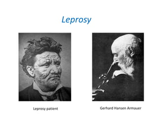
Leprosy
Dr. Kanwal Deep Singh Lyall
M.D. Microbiology
Leprosy
Gerhard Hansen ArmauerLeprosy patient
• Dard me kuch baat hai
• Ask the joy of pain from a leper
• Dates back to ancient Egypt in 4000 BC and
was discussed by Hippocrates in 460 BC.
• The earliest proven human case - 1-50 AD
• Mentioned in Bible
• Kushta in Sushta Samitha – 600 BC in India
• Hansen's disease
• Chronic infection caused Mycobacterium leprae
• Latin word Lepra, which means "scaly"
• Discovered by G. Hansen Armauer in Norway in
1873
• 1st bacterium to be identified as causing disease
in humans
• Non- cultivable
• Man alone gets leprosy and is reservoir of infection
• But in Americans – 9 banded armadillo
• Extremely slow generation time-12 to 13 days.
• Only bacillary diseases with predilection for nerves.
• Disease spectrum ranges from complete absence of
resistance to effective immunity.
• No satisfactory way of detecting past or unapparent
present infection.
Epidemiology
• One of the major health problems of
developing countries.
• Estimated prevalence is >1 case per 1000 of
population.
• High prevalence – India (60% of annual cases),
China, Myanmar, Indonesia, Brazil, Nigeria,
Madagascar, and Nepal.
• In India - Orissa and Bihar – highest prevalence
• Nasal droplets, contact with infected soil, &
even insect vectors
• Through upper resp. tract or skin
• Not highly contagious
• 95% population has innate immunity against it
• Not associated with AIDS (long incubation
period – 2 – 40 years)
Gen. Characteristics
• Obligate intracellular parasite,
• Usually present in parallel bundles of 50 or more
organisms bound by lipid like substances GLIA, called
GLOBI.
• The parallel rows of Globi present a cigar bundle
appearance. Seen inside the histiocytes.
• Bacteria divides by binary fission.
Morphology
• Slender, slightly curved or straight.
• Non motile, non sporing, gram +ve
• Acid fast bacilli , resist decolourisation with 5% H2SO4
• Bacteria are seen singly or in groups, intracellularly
and lying free outside the cell.
Antigenic structure
• Cell wall – 4 layers
1. Innermost – peptidogylcan – shape & rigidity
2. Lipoarabinomanan – B (LAM – B) – highly
immunogenic – serodiagnosis
3. Mycolic acid
4. Outermost – mycosides (has phenolic glycolipid 1
- PGL-1) – protects against enzymes and
suppresses CMI – prediliction to Schwann cells
Cultivation
• No genes for catabolic & respiratory pathways;
transport systems; purine, methionine, &
glutamine synthesis; & nitrogen regulation.
• Non-cultivable
• Non-confirmed reports of growth
• ICRC – ICRC bacillus
• Animals: Mouse, Armadillo, Chimpanzees,
Monkeys, etc.
Mouse
• Uses:
– to do susceptibility to chemotherapeutic agents
– determining the viability of organism
• Disadvantages:
– limited multiplication – insufficient yield
– Short life span – can not study pathogenesis
Armadillo
• Highly susceptible
• Long life span (12 – 15 yrs)
• Low body t°
• High yield
• Disadvantages :
– Only 40% susceptible
– High cost
– Wild armadillos – naturally infected
Pathogenesis
• Chronic granulomatous disease
• Humans – only source
• Localise primarily in skin, peripheral nerves &
nasal mucosa
• Has preference for low t°
• Long incubation & prolonged contact required
• Four types : Lepromatous, Tuberculoid,
Dimorphous, Intermediate
Lepromatous :
• Generalized form
• In people with low immunity
• More infectious
• Lepromin test is negative
• High level of Abs
• Nodular lesions, slow
thickening of peripheral
nerves
• Loss of sensation – trauma –
ulcers - 2° infections
• Continuous bacterimia
Tuberculoid
• Localized form
• In people with high immunity
• Lepromin test is positive
• Low level of Abs
• few non-elevated hypo- or
hyperpigmented macular
patches
• Initially peripherla nerve
involvement
• Later bigger nerves
• Nerves – thickened ,
hardened, tender
• Dimorphous :
– Clinically tuberculoid, but bacteriological &
immunologically lepromatous
– May shift to any extreme
• Intermediate :
– Unstable tissue reaction
– Neither tuberculoid, nor lepromatous
– Regress spontaneously or May shift to any extreme
Reversal (type 1) reactions. Erythematous, oedematous lesions.
Immune reactions
Type 1 (Downgrading &
reversal)
– Borderline leprosy patients→
CMI hypersensitivity →
tuberculoid shift
– Downgrading – pre Rx
(histological become
lepromatous)
– Reversal – post Rx
– Painful tender nerves, loss of
function, Swollen skin lesions,
New skin lesions
– Prednisolone 40 mg, reducing
over 3–6 months
Type 2 (Erythema nodosum leprosum)
– Lepromatous patients undergoing
treatment
– Bacilli die → release Ags → Ag-Ab
complexes
• Tender papules & nodules; may
ulcerate, painful tender nerves,
loss of function, iritis, orchitis,
myositis, lymphadenitis, Fever,
oedema
• Moderate: prednisolone 40mg od
• Severe: thalidomide 2 or
prednisolone 40–80 mg od,
reducing over 1–6 months
Ridley & Jopling’s classification
• Tuberculoid tuberculoid (TT)
• Borderline tuberculoid (BT)
• Borderline borderline (BB)
• Borderline lepromatous (BL)
• Lepromatous lepromatous (LL)
Tuberculoid
Lepromatous
Lepromin test
• Integral lepromin
• Bacillary lepromin
• 0.5 ml ID injection
• Early reaction of Fernandez: erythema &
induration within 24 - 48 hrs, remains 3 – 5
days
• Late reaction of Mitsuda: 1 -2 weeks after inj. ,
peak 4 weeks , nodule – ulecration – healing
Uses
1. Classification of leprosy: +ve in tuberculoid &
-ve in lepromatous
2. Assessment of prognosis : +ve = good
prognosis
3. Assessment of resistance : +ve = resistance
Field workers should be +ve
Laboratory diagnosis
Specimens
• From nasal mucosa, skin lesions, ear lobules
• Slit & scrape method
• Skin biopsy
• Nerve biopsy
• 6-7 sites sampled
Staining
• ZN stain (5% H2SO4)
• AFB arranged in parallel bundles within macrophages
(Lepra cell)
Grading
1 – 10 bacilli in 100 fields 1+
1 – 10 bacilli in 10 fields 2+
1 – 10 bacilli per field 3+
10 - 100 bacilli per field 4+
100 – 1000 bacilli per field 5+
> 1000 bacilli, clumps & Globi in every field 6+
Procedure : slit and scrape method
• Clean selected portion of skin with spirit
• Hold the skin pinched up & raised b/w thumb &
index finger of left hand
• Make an incision (5 mm long, 3 mm deep )
• Blade of scalpel turned at right angle to slit
• Bottom & sides of slit scraped
• Make smear → stain → examine
Interpretation
• LL : 6+ or 5+
• BT : 0 - 2+
• TT : 0
• Detects bacilli present at a conc. > 104/gm of skin
• In untreated patients earlobes yield the greatest
number.
Nasal scrapings
• Scrapings from nasal septum
• Nasal secretions collected by blowing nose into
polyethylene handkerchief
Interpretation :
• +ve in LL,BL type
• -ve in BB, BT, TT
• In patients of LL on chemotherapy: -ve earlier
than skin smears
• To decide whether patient is infectious or not
Skin and nerve biopsy
• Skin biopsy - active edge of patches
• Nerve biopsy - thickened nerve
Indications :
• When diagnosis is uncertain
• For accurate classification
• In TT or indeterminate when sensory impairment
is not marked, as in children
• To assess progress of treatment
• To differentiate between downgrading and
reversal reaction
Interpretation
• TT : Typical tubercles in dermis comprising of
epithelioid cells, langrhan giant cells and
surrounding zone of lymphocytes
• LL : Diffuse highly bacilliferous granuloma
(leproma) in dermis mainly consist of
macrophages.
• Live forms appear solid and uniformly stained.
• Dead or dying forms appear fragmented,
beaded and granular
• Bacteriological index
– BI = Total pluses / no. of smears examined
• Morphological index
– MI = % of uniformly stained bacilli out of total
bacilli counted
• Skin & nerve biopsy – histological
confirmation
• Animal inoculation
• Lepromin test
• Serological tests
Treatment
• Pucibacillary (TT,BT):
– Rifampicin 600 mg once a month +
– Dapsone 100 mg OD
• Multibacillary (BB, BL, LL)
– Rifampicin 600 mg once a month +
– Dapsone 100 mg OD +
– Clofazimine 50 mg OD
• Immunotherapy
• Vaccines
x 6 months
x 2 years or
skin smears –ve
Prevention
• Early diagnosis & treatment
• Surveillance of contacts
• Health education
• Vaccines
X

M leprae

  • 1.
    Leprosy Dr. Kanwal DeepSingh Lyall M.D. Microbiology
  • 2.
  • 3.
    • Dard mekuch baat hai • Ask the joy of pain from a leper
  • 4.
    • Dates backto ancient Egypt in 4000 BC and was discussed by Hippocrates in 460 BC. • The earliest proven human case - 1-50 AD • Mentioned in Bible • Kushta in Sushta Samitha – 600 BC in India
  • 5.
    • Hansen's disease •Chronic infection caused Mycobacterium leprae • Latin word Lepra, which means "scaly" • Discovered by G. Hansen Armauer in Norway in 1873 • 1st bacterium to be identified as causing disease in humans • Non- cultivable
  • 6.
    • Man alonegets leprosy and is reservoir of infection • But in Americans – 9 banded armadillo • Extremely slow generation time-12 to 13 days. • Only bacillary diseases with predilection for nerves. • Disease spectrum ranges from complete absence of resistance to effective immunity. • No satisfactory way of detecting past or unapparent present infection.
  • 7.
    Epidemiology • One ofthe major health problems of developing countries. • Estimated prevalence is >1 case per 1000 of population. • High prevalence – India (60% of annual cases), China, Myanmar, Indonesia, Brazil, Nigeria, Madagascar, and Nepal. • In India - Orissa and Bihar – highest prevalence
  • 8.
    • Nasal droplets,contact with infected soil, & even insect vectors • Through upper resp. tract or skin • Not highly contagious • 95% population has innate immunity against it • Not associated with AIDS (long incubation period – 2 – 40 years)
  • 9.
    Gen. Characteristics • Obligateintracellular parasite, • Usually present in parallel bundles of 50 or more organisms bound by lipid like substances GLIA, called GLOBI. • The parallel rows of Globi present a cigar bundle appearance. Seen inside the histiocytes. • Bacteria divides by binary fission.
  • 10.
    Morphology • Slender, slightlycurved or straight. • Non motile, non sporing, gram +ve • Acid fast bacilli , resist decolourisation with 5% H2SO4 • Bacteria are seen singly or in groups, intracellularly and lying free outside the cell.
  • 11.
    Antigenic structure • Cellwall – 4 layers 1. Innermost – peptidogylcan – shape & rigidity 2. Lipoarabinomanan – B (LAM – B) – highly immunogenic – serodiagnosis 3. Mycolic acid 4. Outermost – mycosides (has phenolic glycolipid 1 - PGL-1) – protects against enzymes and suppresses CMI – prediliction to Schwann cells
  • 12.
    Cultivation • No genesfor catabolic & respiratory pathways; transport systems; purine, methionine, & glutamine synthesis; & nitrogen regulation. • Non-cultivable • Non-confirmed reports of growth • ICRC – ICRC bacillus • Animals: Mouse, Armadillo, Chimpanzees, Monkeys, etc.
  • 13.
    Mouse • Uses: – todo susceptibility to chemotherapeutic agents – determining the viability of organism • Disadvantages: – limited multiplication – insufficient yield – Short life span – can not study pathogenesis
  • 14.
    Armadillo • Highly susceptible •Long life span (12 – 15 yrs) • Low body t° • High yield • Disadvantages : – Only 40% susceptible – High cost – Wild armadillos – naturally infected
  • 15.
    Pathogenesis • Chronic granulomatousdisease • Humans – only source • Localise primarily in skin, peripheral nerves & nasal mucosa • Has preference for low t° • Long incubation & prolonged contact required • Four types : Lepromatous, Tuberculoid, Dimorphous, Intermediate
  • 16.
    Lepromatous : • Generalizedform • In people with low immunity • More infectious • Lepromin test is negative • High level of Abs • Nodular lesions, slow thickening of peripheral nerves • Loss of sensation – trauma – ulcers - 2° infections • Continuous bacterimia Tuberculoid • Localized form • In people with high immunity • Lepromin test is positive • Low level of Abs • few non-elevated hypo- or hyperpigmented macular patches • Initially peripherla nerve involvement • Later bigger nerves • Nerves – thickened , hardened, tender
  • 17.
    • Dimorphous : –Clinically tuberculoid, but bacteriological & immunologically lepromatous – May shift to any extreme • Intermediate : – Unstable tissue reaction – Neither tuberculoid, nor lepromatous – Regress spontaneously or May shift to any extreme
  • 18.
    Reversal (type 1)reactions. Erythematous, oedematous lesions.
  • 19.
    Immune reactions Type 1(Downgrading & reversal) – Borderline leprosy patients→ CMI hypersensitivity → tuberculoid shift – Downgrading – pre Rx (histological become lepromatous) – Reversal – post Rx – Painful tender nerves, loss of function, Swollen skin lesions, New skin lesions – Prednisolone 40 mg, reducing over 3–6 months Type 2 (Erythema nodosum leprosum) – Lepromatous patients undergoing treatment – Bacilli die → release Ags → Ag-Ab complexes • Tender papules & nodules; may ulcerate, painful tender nerves, loss of function, iritis, orchitis, myositis, lymphadenitis, Fever, oedema • Moderate: prednisolone 40mg od • Severe: thalidomide 2 or prednisolone 40–80 mg od, reducing over 1–6 months
  • 20.
    Ridley & Jopling’sclassification • Tuberculoid tuberculoid (TT) • Borderline tuberculoid (BT) • Borderline borderline (BB) • Borderline lepromatous (BL) • Lepromatous lepromatous (LL) Tuberculoid Lepromatous
  • 22.
    Lepromin test • Integrallepromin • Bacillary lepromin • 0.5 ml ID injection • Early reaction of Fernandez: erythema & induration within 24 - 48 hrs, remains 3 – 5 days • Late reaction of Mitsuda: 1 -2 weeks after inj. , peak 4 weeks , nodule – ulecration – healing
  • 23.
    Uses 1. Classification ofleprosy: +ve in tuberculoid & -ve in lepromatous 2. Assessment of prognosis : +ve = good prognosis 3. Assessment of resistance : +ve = resistance Field workers should be +ve
  • 24.
  • 25.
    Specimens • From nasalmucosa, skin lesions, ear lobules • Slit & scrape method • Skin biopsy • Nerve biopsy • 6-7 sites sampled
  • 26.
    Staining • ZN stain(5% H2SO4) • AFB arranged in parallel bundles within macrophages (Lepra cell) Grading 1 – 10 bacilli in 100 fields 1+ 1 – 10 bacilli in 10 fields 2+ 1 – 10 bacilli per field 3+ 10 - 100 bacilli per field 4+ 100 – 1000 bacilli per field 5+ > 1000 bacilli, clumps & Globi in every field 6+
  • 27.
    Procedure : slitand scrape method • Clean selected portion of skin with spirit • Hold the skin pinched up & raised b/w thumb & index finger of left hand • Make an incision (5 mm long, 3 mm deep ) • Blade of scalpel turned at right angle to slit • Bottom & sides of slit scraped • Make smear → stain → examine
  • 28.
    Interpretation • LL :6+ or 5+ • BT : 0 - 2+ • TT : 0 • Detects bacilli present at a conc. > 104/gm of skin • In untreated patients earlobes yield the greatest number.
  • 29.
    Nasal scrapings • Scrapingsfrom nasal septum • Nasal secretions collected by blowing nose into polyethylene handkerchief Interpretation : • +ve in LL,BL type • -ve in BB, BT, TT • In patients of LL on chemotherapy: -ve earlier than skin smears • To decide whether patient is infectious or not
  • 30.
    Skin and nervebiopsy • Skin biopsy - active edge of patches • Nerve biopsy - thickened nerve Indications : • When diagnosis is uncertain • For accurate classification • In TT or indeterminate when sensory impairment is not marked, as in children • To assess progress of treatment • To differentiate between downgrading and reversal reaction
  • 31.
    Interpretation • TT :Typical tubercles in dermis comprising of epithelioid cells, langrhan giant cells and surrounding zone of lymphocytes • LL : Diffuse highly bacilliferous granuloma (leproma) in dermis mainly consist of macrophages.
  • 32.
    • Live formsappear solid and uniformly stained. • Dead or dying forms appear fragmented, beaded and granular • Bacteriological index – BI = Total pluses / no. of smears examined • Morphological index – MI = % of uniformly stained bacilli out of total bacilli counted
  • 33.
    • Skin &nerve biopsy – histological confirmation • Animal inoculation • Lepromin test • Serological tests
  • 34.
    Treatment • Pucibacillary (TT,BT): –Rifampicin 600 mg once a month + – Dapsone 100 mg OD • Multibacillary (BB, BL, LL) – Rifampicin 600 mg once a month + – Dapsone 100 mg OD + – Clofazimine 50 mg OD • Immunotherapy • Vaccines x 6 months x 2 years or skin smears –ve
  • 35.
    Prevention • Early diagnosis& treatment • Surveillance of contacts • Health education • Vaccines
  • 36.