Downloaded 191 times
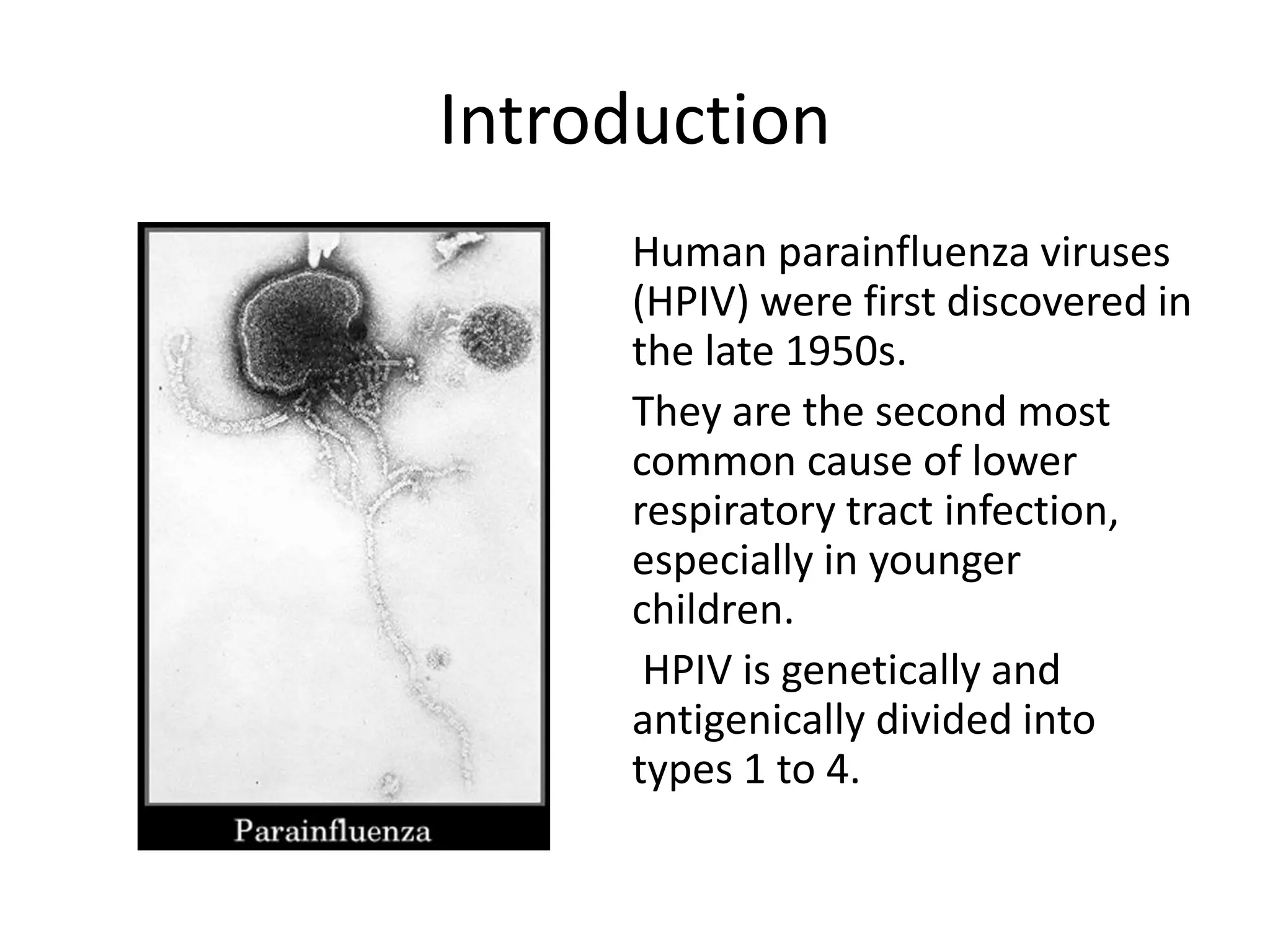


Human Parainfluenza Virus (HPIV) is a common cause of respiratory infections, especially in children. There are 4 types of HPIV that can cause illnesses ranging from mild upper respiratory infections to severe croup. HPIV is an enveloped RNA virus that enters host cells and replicates through transcription and translation of viral proteins. Treatment focuses on managing symptoms like cough and stridor. Precautions like masks and handwashing are recommended to prevent transmission through respiratory droplets.


