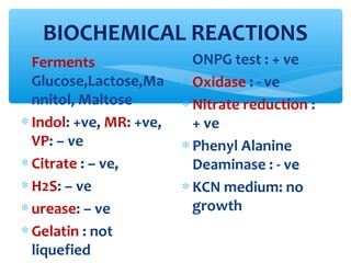
ENTEROBACTERIACEAE
Dr V S Vatkar
Associate Professor
Microbiology Department
D Y Patil Medical College, Kolhapur
∗ Found in soil & water.
∗ Some of them are predominant
bacterial flora of human intestine &
animals.
∗ Gram negative, non sporing, motile or
non motile bacilli, capsulated or non
capsulated
∗ Grows aerobically or anaerobically.
∗ Oxidase negative.
∗ Catalase positive.
∗ Ferment glucose & other carbohydrates:
produce acid or acid & gas.
∗ Reduces nitrates to nitrites.
DEFINITION
∗ In 1972 Edward & Ewing described 11
genera & 26 species.
∗ In 1985 Farmer & associates described
22 genera & 69 species & 29 enteric
groups.
∗ Now 31 genera & 139
species,bigroups,unnamed enteric
groups.
TAXONOMY
Lactose fermenters: E.coli,Klebsiella
Late lactose fermenters:
Shigella sonei,paracolons
Non lactose fermenters: Salmonella &
Shigella
All LF enteric bacilli : known as COLIFORM BACILLI,
majority of intestinal commensals are LF(colon bacilli
& E.coli).
OLD CLASSIFICATION
Lactose fermentation on MacConkey Agar
EDWARDS & EWING CLASSIFICATION
∗ TRIBE I : ESCHERICHIAE
Genus :
• Escherichia
• Shigella
• TRIBE III :
• SALMONELLAE
• Genus :
• Salmonella
∗ TRIBE II :
EDWARDSIELLAE
Genus:
• Edwardsiella
• TRIBE IV :
• CITRIBACTERIACEAE
• Genus :
• Citrobacter
Classification ( contd )
∗ TRIBE V
KLEBSIELLEAE :
Genus:
Klebsiella,
Enterobacter, Serratia,
Hafnia & Pantoea
TRIBE VII
YERSINIEAE :
Genus
Yersinia
∗ TRIBE VI :
PROTEAE :
Genus:
•Proteus, Providencia,
Morganella
TRIBE VIII
ERVINEAE :
Genus
Ervinia
N.A. Low,convex,smooth/muciod,grey colonies.
B.A. Same as N.A. some show hemolysis,
swarming(Proteus)
∗ M.A. LF or NLF colonies
Special Media like:
- DCA LF or NLF colonies( salmonella & Shigella)
- Eosin Methylene Blue Agar(EMB): Green sheen
( E.coli)
CHARACTERISTICS
FOR PRESUMPTIVE IDENTIFICATION
∗Mainly by Biochemical reactions
•Glucose fermentation
•Cytochrome oxidase NEGATIVE
•Nitrates : reduced to Nitrites
•IMViC tests
•Motility test
SCREENING CHARACTERISTICS
CARBOHYDRATE UTILISATION
∗Glucose
fermentation
∗Lactose
fermentation
mainly
ACIDS: Lactic acid,
Acetic acid, carbonic
acid or Formic acid
GAS : Hydrogen, CO2,
N2.
OXYDASE TEST
∗Enterobacteriac
eae bacilli give
test
NEGATIVE(E.coli)
∗ Reagents :
tetramethyl-para-
phenyl diamine or
dimethyl para
phenyl diamine
hydrochloride.
NITRATE REDUCTION TEST
∗ All Enterobacteriaceae
gr.give test +ve
except : Enterobacter
aggtomerans,some
Serratia spp. Yersinia.
∗ Indicators:
∗ ά-nephthalamine &
sulfanilic acid.
∗ RED color +ve E.coli
∗ Yellow color – ve
Acinetobacter
PHENYLALANINE DEAMINASE
PRODUCTION TEST
∗Differentiation of
proteus from
other GNB.
∗Indicator : Ferric
chloride
∗GREEN color +ve
test
TSI & H2S TEST
∗L F: Yellow slant &
butt.
∗NLF: Red slant &
Yellow butt.
∗Non fermenters:
Red slant & butt.
∗H2S: Yellow butt,
Red slant, black
color on slant.
INDOLE TEST
∗Kovac’s reagent
or Ehrlich’
reagent used as a
indicator.
∗Red color ring +ve
test
METHYL RED TEST
∗Ferment glucose
to form pyruvate
∗Indicator : Methyl
red
∗Red color +ve test
V-P TEST
∗ Ferment glucose to
form Glycol
∗ Indicators : alc soln
of ά-nephthol &
KOH
∗ Red / Pink color +
ve test
CITREATE TEST
∗ Simmon’s citrate
agar
∗ Indicator :
Bromothymol blue
∗ Blue color + ve test
UREASE TEST
∗Christensen
medium / Stuart’s
urea broth
∗Indicator : Phenol
red
∗Purple pink color
+ve
∗ Help to identify Late Lactose Fermenting
organisms.( colorless +ve,yellow – ve)
∗ E.coli & Shigella sonei, Salmonella spp.&
Citrobacter spp. ( E.coli +ve & proteus –
ve,control)
DECARBOXYLATION OF
LYSINE,ORNITHIN,ARGININE :
Variable
ONPG & β GALACTOCIDASE TEST
Escherichia coli
∗ Named after the
scientist ESCHERICH
(1885)
∗ Present in human &
animal GIT.
∗ Detection of E.coli in
drinking water shows
recent pollution of water
with human or animal
faeces.
∗ E fergusonii, E hermanii, E
vulneris: little medical
importance
MORPHOLOGY
∗ GNB , STRAIGHT
RODS
∗ SIZE 1-3×0.4-0.7µm
∗ MOTILE :
peritrichous flagella
∗ Non-sporing
∗ Capsule & fimbria in
some strains.
Gram stain
∗ AEROBES & FACULTATIVE ANAEROBES
∗ TEMP 10 – 40ºC , optimum temp 37ºC
∗ Colony characters : large, whitish, smooth,
opaque, emulsify in saline ( S ). Rough
colonies : irregular, dull, autoagglutinable in
saline ( R ). S – R variation : repeated
subcultures, loss of virulence factor( surface
Ag ).
∗ Broth : general turbidity.
∗ DCA & SS media : Inhibition of growth
CULTURAL CHARACTERISTICS
Colonies on M.A. & CLED
Colonies on EMB medium
XLD MEDIUM
( Xylose Lysine Deoxycholate agar )
∗ Yellow colonies of
E.coli.
BIOCHEMICAL REACTIONS
∗ Ferments
Glucose,Lactose,Ma
nnitol, Maltose
∗ Indol: +ve, MR: +ve,
VP: – ve
∗ Citrate : – ve,
∗ H2S: – ve
∗ urease: – ve
∗ Gelatin : not
liquefied
∗ ONPG test : + ve
∗ Oxidase : - ve
∗ Nitrate reduction :
+ ve
∗ Phenyl Alanine
Deaminase : - ve
∗ KCN medium: no
growth
* O’ Ag (somatic) : 170 Ag recorded, inhibit phagocytosis
∗F Ag : fimbrial Ag, heat labile, detached when
heated to 100 o C
∗‘K’ Ag (capsular): 100 , acidic polysaccharide, located
in the ENVELOPE/ Micro capsule. 3 parts : L Ag, A Ag, B Ag.
Later shown that B Ag is not a separate entity.
∗‘H’ Ag (flagellar) : 75
* Normal intestinal strains : K Ag absent, has Early ‘O’ gr.
(1,2,3,4 etc)
*Pathogenic strains : Later ‘O’ gr (26,55,86,111 etc)
ANTIGENIC STRUCTURE
∗ Surface Ag :
a) Somatic Lipo-polysaccharides O Ag :
- resist PHAGOCYTOSIS
- Endotoxic activity
- Prevent bactericidal effect of complement
b) Envelope or K Ag :
- resist PHAGOCYTOSIS
- Prevent antibacterial effect in serum
VIRULENCE FACTORS ( TWO TYPES)
c) Fimbriae :
- Chromosomally determined : non
pathogenic
- Plasmid coded : small in no. & acts as
virulence factor
- OR colonization factor Ag (CFA) : in
enterotoxogenic E.coli causes Diarrhoea.
- In UTI: P fimbria binds specifically to P
bl.gr.substance on human RBCs &
uroepithelial cells.
SURFACE Ag (cond)
∗EXOTOXINS:
- Hemolysin: produce by most of the virulent
strains than avirulent strains.
- Enterotoxin: a) Heat Labile Toxin (LT):
complex polysaccharide subunits : 1 A
subunit: active
5 B subunits : binding. Produced by ETEC
b) Heat Stable Toxin : STa(STI) & STb(STII) ETEC
c) Verotoxin (VT) also called Shiga like toxin
,produced by EHEC.
TOXINS
Toxin binds to Gm1 receptors of intestinal epithelium
with subunit B subunit A activated
activates Adenyl
cAMP cyclase.
increase outflow of
water & electrolytes leads to DIARRHOEA
in intestinal lumen
*
MOA of Cholera toxin is same as LT
Mode Of Action of LT
ST I binds to cGMP activation of
cGMP
in intestine.
fluid accumulation in
intestine.
Mode Of Action of ST
∗ Cytotoxic effect on Vero cells , HeLa
cells
∗ Similar to Shigella dysenteriae type 1
toxin so called Shiga like toxin
∗ VT genes : phage coded
∗ A & B subunits
∗ Antigenically different VT is called VT2,
not neutralized by Shiga like toxin VT1.
Verotoxin (VT)
∗Urinary Tract Infection (UTI)
∗DIARRHOEA
∗PYOGENIC INFECTIONS
∗SEPTICEMIA
CLINICAL INFECTIONS
∗Acquired after Diagnostic
instrumentation, prostate
enlargement, renal calculi,
pregnancy ( Asymptomatic
bacteriuria)
∗Serotypes : ‘O’ gr 1,2,4,6,7.
Urinary Tract Infection
∗ Asymptomatic bacteriuria : asymptomatic inf
during pregnancy may cause PYELONEPHRITIS later
in pregnancy, HT leads to prematurity & perinatal
death of fetus.
∗ Lower UTI mainly due to fecal contamination &
may not have K Ag ( organism causing cystitis).
Isolates from pyelonephritis carry K Ag
(ascending inf)
∗ DIAGNOSIS OF UTI :
- Normal urine is sterile, but during voiding it may
contaminate by genital commensals.
UTI
∗ Sample collection
- Collect urine by catheterizations to avoid
contamination (sometimes lower UTI may ascend
to cause ascending inf.) .
- CLEANE VOIDED MIDSTREAM sample of urine is
collected.
- Transport to lab without any delay. Delay more
than 1-2 hrs REFRIGERATE the sample.
- Quantitative culture : most of the specimen
contain 10,000 to 1,00,000 bact / ml. Active inf may
contain 1,00,000 bact / ml. this level is SIGNIFICANT
BACTERIURIA. Patient on antibiotics, diuretic drugs,
bact like staph , low count may be significant.
∗ Griess Nitrate test :
absence of nitrate in normal urine. Presence
of nitrates: nitrate reducing bact.
∗ Catalase test :
effervescence on adding H2O2. +ve result
seen in hematuria
∗ Triphenyl tetrazolium chloride(TTC) test : detects
respiratory activity of growing bacteria.
Pink ppt
∗ Microscopy: GNB seen
∗ Glucose test paper :
utilization of minute amt of glucose present
in urine by bact.
SCREENING TESTS for UTI for presumptive
diagnosis
∗Deep slide culture method: agar
coated glass slide is immerged in urine
sample , incubate, growth is estimated by
colony counter or by color change of
indicator.
(none of the screening test is as sensitive /
reliable as culture).
CULTURE : Two methods
- Pour Plate method
- semiquantitative culture method : standard
loop method
∗ Isolates are identified by biochemical
reactions
∗AST : development of drug resistance
frequently with E.coli inf.
∗ First isolated from infant suffering from enteritis.
∗ Bary 1945: found the role E.coli (sp.type) during
hospital out breaks of childhood diarrhoea in London.
∗ TYPES OF DIARRHOGENIC E.coli :
a) Enteropathogenic E.coli (EPEC)
b) Enterotoxogenic E.coli (ETEC)
c) Enteroinvasive E.coli (EIEC)
d) Enterohaemorrhagic E.coli (EHEC)
e) Enteroaggregative E.coli (EAEC)
DIARRHOEA
Pathogenesis of EPEC
∗ Bacteria attaches to
the mucosa of S.I.
∗ Then attaches to the
cup like projections
(pedestals) of host cell
membrane.
∗ Distruption of brush
border (microvilli)
Pathogenesis of ETEC
Pathogenesis of E.coli
ETEC O sero
gr
6,8,15,25,27
,63,78,115,
148,153,159
Infant &
adult
watery
diarrhoea
traveler's
diarrhoea
Bact adhere
to intestinal
mucosa by
CFA(colonis
ation factor
Ag),
releases
LT / ST or
both
Typing with
sp antisera,
demo of
toxin in
tissue by LA
ELISA,
genetic
probes for
ST & LT
EIEC 28ac,112ac
,
124,136,143
,144,152,16
4
Dysente
ry like
disease
all ages
Epith cell
invasion
Typing with
sp antisera.
definative:He
La/HEp-2 cell
inv assay.
Virulence
Marker
Ag(plasmid
coded)
detected by
ELISA.
Sereny test
VCEC
Food
poisoning
(canned
food)
O 157:H 7
O 26: H 1
Bloody
diarrhoea,
HC,HUS
VT1 & VT 2
or Shiga like
toxin, affects
vascular
endothelium,
renal lesion
& capillary
microangiop
athy.
Typing with
sp
antiserum,VT
1 & 2 probes,
demo of VT 1
& 2 in Vero
cells(cytotoxi
c effect)
EPEC O gr.
18,26,44,55,
86,111,114,
119,125,126
,127,128,14
2
Diarrhoea
in infants,
rarely in
adults
Pathogen
esis
Unknown
Typing with
sp
antisera,EAF
probes,
demo of
adhesion in
HeLa,HEp-2
cell
∗ Persistent diarrhoea, in developing countries
∗ Produces heat stable enterotoxin 1(EAST 1)
∗ O : untypeble H : typable
∗ Aggregate in ‘Stacked Brick’ form on HEp-2 cells
∗Diffusely Adherent E coli: less well
established as pathogen
EAEC
∗Intra abdominal infections :
Peritonitis, abscesses from
spillage of bowel contents.
∗Perianal inf
∗Neonatal meningitis
∗Wound inf
PYOGENIG INFECTIONS
∗SEPTICEMIA :
Blood stream
invasion causes
septic
shock(endotoxic
shock), systemic
inflammatory
response
syndrome (SIDS)
∗Antibiotic
sensitivity test :
- organism shows
resistance to
multiple drugs like
Penicillin,
Cephalosporins,
Quinolone gr etc,
- AST is most imp for
the treatment.
Detection of O157: H 7
∗ Detection of Shiga like toxin
directly from stool.
∗ Direct plating on SMAC
(Sorbitol MacConkey Agar).
OR
∗ Cefixim SMAC OR
∗ Cefixim & Tellurite SMAC OR
media containing 5 bromo-5
chloro-3 indoxyl-β-D
glucuronide OR 4
methylumbelliferyl-β-D
glucuronide.
∗ Immunogenic separation by
using O 157 sp Ab coated
beads after culture.
∗ E.fergusonii : isolated from stool
∗ E.hermanii : isolated from wd, sputum,
stool. Agglutinate with E.coli O 157
antiserum
∗ E.vulneris
∗ E.blatte
Other Escherichia spp
Enterobacteriasese seminar

Enterobacteriasese seminar

  • 1.
    ENTEROBACTERIACEAE Dr V SVatkar Associate Professor Microbiology Department D Y Patil Medical College, Kolhapur
  • 2.
    ∗ Found insoil & water. ∗ Some of them are predominant bacterial flora of human intestine & animals.
  • 3.
    ∗ Gram negative,non sporing, motile or non motile bacilli, capsulated or non capsulated ∗ Grows aerobically or anaerobically. ∗ Oxidase negative. ∗ Catalase positive. ∗ Ferment glucose & other carbohydrates: produce acid or acid & gas. ∗ Reduces nitrates to nitrites. DEFINITION
  • 4.
    ∗ In 1972Edward & Ewing described 11 genera & 26 species. ∗ In 1985 Farmer & associates described 22 genera & 69 species & 29 enteric groups. ∗ Now 31 genera & 139 species,bigroups,unnamed enteric groups. TAXONOMY
  • 5.
    Lactose fermenters: E.coli,Klebsiella Latelactose fermenters: Shigella sonei,paracolons Non lactose fermenters: Salmonella & Shigella All LF enteric bacilli : known as COLIFORM BACILLI, majority of intestinal commensals are LF(colon bacilli & E.coli). OLD CLASSIFICATION Lactose fermentation on MacConkey Agar
  • 6.
    EDWARDS & EWINGCLASSIFICATION ∗ TRIBE I : ESCHERICHIAE Genus : • Escherichia • Shigella • TRIBE III : • SALMONELLAE • Genus : • Salmonella ∗ TRIBE II : EDWARDSIELLAE Genus: • Edwardsiella • TRIBE IV : • CITRIBACTERIACEAE • Genus : • Citrobacter
  • 7.
    Classification ( contd) ∗ TRIBE V KLEBSIELLEAE : Genus: Klebsiella, Enterobacter, Serratia, Hafnia & Pantoea TRIBE VII YERSINIEAE : Genus Yersinia ∗ TRIBE VI : PROTEAE : Genus: •Proteus, Providencia, Morganella TRIBE VIII ERVINEAE : Genus Ervinia
  • 8.
    N.A. Low,convex,smooth/muciod,grey colonies. B.A.Same as N.A. some show hemolysis, swarming(Proteus) ∗ M.A. LF or NLF colonies Special Media like: - DCA LF or NLF colonies( salmonella & Shigella) - Eosin Methylene Blue Agar(EMB): Green sheen ( E.coli) CHARACTERISTICS FOR PRESUMPTIVE IDENTIFICATION
  • 9.
    ∗Mainly by Biochemicalreactions •Glucose fermentation •Cytochrome oxidase NEGATIVE •Nitrates : reduced to Nitrites •IMViC tests •Motility test SCREENING CHARACTERISTICS
  • 10.
    CARBOHYDRATE UTILISATION ∗Glucose fermentation ∗Lactose fermentation mainly ACIDS: Lacticacid, Acetic acid, carbonic acid or Formic acid GAS : Hydrogen, CO2, N2.
  • 11.
    OXYDASE TEST ∗Enterobacteriac eae bacilligive test NEGATIVE(E.coli) ∗ Reagents : tetramethyl-para- phenyl diamine or dimethyl para phenyl diamine hydrochloride.
  • 12.
    NITRATE REDUCTION TEST ∗All Enterobacteriaceae gr.give test +ve except : Enterobacter aggtomerans,some Serratia spp. Yersinia. ∗ Indicators: ∗ ά-nephthalamine & sulfanilic acid. ∗ RED color +ve E.coli ∗ Yellow color – ve Acinetobacter
  • 13.
    PHENYLALANINE DEAMINASE PRODUCTION TEST ∗Differentiationof proteus from other GNB. ∗Indicator : Ferric chloride ∗GREEN color +ve test
  • 14.
    TSI & H2STEST ∗L F: Yellow slant & butt. ∗NLF: Red slant & Yellow butt. ∗Non fermenters: Red slant & butt. ∗H2S: Yellow butt, Red slant, black color on slant.
  • 15.
    INDOLE TEST ∗Kovac’s reagent orEhrlich’ reagent used as a indicator. ∗Red color ring +ve test
  • 16.
    METHYL RED TEST ∗Fermentglucose to form pyruvate ∗Indicator : Methyl red ∗Red color +ve test
  • 17.
    V-P TEST ∗ Fermentglucose to form Glycol ∗ Indicators : alc soln of ά-nephthol & KOH ∗ Red / Pink color + ve test
  • 18.
    CITREATE TEST ∗ Simmon’scitrate agar ∗ Indicator : Bromothymol blue ∗ Blue color + ve test
  • 19.
    UREASE TEST ∗Christensen medium /Stuart’s urea broth ∗Indicator : Phenol red ∗Purple pink color +ve
  • 20.
    ∗ Help toidentify Late Lactose Fermenting organisms.( colorless +ve,yellow – ve) ∗ E.coli & Shigella sonei, Salmonella spp.& Citrobacter spp. ( E.coli +ve & proteus – ve,control) DECARBOXYLATION OF LYSINE,ORNITHIN,ARGININE : Variable ONPG & β GALACTOCIDASE TEST
  • 21.
    Escherichia coli ∗ Namedafter the scientist ESCHERICH (1885) ∗ Present in human & animal GIT. ∗ Detection of E.coli in drinking water shows recent pollution of water with human or animal faeces. ∗ E fergusonii, E hermanii, E vulneris: little medical importance
  • 22.
    MORPHOLOGY ∗ GNB ,STRAIGHT RODS ∗ SIZE 1-3×0.4-0.7µm ∗ MOTILE : peritrichous flagella ∗ Non-sporing ∗ Capsule & fimbria in some strains.
  • 23.
  • 24.
    ∗ AEROBES &FACULTATIVE ANAEROBES ∗ TEMP 10 – 40ºC , optimum temp 37ºC ∗ Colony characters : large, whitish, smooth, opaque, emulsify in saline ( S ). Rough colonies : irregular, dull, autoagglutinable in saline ( R ). S – R variation : repeated subcultures, loss of virulence factor( surface Ag ). ∗ Broth : general turbidity. ∗ DCA & SS media : Inhibition of growth CULTURAL CHARACTERISTICS
  • 25.
  • 26.
  • 27.
    XLD MEDIUM ( XyloseLysine Deoxycholate agar ) ∗ Yellow colonies of E.coli.
  • 28.
    BIOCHEMICAL REACTIONS ∗ Ferments Glucose,Lactose,Ma nnitol,Maltose ∗ Indol: +ve, MR: +ve, VP: – ve ∗ Citrate : – ve, ∗ H2S: – ve ∗ urease: – ve ∗ Gelatin : not liquefied ∗ ONPG test : + ve ∗ Oxidase : - ve ∗ Nitrate reduction : + ve ∗ Phenyl Alanine Deaminase : - ve ∗ KCN medium: no growth
  • 29.
    * O’ Ag(somatic) : 170 Ag recorded, inhibit phagocytosis ∗F Ag : fimbrial Ag, heat labile, detached when heated to 100 o C ∗‘K’ Ag (capsular): 100 , acidic polysaccharide, located in the ENVELOPE/ Micro capsule. 3 parts : L Ag, A Ag, B Ag. Later shown that B Ag is not a separate entity. ∗‘H’ Ag (flagellar) : 75 * Normal intestinal strains : K Ag absent, has Early ‘O’ gr. (1,2,3,4 etc) *Pathogenic strains : Later ‘O’ gr (26,55,86,111 etc) ANTIGENIC STRUCTURE
  • 30.
    ∗ Surface Ag: a) Somatic Lipo-polysaccharides O Ag : - resist PHAGOCYTOSIS - Endotoxic activity - Prevent bactericidal effect of complement b) Envelope or K Ag : - resist PHAGOCYTOSIS - Prevent antibacterial effect in serum VIRULENCE FACTORS ( TWO TYPES)
  • 31.
    c) Fimbriae : -Chromosomally determined : non pathogenic - Plasmid coded : small in no. & acts as virulence factor - OR colonization factor Ag (CFA) : in enterotoxogenic E.coli causes Diarrhoea. - In UTI: P fimbria binds specifically to P bl.gr.substance on human RBCs & uroepithelial cells. SURFACE Ag (cond)
  • 32.
    ∗EXOTOXINS: - Hemolysin: produceby most of the virulent strains than avirulent strains. - Enterotoxin: a) Heat Labile Toxin (LT): complex polysaccharide subunits : 1 A subunit: active 5 B subunits : binding. Produced by ETEC b) Heat Stable Toxin : STa(STI) & STb(STII) ETEC c) Verotoxin (VT) also called Shiga like toxin ,produced by EHEC. TOXINS
  • 33.
    Toxin binds toGm1 receptors of intestinal epithelium with subunit B subunit A activated activates Adenyl cAMP cyclase. increase outflow of water & electrolytes leads to DIARRHOEA in intestinal lumen * MOA of Cholera toxin is same as LT Mode Of Action of LT
  • 34.
    ST I bindsto cGMP activation of cGMP in intestine. fluid accumulation in intestine. Mode Of Action of ST
  • 35.
    ∗ Cytotoxic effecton Vero cells , HeLa cells ∗ Similar to Shigella dysenteriae type 1 toxin so called Shiga like toxin ∗ VT genes : phage coded ∗ A & B subunits ∗ Antigenically different VT is called VT2, not neutralized by Shiga like toxin VT1. Verotoxin (VT)
  • 36.
    ∗Urinary Tract Infection(UTI) ∗DIARRHOEA ∗PYOGENIC INFECTIONS ∗SEPTICEMIA CLINICAL INFECTIONS
  • 37.
    ∗Acquired after Diagnostic instrumentation,prostate enlargement, renal calculi, pregnancy ( Asymptomatic bacteriuria) ∗Serotypes : ‘O’ gr 1,2,4,6,7. Urinary Tract Infection
  • 38.
    ∗ Asymptomatic bacteriuria: asymptomatic inf during pregnancy may cause PYELONEPHRITIS later in pregnancy, HT leads to prematurity & perinatal death of fetus. ∗ Lower UTI mainly due to fecal contamination & may not have K Ag ( organism causing cystitis). Isolates from pyelonephritis carry K Ag (ascending inf) ∗ DIAGNOSIS OF UTI : - Normal urine is sterile, but during voiding it may contaminate by genital commensals. UTI
  • 39.
    ∗ Sample collection -Collect urine by catheterizations to avoid contamination (sometimes lower UTI may ascend to cause ascending inf.) . - CLEANE VOIDED MIDSTREAM sample of urine is collected. - Transport to lab without any delay. Delay more than 1-2 hrs REFRIGERATE the sample. - Quantitative culture : most of the specimen contain 10,000 to 1,00,000 bact / ml. Active inf may contain 1,00,000 bact / ml. this level is SIGNIFICANT BACTERIURIA. Patient on antibiotics, diuretic drugs, bact like staph , low count may be significant.
  • 40.
    ∗ Griess Nitratetest : absence of nitrate in normal urine. Presence of nitrates: nitrate reducing bact. ∗ Catalase test : effervescence on adding H2O2. +ve result seen in hematuria ∗ Triphenyl tetrazolium chloride(TTC) test : detects respiratory activity of growing bacteria. Pink ppt ∗ Microscopy: GNB seen ∗ Glucose test paper : utilization of minute amt of glucose present in urine by bact. SCREENING TESTS for UTI for presumptive diagnosis
  • 41.
    ∗Deep slide culturemethod: agar coated glass slide is immerged in urine sample , incubate, growth is estimated by colony counter or by color change of indicator. (none of the screening test is as sensitive / reliable as culture). CULTURE : Two methods - Pour Plate method - semiquantitative culture method : standard loop method
  • 42.
    ∗ Isolates areidentified by biochemical reactions ∗AST : development of drug resistance frequently with E.coli inf.
  • 43.
    ∗ First isolatedfrom infant suffering from enteritis. ∗ Bary 1945: found the role E.coli (sp.type) during hospital out breaks of childhood diarrhoea in London. ∗ TYPES OF DIARRHOGENIC E.coli : a) Enteropathogenic E.coli (EPEC) b) Enterotoxogenic E.coli (ETEC) c) Enteroinvasive E.coli (EIEC) d) Enterohaemorrhagic E.coli (EHEC) e) Enteroaggregative E.coli (EAEC) DIARRHOEA
  • 44.
    Pathogenesis of EPEC ∗Bacteria attaches to the mucosa of S.I. ∗ Then attaches to the cup like projections (pedestals) of host cell membrane. ∗ Distruption of brush border (microvilli)
  • 45.
  • 46.
  • 47.
    ETEC O sero gr 6,8,15,25,27 ,63,78,115, 148,153,159 Infant& adult watery diarrhoea traveler's diarrhoea Bact adhere to intestinal mucosa by CFA(colonis ation factor Ag), releases LT / ST or both Typing with sp antisera, demo of toxin in tissue by LA ELISA, genetic probes for ST & LT EIEC 28ac,112ac , 124,136,143 ,144,152,16 4 Dysente ry like disease all ages Epith cell invasion Typing with sp antisera. definative:He La/HEp-2 cell inv assay. Virulence Marker Ag(plasmid coded) detected by ELISA. Sereny test
  • 48.
    VCEC Food poisoning (canned food) O 157:H 7 O26: H 1 Bloody diarrhoea, HC,HUS VT1 & VT 2 or Shiga like toxin, affects vascular endothelium, renal lesion & capillary microangiop athy. Typing with sp antiserum,VT 1 & 2 probes, demo of VT 1 & 2 in Vero cells(cytotoxi c effect) EPEC O gr. 18,26,44,55, 86,111,114, 119,125,126 ,127,128,14 2 Diarrhoea in infants, rarely in adults Pathogen esis Unknown Typing with sp antisera,EAF probes, demo of adhesion in HeLa,HEp-2 cell
  • 49.
    ∗ Persistent diarrhoea,in developing countries ∗ Produces heat stable enterotoxin 1(EAST 1) ∗ O : untypeble H : typable ∗ Aggregate in ‘Stacked Brick’ form on HEp-2 cells ∗Diffusely Adherent E coli: less well established as pathogen EAEC
  • 50.
    ∗Intra abdominal infections: Peritonitis, abscesses from spillage of bowel contents. ∗Perianal inf ∗Neonatal meningitis ∗Wound inf PYOGENIG INFECTIONS
  • 51.
    ∗SEPTICEMIA : Blood stream invasioncauses septic shock(endotoxic shock), systemic inflammatory response syndrome (SIDS) ∗Antibiotic sensitivity test : - organism shows resistance to multiple drugs like Penicillin, Cephalosporins, Quinolone gr etc, - AST is most imp for the treatment.
  • 52.
    Detection of O157:H 7 ∗ Detection of Shiga like toxin directly from stool. ∗ Direct plating on SMAC (Sorbitol MacConkey Agar). OR ∗ Cefixim SMAC OR ∗ Cefixim & Tellurite SMAC OR media containing 5 bromo-5 chloro-3 indoxyl-β-D glucuronide OR 4 methylumbelliferyl-β-D glucuronide. ∗ Immunogenic separation by using O 157 sp Ab coated beads after culture.
  • 53.
    ∗ E.fergusonii :isolated from stool ∗ E.hermanii : isolated from wd, sputum, stool. Agglutinate with E.coli O 157 antiserum ∗ E.vulneris ∗ E.blatte Other Escherichia spp