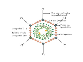
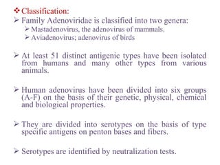
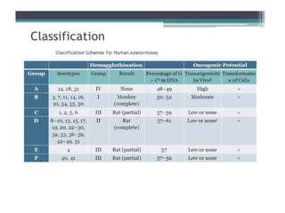
ADENOVIRUS
Introduction:
Adenoviruses can replicate and produce disease in
respiratory, gastrointestinal and urinary tract and
in the eye.
Many adenovirus infection are subclinical and
virus persist in hosts for months.
One third of 51 serotypes are responsible for most
cases of human adenovirus disease.
Whereas a few types serve as models for cancer
induction in animal models.
Morphology
 Are double stranded DNA viruses having icosahedral
symmetry and measures 70-90 nm in diameter.
 Capsid composed of 252 capsomeres; in which 12 are
penton bases and has fibers projecting from each of 12
vertices or penton bases and rest of capsid is composed
of 240 hexon capsomeres.
 No envelope
 The hexon, penton and fiers constitute major antigens
of adenovirus important in viral classification and
disease diagnosis.
 The DNA genome (25-45 Kb) is linear and double
stranded.
The penton bases carry toxin like activity and
causes rapid appearance of cytopathic effects
and detachment of the cells from the surface
on which they are growing.
Fibers contain type specific antigens important
in serotyping and has hemagglutinating
activity.
Classification:
 Family Adenoviridae is classified into two genera:
Mastadenovirus, the adenovirus of mammals.
Aviadenovirus; adenovirus of birds
 At least 51 distinct antigenic types have been isolated
from humans and many other types from various
animals.
 Human adenovirus have been divided into six groups
(A-F) on the basis of their genetic, physical, chemical
and biological properties.
 They are divided into serotypes on the basis of type
specific antigens on penton bases and fibers.
 Serotypes are identified by neutralization tests.
Pathogenesis:
 Infect and replicate in epithelial cells of respiratory
tract, gastrointestinal tract, urinary tract and the eye.
 Do not spread beyond regional lymph nodes.
 Group C persists as latent infections for years in
adenoids and tonsils and are shed in feces for many
months after initial infection.
 Most adenovirus replicate in intestinal epithelium after
ingestion but usually produce sub clinical infection
than overt symptoms.
 About 1/3 of human serotypes are commonly
associated with human illnesses.
 Single serotype may cause different clinical diseases
and conversely more than one serotype can cause same
clinical illness.
 Adenoviruses 1-7 are more common types worldwide
and account for most instances of adenovirus associated
illness.
 Responsible for 5% of respiratory diseases in young
children but account for much less in adults.
 Most infections are mild and self limited.
 Occasionally case disease in other organs particularly
eye and gastrointestinal tract.
o Respiratory Diseases:
 Typical symptoms: cough, nasal congestion, fever and sore
throat.
 Symptoms most commonly manifested in infants and
children and usually involves group C viruses.
 Adenoviruses particularly 3, 7 and 21 are thought to be
responsible for about 10-20% pneumonias in childhood.
 Mortality rate of pneumonia 8-10% in very young.
 Also are cause of acute respiratory disease syndrome among
military recruits.
 Characterised by fever, sorethroat, nasal congestion, cough
and malaise; sometimes leading to pneumonia.
Occurs in epidemic form among young
military recruits under conditions of fatigue,
stress, and crowding soon after induction.
Disease is caused by serotype 4 and 7 and
occasionally by type 3.
Outbreak of severe respiratory disease in 2007
caused by adenovirus 14 affects all age
groups.
• Eye Infections:
 Mild occular involvement may be part of respiratory pharyngeal
syndrome.
 Pharyngoconjunctival fever occurs as outbreak such as in children’s
summer camps (swimming pool conjunctivitis) and caused by type 3
and 7.
 Duration 1-2 weeks and complete recovery with no lasting sequelae
is common outcome.
 Epidemic keratoconjunctivitis more serious disease; occurs mainly
in adults and is highly contagious.
 Source of infection: sinks and hand towel.
 Disease is characterises by acute conjunctivitis followed by keratitis
that usually resolves in two weeks but may leave subepithelial
opacities in the cornea for up to two years.

o Gastrointestinal disease:
 Can often been isolated from feces but their relation to
intestinal disease has not been conclusively established.
 However two serotypes (types 40 and 41) have been
etiologically associated with infantile gastroenteritis.
 May account for 5-15% cases of viral gastroenteritis in
young children.
 Adenoviruses 40 and 41 are abundantly present in
diarrhoeal stools.
 Enteric adenovirus are very difficult to cultivate.
 Stool ELISA is often used for detection of these types.
o Other Diseases:
 Cause variety of casual and severe diseases in
immunocompromised patients.
 Most common problem in transplant patients is
respiratory diseases that may progress to pneumonia
and may be fatal (usually types 1-7).
 Adenovirus hepatitis in children having liver transplant.
 AIDS patients mainly suffer from gastroenteritis due to
adenovirus.
 Types 11 and 21 may cause acute hemorrhagic cystitis
in children esp. boys.
Laboratory Diagnosis:
o Specimen: throat washings, stool, urine and eye for
pharyngoconjunctival fever.
 Samples should be collected from affected sites as early
as possible.
 Duration of excretion of the virus varies among various
clinical illness;
1-3 days from throat of adults with common cold,
3-5 days for throat, stool and eyes, for
pharyngoconjunctival fever,
 2 weeks eye for keratoconjunctivitis,
3-6 weeks, throat and stool of children with respiratory
illness,
2-12 months in stool, throat, urine of immunocompromised
patients.
o Isolation
 Viral isolation in cell culture requires human cells.
 Human epithelial cell lines such as Hep-2, HeLa and
BK are sensitive but difficult to maintain.
 Isolates grown in these cells are identified by noting
cytopathic effects or by immunofluorescence staining
using anti hexon antibody , heamagglutination
inhibition test and neutralization test.
 Shell vial techniques can also be employed for isolation
of the virus.
 Can also be detected by electron microscopy, ELISA or
by latex agglutination tests.
o Serology
Paired sera should be used to detect rise in
antibody titre.
Antibodies in patients sera can be detected by
ELISA, complement fixatation test,
heamagglutination inhibition and neutralization
test.
o Molecular techniques:
PCR, restriction endonuclease digestion, DNA
hybridization.
o Treatment: no specific treatment
Rotavirus
• Belongs to reoviridae family.
• First identified as cause of diarrhea in 1973.
• Double stranded RNA virus having icosahedral symmetry.
• 60-80 nm in diameter and have double capsid shell
• Non enveloped virus.
• Rota in Latin means wheel.
• Under electron microscope resembles as little wheel with short spokes
radiating from wide hub to a clearly defined outer rim.
• Complete or double shelled virus measures 70nm and incomplete single
shelled virus is smaller about 60 nm.
• Double stranded RNA as genome with 10-12 discrete segments.
• Contains nine structural proteins; core contains several enzymes.
• Major cause of infantile diarrhoea.
Classification and Antigenic Properties:
 Classified into 5 species (A-E) plus two tentative species (F and G)
based on antigenic epitopes on the internal structural protein VP6.
 These can be detected by ELISA, immune electron microscopy.
 Group A major human pathogen.
 Outer capsid proteins VP4 and VP7 carry epitopes important in
neutralizing activity with VP7 predominant antigen.
 Group A have been divided into subgroups I and II by ELISA, CF
or immune adherence agglutination and into serotypes (1,2,3 etc.) by
Pathogenesis:
The virus enters the body through the mouth.
Infects cells in the villi of small intestine (gastric
and colonic mucosa are spared).
Multiply in the cytoplasm of the enterocytes and
damage their transport mechanisms.
Rotavirus encoded protein NSP4, is viral
enterotoxin and induces secretion by triggering
signal transduction pathway.
Damaged cells may slough off into the lumen of
the intestine and release large quantities of virus.
Viral excretion lasts from 2 to 12 days.
Diarrhoea caused by rotavirus is due to
impaired sodium and glucose absorption as
damaged cells on villi are replaced by
nonabsorbing immature crypt cells.
It may take 3-8 weeks for normal function to
be restored.
Clinical Findings:
 Cause major portion of diarrhoeal illness in infants and
children world wide but not in adults.
 Incubation period:1-3 days
 Typical symptoms include watery diarrhoea, fever,
abdominal pain and vomiting leading to dehydration.
 Stools usually greenish yellow or pale with no blood or
mucus.
 Group B rotavirus have been implicated in large
outbreaks of severe gastroenteritis in adults in China.
Laboratory Diagnosis:
 The most widely available method for confirmation of
rotavirus infection is detection of rotavirus antigen in
stool by enzyme-linked immunoassay (EIA) or by
immune electron microscopy.
 Polymerase chain reaction most sensitive method.
 ELISA can be employed for detection of rise in
antibody titre.
treatment: no specific treatment; supportive rehydration
therapy
Prophylaxis:RV5 (RotaTeq); a live oral vaccine and
RV1 (Rotarix), a live oral vaccine
Norwalk virus
• The Norwalk virus was first identified in 1968 after a severe
outbreak of “winter vomiting” in a school in Norwalk Ohio.
• The descendents of Norwalk virus are now called noroviruses
because these viruses change shape slightly about every 4 years so
there is no virus that is exactly the same as the one discovered in
1968, but still related.
 Belongs to Calciviridae family.
 Single stranded RNA viruses having icosahedral symmetry.
 Size: 27-40 nm in diameter, non enveloped virus.
 genome: single stranded, linear, positive sense, non segmented
RNA.
• Major cause of nonbacterial epidemic gastroenteritis.
• Epidemic nonbacterial gastroenteritis is characterised by:
I. Absence of bacterial pathogens
II. Gastroenteritis with rapid onset and recovery and relatively
mild systemic signs;
III. An epidemiological pattern of a highly communicable disease
that spreads rapidly with no particular predilection in terms of
age and geography.
 Various descriptive terms used: epidemic viral
gastroenteritis, winter vomiting disease, viral diarrhoea
depending on predominant clinical feature.
• Mode of transmission: through food, indirect contact or direct
contact.
• Incubation period: 24-48 hours
• The onset is rapid and clinical course is brief, lasting 12-48 hours.
• symptoms: diarrhoea, nausea, vomiting, low-grade fever, abdominal
cramps, headache and malaise.
• Dehydration is the most common complication in young and adults.
• Most people will recover from this sickness in about 24-72 hours
without any long term problems.
• Viral shedding persists for as long as 1 month.
• No sequelae have been reported.
Laboratory Diagnosis:
 Specimen: feces and vomitus
 Electron Microscopy and Immunoelectron Microscopy
can be used for detection of virus in stool samples.
 ELISA is used to detect rise in antibody titre in patient
serum.
 Real-Time reverse transcriptase-polymerase chain
reaction (RT-qPCR) is the most common way of testing
samples for the virus in stool and vomitus and also in
environmental samples (contaminated food and water).
 treatment: no specific treatment; only supportive
treatment
Astrovirus:
• 28-38nm in diameter, exhibit distinctive starlike
morphology.
• Contain single stranded positive sense RNA.
• Astroviridae consists two genera.
• All human viruses are classified in Mamastrovirus
genus.
• At least 8 serotypes of human viruses are
Cause diarrhoeal illness and may shed in
extraordinarily in large quantities in feces.
The virus is transmitted by fecal-oral route
through contaminated food and water, person-
person contact, or contaminated surfaces.
They are recognised as pathogens for infants and
children, elderly institutionalized patients and
immunocompromised persons.
Shed for prolonged periods in feces by
immunocompromised hosts.

Adenovirus Microbiology

  • 1.
  • 2.
    Introduction: Adenoviruses can replicateand produce disease in respiratory, gastrointestinal and urinary tract and in the eye. Many adenovirus infection are subclinical and virus persist in hosts for months. One third of 51 serotypes are responsible for most cases of human adenovirus disease. Whereas a few types serve as models for cancer induction in animal models.
  • 3.
    Morphology  Are doublestranded DNA viruses having icosahedral symmetry and measures 70-90 nm in diameter.  Capsid composed of 252 capsomeres; in which 12 are penton bases and has fibers projecting from each of 12 vertices or penton bases and rest of capsid is composed of 240 hexon capsomeres.  No envelope  The hexon, penton and fiers constitute major antigens of adenovirus important in viral classification and disease diagnosis.  The DNA genome (25-45 Kb) is linear and double stranded.
  • 4.
    The penton basescarry toxin like activity and causes rapid appearance of cytopathic effects and detachment of the cells from the surface on which they are growing. Fibers contain type specific antigens important in serotyping and has hemagglutinating activity.
  • 6.
    Classification:  Family Adenoviridaeis classified into two genera: Mastadenovirus, the adenovirus of mammals. Aviadenovirus; adenovirus of birds  At least 51 distinct antigenic types have been isolated from humans and many other types from various animals.  Human adenovirus have been divided into six groups (A-F) on the basis of their genetic, physical, chemical and biological properties.  They are divided into serotypes on the basis of type specific antigens on penton bases and fibers.  Serotypes are identified by neutralization tests.
  • 8.
    Pathogenesis:  Infect andreplicate in epithelial cells of respiratory tract, gastrointestinal tract, urinary tract and the eye.  Do not spread beyond regional lymph nodes.  Group C persists as latent infections for years in adenoids and tonsils and are shed in feces for many months after initial infection.  Most adenovirus replicate in intestinal epithelium after ingestion but usually produce sub clinical infection than overt symptoms.  About 1/3 of human serotypes are commonly associated with human illnesses.
  • 9.
     Single serotypemay cause different clinical diseases and conversely more than one serotype can cause same clinical illness.  Adenoviruses 1-7 are more common types worldwide and account for most instances of adenovirus associated illness.  Responsible for 5% of respiratory diseases in young children but account for much less in adults.  Most infections are mild and self limited.  Occasionally case disease in other organs particularly eye and gastrointestinal tract.
  • 10.
    o Respiratory Diseases: Typical symptoms: cough, nasal congestion, fever and sore throat.  Symptoms most commonly manifested in infants and children and usually involves group C viruses.  Adenoviruses particularly 3, 7 and 21 are thought to be responsible for about 10-20% pneumonias in childhood.  Mortality rate of pneumonia 8-10% in very young.  Also are cause of acute respiratory disease syndrome among military recruits.  Characterised by fever, sorethroat, nasal congestion, cough and malaise; sometimes leading to pneumonia.
  • 11.
    Occurs in epidemicform among young military recruits under conditions of fatigue, stress, and crowding soon after induction. Disease is caused by serotype 4 and 7 and occasionally by type 3. Outbreak of severe respiratory disease in 2007 caused by adenovirus 14 affects all age groups.
  • 12.
    • Eye Infections: Mild occular involvement may be part of respiratory pharyngeal syndrome.  Pharyngoconjunctival fever occurs as outbreak such as in children’s summer camps (swimming pool conjunctivitis) and caused by type 3 and 7.  Duration 1-2 weeks and complete recovery with no lasting sequelae is common outcome.  Epidemic keratoconjunctivitis more serious disease; occurs mainly in adults and is highly contagious.  Source of infection: sinks and hand towel.  Disease is characterises by acute conjunctivitis followed by keratitis that usually resolves in two weeks but may leave subepithelial opacities in the cornea for up to two years. 
  • 13.
    o Gastrointestinal disease: Can often been isolated from feces but their relation to intestinal disease has not been conclusively established.  However two serotypes (types 40 and 41) have been etiologically associated with infantile gastroenteritis.  May account for 5-15% cases of viral gastroenteritis in young children.  Adenoviruses 40 and 41 are abundantly present in diarrhoeal stools.  Enteric adenovirus are very difficult to cultivate.  Stool ELISA is often used for detection of these types.
  • 14.
    o Other Diseases: Cause variety of casual and severe diseases in immunocompromised patients.  Most common problem in transplant patients is respiratory diseases that may progress to pneumonia and may be fatal (usually types 1-7).  Adenovirus hepatitis in children having liver transplant.  AIDS patients mainly suffer from gastroenteritis due to adenovirus.  Types 11 and 21 may cause acute hemorrhagic cystitis in children esp. boys.
  • 15.
    Laboratory Diagnosis: o Specimen:throat washings, stool, urine and eye for pharyngoconjunctival fever.  Samples should be collected from affected sites as early as possible.  Duration of excretion of the virus varies among various clinical illness; 1-3 days from throat of adults with common cold, 3-5 days for throat, stool and eyes, for pharyngoconjunctival fever,  2 weeks eye for keratoconjunctivitis, 3-6 weeks, throat and stool of children with respiratory illness, 2-12 months in stool, throat, urine of immunocompromised patients.
  • 16.
    o Isolation  Viralisolation in cell culture requires human cells.  Human epithelial cell lines such as Hep-2, HeLa and BK are sensitive but difficult to maintain.  Isolates grown in these cells are identified by noting cytopathic effects or by immunofluorescence staining using anti hexon antibody , heamagglutination inhibition test and neutralization test.  Shell vial techniques can also be employed for isolation of the virus.  Can also be detected by electron microscopy, ELISA or by latex agglutination tests.
  • 17.
    o Serology Paired serashould be used to detect rise in antibody titre. Antibodies in patients sera can be detected by ELISA, complement fixatation test, heamagglutination inhibition and neutralization test. o Molecular techniques: PCR, restriction endonuclease digestion, DNA hybridization. o Treatment: no specific treatment
  • 18.
    Rotavirus • Belongs toreoviridae family. • First identified as cause of diarrhea in 1973. • Double stranded RNA virus having icosahedral symmetry. • 60-80 nm in diameter and have double capsid shell • Non enveloped virus. • Rota in Latin means wheel. • Under electron microscope resembles as little wheel with short spokes radiating from wide hub to a clearly defined outer rim. • Complete or double shelled virus measures 70nm and incomplete single shelled virus is smaller about 60 nm.
  • 19.
    • Double strandedRNA as genome with 10-12 discrete segments. • Contains nine structural proteins; core contains several enzymes. • Major cause of infantile diarrhoea. Classification and Antigenic Properties:  Classified into 5 species (A-E) plus two tentative species (F and G) based on antigenic epitopes on the internal structural protein VP6.  These can be detected by ELISA, immune electron microscopy.  Group A major human pathogen.  Outer capsid proteins VP4 and VP7 carry epitopes important in neutralizing activity with VP7 predominant antigen.  Group A have been divided into subgroups I and II by ELISA, CF or immune adherence agglutination and into serotypes (1,2,3 etc.) by
  • 21.
    Pathogenesis: The virus entersthe body through the mouth. Infects cells in the villi of small intestine (gastric and colonic mucosa are spared). Multiply in the cytoplasm of the enterocytes and damage their transport mechanisms. Rotavirus encoded protein NSP4, is viral enterotoxin and induces secretion by triggering signal transduction pathway. Damaged cells may slough off into the lumen of the intestine and release large quantities of virus.
  • 22.
    Viral excretion lastsfrom 2 to 12 days. Diarrhoea caused by rotavirus is due to impaired sodium and glucose absorption as damaged cells on villi are replaced by nonabsorbing immature crypt cells. It may take 3-8 weeks for normal function to be restored.
  • 23.
    Clinical Findings:  Causemajor portion of diarrhoeal illness in infants and children world wide but not in adults.  Incubation period:1-3 days  Typical symptoms include watery diarrhoea, fever, abdominal pain and vomiting leading to dehydration.  Stools usually greenish yellow or pale with no blood or mucus.  Group B rotavirus have been implicated in large outbreaks of severe gastroenteritis in adults in China.
  • 24.
    Laboratory Diagnosis:  Themost widely available method for confirmation of rotavirus infection is detection of rotavirus antigen in stool by enzyme-linked immunoassay (EIA) or by immune electron microscopy.  Polymerase chain reaction most sensitive method.  ELISA can be employed for detection of rise in antibody titre. treatment: no specific treatment; supportive rehydration therapy Prophylaxis:RV5 (RotaTeq); a live oral vaccine and RV1 (Rotarix), a live oral vaccine
  • 25.
    Norwalk virus • TheNorwalk virus was first identified in 1968 after a severe outbreak of “winter vomiting” in a school in Norwalk Ohio. • The descendents of Norwalk virus are now called noroviruses because these viruses change shape slightly about every 4 years so there is no virus that is exactly the same as the one discovered in 1968, but still related.  Belongs to Calciviridae family.  Single stranded RNA viruses having icosahedral symmetry.  Size: 27-40 nm in diameter, non enveloped virus.  genome: single stranded, linear, positive sense, non segmented RNA.
  • 27.
    • Major causeof nonbacterial epidemic gastroenteritis. • Epidemic nonbacterial gastroenteritis is characterised by: I. Absence of bacterial pathogens II. Gastroenteritis with rapid onset and recovery and relatively mild systemic signs; III. An epidemiological pattern of a highly communicable disease that spreads rapidly with no particular predilection in terms of age and geography.  Various descriptive terms used: epidemic viral gastroenteritis, winter vomiting disease, viral diarrhoea depending on predominant clinical feature.
  • 28.
    • Mode oftransmission: through food, indirect contact or direct contact. • Incubation period: 24-48 hours • The onset is rapid and clinical course is brief, lasting 12-48 hours. • symptoms: diarrhoea, nausea, vomiting, low-grade fever, abdominal cramps, headache and malaise. • Dehydration is the most common complication in young and adults. • Most people will recover from this sickness in about 24-72 hours without any long term problems. • Viral shedding persists for as long as 1 month. • No sequelae have been reported.
  • 29.
    Laboratory Diagnosis:  Specimen:feces and vomitus  Electron Microscopy and Immunoelectron Microscopy can be used for detection of virus in stool samples.  ELISA is used to detect rise in antibody titre in patient serum.  Real-Time reverse transcriptase-polymerase chain reaction (RT-qPCR) is the most common way of testing samples for the virus in stool and vomitus and also in environmental samples (contaminated food and water).  treatment: no specific treatment; only supportive treatment
  • 30.
    Astrovirus: • 28-38nm indiameter, exhibit distinctive starlike morphology. • Contain single stranded positive sense RNA. • Astroviridae consists two genera. • All human viruses are classified in Mamastrovirus genus. • At least 8 serotypes of human viruses are
  • 32.
    Cause diarrhoeal illnessand may shed in extraordinarily in large quantities in feces. The virus is transmitted by fecal-oral route through contaminated food and water, person- person contact, or contaminated surfaces. They are recognised as pathogens for infants and children, elderly institutionalized patients and immunocompromised persons. Shed for prolonged periods in feces by immunocompromised hosts.