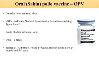
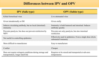
2. INTRODUCTION
• Acute viral infection caused by an RNA virus
• Primarily an infection of the human
alimentary tract
• May infect central nervous system in 1% -
resulting in varying degrees of paralysis and
death
3. PROBLEM STATEMENT
• In pre-vaccination era, poliomyelitis was found in all countries of the world
• Extensive use of polio vaccines since 1954 eliminated the disease in developed countries
• In 1988, World Health Assembly resolved to eradicate poliomyelitis globally
• Number of endemic countries reduced from 125 in 1988 to 3 in 2014 – Afghanistan,
Pakistan, Nigeria
• India has not reported any polio case since January 2011, and has been declared polio
free since 27th March, 2014
• Of the 3 strains of wild poliovirus (type 1, 2 and 3), wild poliovirus Type 2 and 3 have
been eradicated
4. EPIDEMIOLOGICAL DETERMINANTS
Agent factors
• Causative agent – Poliovirus. It has 3 serotypes 1,2 and 3. Most outbreaks of
paralytic polio are due to Type 1. It can survive for long periods in the external
environment. In a cold environment, can live in water for 4 months and in faeces
for 6 months.
• Reservoir of infection – Man is the only known reservoir. Most infections are
subclinical which play a dominant role in the spread of infection.
• Infectious material – Faeces and oropharyngeal secretions of an infected person
• Period of communicability – Most infectious 7 to 10 days before and after onset of
symptoms. In the faeces, the virus is excreted commonly for 2 to 3 weeks,
sometimes as long as 3-4 months
5. Host factors:
• Age – All age groups, but children are more susceptible because adults have
acquired immunity. Most vulnerable age is between 6 months and 3 years of
age
• Risk factors – Fatigue, trauma, intramuscular injections, operatives
procedures such as tonsillectomy
• Immunity – Maternal antibodies gradually disappear during first 6 months of
life. Immunity following infection is fairly solid. Infection with one type
does not protect completely against the other two types of viruses.
Environmental factors:
• More likely to occur during rainy season
• Contaminated water, food, flies
• Overcrowding, poor sanitation
6. MODE OF TRANSMISSION
a) Faeco–oral route: Main route of spread. Directly through contaminated
fingers or indirectly through contaminated water, milk, foods, flies and
articles of daily use
b) Droplet infection: Occurs in acute phase of disease when the virus occurs in
the throat through Close personal contact
INCUBATION PERIOD
Usually 7 to 14 days (range 3 to 35 days)
9. a) Inapparent (Sub-Clinical) Infection: No Presenting symptoms.
Diagnosis only by virus isolation or rising antibody titres.
b) Abortive Polio or Minor Illness: Mild or self-limiting illness
illness due to viraemia. Patient recovers quickly. Diagnosis only by
virus isolation or rising antibody titres.
c) Non–paralytic Polio: Stiffness and pain in the neck and back. The
disease lasts for 2 to 10 days. Recovery is rapid. Synonymous with
aseptic meningitis.
10. d) Paralytic Polio:
• Virus invades CNS and causes varying degrees of paralysis
• History of fever at the time of onset of paralysis
• Associated symptoms – malaise, anorexia, nausea, vomiting, headache, sore
throat, constipation and abdominal pain.
• Signs of meningeal irritation, i.e., stiffness of neck and back muscles. Tripod
sign may be present, i.e. the child finds difficulty in sitting and sits by
supporting hands at the back and by partially flexing the hips and knees.
• Paralysis – maximum in less than 4 days (may take 4-7 days). Descending
type i.e. starting at the hip and then moving down to the distal parts of the
extremity, asymmetrical and patchy, proximal > distal, no sensory loss, facial
asymmetry, difficulty in swallowing, weakness or loss of voice, respiratory
insufficiency
• After the acute phase, atrophy of affected muscles lead to residual paralysis
11.
12. TREATMENT
• No specific treatment
• Good nursing the beginning can minimize or even prevent crippling
• Physiotherapy is of vital importance. Can be initiated in the affected
limb immediately
13. PREVENTION
• Immunization is the only effective way
• Two types of vaccines are available worldwide
1. Inactivated (Salk) polio vaccine – IPV
2. Oral (Sabin) polio vaccine – OPV
14. Inactivated (Salk) polio vaccine – IPV
• Contains Wild Polio Virus (WPV) strains – 1, 2, 3
• Route of administration – intradermal
• Dose – 0.1 ml
• Site of administration – Right upper arm
• Schedule – 6 weeks and 14 weeks
15. • Advantages:
• Since it does not contain live virus, is safe to administer
• To persons with immune deficiency diseases
• To persons undergoing corticosteroid and radiation therapy
• To those over 50 years who are receiving vaccines for the first time
• During pregnancy
• Disadvantages:
• Does not induce intestinal or local immunity, so do not prevent reinfection of the
gut
• Does not produce herd immunity
• Not helpful during epidemics because immunity is not rapidly achieved, more
than one dose required and injections are to be avoided during epidemic times
• No serious adverse reactions
16. Oral (Sabin) polio vaccine – OPV
• Contains live attenuated virus
• bOPV used in the National Immunisation Schedule containing
Types 1 and 3
• Route of administration – oral
• Dose – 2 drops
• Schedule – At birth; 6, 10 and 14 weeks, Booster doses at 16-24
months and 5-6 years
17. • Advantages -
• Easy to administer
• Induces both humoral and intestinal immunity
• Antibody is quickly produced in a large proportion of vaccinees
• Vaccinee excretes the virus and infects others who are also immunized
• Useful in controlling epidemics
• Relatively inexpensive
18. • Complications -
• Remarkably free from complications
• Rare cases of Vaccine associated paralytic polio
• Caused by Vaccine derived poliovirus (VDPV)
• Clinically indistinguishable, only by laboratory analysis
• VDPVs are divided into three categories
1) cVDPV – when evidence of person to person transmission in the
community exists
2) Immunodeficiency associated VDPV (iVDPV) – isolated from
persons with primary immunodeficiencies
3) ambiguos VDPV (aVDPV) – which are either clinical isolates from
person with no known immunodeficiency or sewage isolated whose
source is unknown
19. • Contraindications –
• Immunocompromised individuals
• Leukaemia, malignancy and those receiving corticosteroids
• Should be delayed until after pregnancy
20. Differences between IPV and OPV
IPV (Salk type) OPV (Sabin type)
Killed formolised virus Live attenuated virus
Given intradermally or IM Given orally
Induces circulating antibody, but no local (intestinal)
immunity
Immunity is both humoral and intestinal. Induces
antibody quickly
Prevents paralysis, but does not prevent reinfection by
WPV
Prevents not only paralysis, but also intestinal
reinfection
Not useful in controlling epidemics
Effectively used in epidemics. Even a single dose elicits
substantial immunity
More difficult to manufacture Easy to manufacture
Costlier Cheaper
Does not require stringent conditions during storage and
transportation, longer shelf-life
Requires to be stored and transported at sub-zero
temperatures
21. PULSE POLIO IMMUNISATION
• Sudden, simultaneous, mass administration of OPV on a single day to all children
0-5 years of age, regardless to previous immunization.
• First round of PPI consisted of two immunization days 6 weeks apart on 9th
December 1995 and 20th January 1996. It targeted all children under 3 years of age.
Later, on WHO recommendation, age group increased from under 3 to under 5 years.
• Usually two rounds conducted about 4-6 weeks apart during low transmission season
i.e. between November to February. Depended on extent of poliovirus transmission
in the community
22. • The dose of OPV during PPIs are extra doses which supplement and do not replace
the doses received during routine immunization. No minimum interval between PPI
and scheduled OPV doses
• It ‘floods’ the community with OPV within a very short period of time, thereby
interrupting transmission of virus throughout the community
• Intensification – house to house, mop – up campaign, search and vaccinate
• In 1998, vaccine vial monitor introduced
23. POLIO SURVEILLANCE
• Most important part of the polio eradication initiative
• Without surveillance, it would be impossible to pinpoint where and how WPV is
still circulating, or to verify that the virus has been eradicated
• It identifies new cases and detects importation of WPV
• Acute flaccid paralysis (AFP) – It is defined as any child under 15 years of age
with acute, flaccid paralysis or any person with paralytic illness at any age when
polio is suspected
24. • Steps of AFP Surveillance:
1. Finding and reporting children with acute flaccid paralysis
2. Transporting stool samples for analysis
3. Isolating poliovirus
4. Mapping the virus
• Environmental surveillance – testing sewage or other environmental samples
for the presence of poliovirus. It can confirm WPV infections in the absence of
cases of paralysis.
25. SURVEILLANCE INDICATORS
Indicator
Minimum levels for certification of standard
surveillance
Completeness of reporting At least 80% of expected routine AFP surveillance reports should
be received on time, including zero reports where no AFP cases are
seen
Sensitivity of surveillance At least once case of non-polio AFP should be detected annually per
1.00.000 population aged less that 15 years. In endemic regions,
should be 2 per 1,00,000
Completeness of case
investigation
All AFP cases should have a full clinical and virological investigation
with at least 80% of AFP cases having ‘adequate’* stool specimens
collected
Completeness of follow – up At least 80% of AFP cases should have a follow – up examination for
residual paralysis at 60 days after the onset of paralysis
Laboratory performance All AFP case specimens must be processed in a WHO accredited
laboratory within the Global Polio Laboratory Network (GPLN)
26. • Adequate stool sample means:
• Two stool specimens
• Sufficient quantity – 8 grams, roughly size of thumb
• Collected at least 24 hours apart
• Within 14 days after the onset of paralysis
• Arriving in the laboratory by reverse cold chain and with proper
documentation