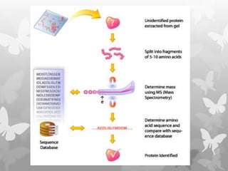
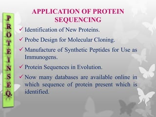
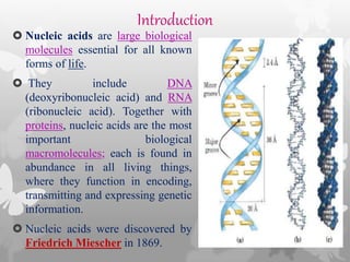
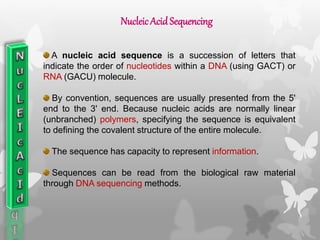
By
KAUSHAL KUMAR SAHU
Assistant Professor (Ad Hoc)
Department of Biotechnology
Govt. Digvijay Autonomous P. G. College
Raj-Nandgaon ( C. G. )
SYNOPSIS
Protein Sequencing
Introduction
Protein Sequencing
History of Protein Sequencing
Determining Amino Acid Composition
N-terminal amino acid analysis
C-terminal amino acid analysis
Edman degradation
The Edman degradation reaction
Limitations of the Edman degradation
Mass spectrometry
Synopsis
Nucleic Acid Sequencing
Introduction
Nucleic Acid Sequencing
Type of Nucleic Acid Sequencing
DNA Sequencing
Method of DNA Sequencing
Application of DNA Sequencing
DNA Sequencing Institutes
Conclusion
Reference
Introduction
Proteins are large biological molecules
consisting of one or more chains of amino acids.
Proteins differ from one another primarily in
their sequence of amino acids, which is dictated
by the nucleotide sequence of their genes, and
which usually results in folding of the protein
into a specific three-dimensional structure that
determines its activity.
PROTEIN Sequencing
Protein sequencing is a technique to
determine the amino acid sequence of
a protein.
Proteins are very long polypeptide chains
of 100 to several thousand amino acid
residues.
Amino acids can be joined covalently
through peptide bonds to form peptides
and proteins.
Protein sequencing denotes the process
of finding the amino acid sequence, or
primary structure of a protein.
History
The first protein sequencing was achieved
by Frederic Sanger in 1953. He determined
the amino acid sequence of bovine insulin.
For this achievement Sanger was awarded
the Nobel Prize in1958.
Pehr Edman, developed a machine,
determining peptide sequences automatically,
called a Sequenator.
DETERMINIG AMINO ACID COMPOSITION
Essential steps for protein sequencing
 Separation
 Hydrolysis
Hydrolysis is done by heating a sample of the
protein in 6 Molar HCl to 100-110 C° for 24
hours or longer.
However, these conditions are so vigorous that
some amino acids (serine, threonine, tyrosine,
tryptophan, glutamine and cystine) are
degraded.
N-terminal Residue Identification
C – Terminal amino acid analysis
Edman degradation method
SEQUENCING METHODS
N-terminal Residue Identification
This procedure is often used in conjugation with hydrolysis
is to label & identify the amino terminal amino acid residue.
For this purpose Fredrick Sanger developed the reagent
1-fluoro- 2,4 dinitrobenzene (FDNB).
After the amino terminal residue is labeled, the polypeptide
is hydrolyzed to its constituent amino acid and the labeled
amino acid identified.
SEQUENCINGMETHODS
 Some other reagents are also used to label the amino
terminal residue such as Dansyl chloride and Dabsyl
chloride.
 Dansyl chloride derivatives is highly fluorescent
sulfonamide derivatives and can be detected and measured in
much lower concentration than dinitrophenyl derivative.
 Dabsyl chloride, is now commonly used because it forms
intensively coloured derivatives.
Sanger's method
C – Terminal amino acid analysis
The most common method is to add Carboxypeptidase.
Carboxypeptidase are enzyme that cleaves amino acid
residues from C – terminus of polypeptide in a successive
fashion.
Another method of C – terminal amino acid analysis is
treated with hydrazine, when a peptide groups split and the
carbonyl component is converted to corresponding
hydrazine.
Edman degradation method
 To sequence the entire polypeptide a chemical method devised by
Pehr Edman is usually employed.
 The Edman degradation procedure labels and removes only the
amino-terminal residue from a peptide without disrupting the other
peptide bonds between the other amino acid residues.
 The Edman degradation sequentially removes one residue at a time
from the amino end of a peptide.
 Phenylisothiocyanate (PTC) reagent is used for the Edman
degradation.
The peptide is reacted with phenylisothiocyanate
under mildly alkaline conditions, which converts
the amino terminal amino acid to a
phenylthiocarbamoyl (PTC) adduct.
The peptide bond next to the PTC adduct is then
cleaved in a step carried out in anhydrous
trifluoroacetic acid, with removal of the amino-
terminal amino acid as an anilinothiazolinone
derivative.
The derivatized amino acid is extracted with
organic solvents, converted to the more stable
phenylthiohydantoin derivative by treatment with
aqueous acid, and then identified.
Edman Degradation method
Limitation of Edman degradation
Because the Edman degradation proceeds
from the N-terminus of the protein, it will not
work if the N-terminal amino acid has been
chemically modified .
Only 50 amino acid is identified by this
method, if protein chain is longer than 50
amino acid than the protein chain cut by
different method.
SEQUENCING OF LARGE PROTEIN
There are some steps in this process:-
Breaking of disulfide bonds.
 Disulfide bonds interfere with the sequencing
procedure. A cystein residue that has one of its
peptide bonds cleaved by the Edman procedure
will remain attached to another polypeptide.
 Disulfide bonds also interfere with the
enzymatic or chemical cleavage of the
polypeptide.
 Two approaches to irreversible breakage of
disulfide bonds are –
Oxidation by performic acid.
Reduction by dithiothreitol.
Cleaving the polypeptide chain
Protein is cleaved into a set of specific
fragments by chemical or enzymatic
methods.
Several methods can be used for
fragmenting the polypeptide chain. Enzymes
called proteases catalyze the hydrolytic
cleavage of peptide bonds.
Each fragment is then purified and
sequenced by the Edman
procedure.
Finally the order in which the
fragments appear in the original
protein is determined.
Mass spectroscopy
 Mass spectrometers explicit the difference
in the mass to charge (m/z) ratio of ionized
atoms or molecules to separate them from
each others.
 The (m/z) ratio of molecules is also a highly
characteristic properly than can be used for
determining chemical and structural
information.
The basic operation of mass spectrometer is
 Evaporation and ionize molecules in vacuum, creating
a gas phase ions.
 Separate the ions in space and/or time based on their
m/z ratio.
 Measure the amount with specific m/z ratio.
The heating or other treatment needed to transfer a
macromolecule to the gas phase usually caused its
rapid decomposition. So different techniques were
developed to overcome this problem.
i. Electrospray ionization mass spectrometry or
ESI MS.
ii. Tandem mass spectrometry.
Macromolecules in solution are
forced directly from the liquid to
gas phase.
A solution of analytes is passed
through a charged needle that is
kept at a high electrical potential,
dispersing the solution into a fine
mist of charged microdroplets.
The solvent surrounding the
macromolecules rapidly
evaporates, and the resulting
multiply charged macromolecular
ions are thus introduced
nondestructively into the gas
phase. This technique is called
electrospray ionization mass
spectrometry, or ESI MS.
Electrosprayionization massspectrometryor ESIMS.
Tandem mass spectrometry
A solution containing the protein under
investigation is first treated with a protease or
chemical reagent to hydrolyze it to a mixture
of shorter peptides then the mixture is
injected .
In the first, the peptide mixture is sorted and
the ionized fragments are manipulated so that
only one of the several types of peptides
produced by cleavage emerges at the other
end.
Then the selected peptides travel through a vacuum chamber
between the two mass spectrometers. In this collision cell, the
peptide is further fragmented by high-energy impact with a
“collision gas,” a small amount of a noble gas such as helium
or argon.
The second mass spectrometer then measures the m/z ratios
of all the charged fragments.
APPLICATION OF PROTEIN
SEQUENCING
 Identification of New Proteins.
 Probe Design for Molecular Cloning.
 Manufacture of Synthetic Peptides for Use as
Immunogens.
 Protein Sequences in Evolution.
 Now many databases are available online in
which sequence of protein present which is
identified.
Introduction
 Nucleic acids are large biological
molecules essential for all known
forms of life.
 They include DNA
(deoxyribonucleic acid) and RNA
(ribonucleic acid). Together with
proteins, nucleic acids are the most
important biological
macromolecules; each is found in
abundance in all living things,
where they function in encoding,
transmitting and expressing genetic
information.
 Nucleic acids were discovered by
Friedrich Miescher in 1869.
NucleicAcid Sequencing
A nucleic acid sequence is a succession of letters that
indicate the order of nucleotides within a DNA (using GACT) or
RNA (GACU) molecule.
By convention, sequences are usually presented from the 5'
end to the 3' end. Because nucleic acids are normally linear
(unbranched) polymers, specifying the sequence is equivalent
to defining the covalent structure of the entire molecule.
The sequence has capacity to represent information.
Sequences can be read from the biological raw material
through DNA sequencing methods.
Type of Nucleic Acid Sequencing
DNA Sequencing
RNA Sequencing
There are two type of Nucleic Acid
Sequencing-
DNA Sequencing
DNA sequencing is the process of determining the precise order of
nucleotides within a DNA molecule.
It includes any method or technology that is used to determine the
order of the four bases— adenine, guanine, cytosine, and thymine in a
strand of DNA.
The advent of rapid DNA sequencing methods has greatly accelerated
biological and medical research and discovery.
Knowledge of DNA sequences has become indispensable for basic
biological research, and in numerous applied fields such as diagnostic,
biotechnology, forensic biology, and biological systematics.
DNA sequencing is often talked about in the context of the Human
Genome Project, in which, the complete human genome was
successfully sequenced in 2001.
HISTORY
 James Watson and Francis Crick published the
first description of the crystallographic double-
helix DNA structure in 1953.
 In 1977, Maxam and Gilbert was first
developed DNA sequencing technique by
using chemical reagents.
 In 1980, Fredrick Sanger developed most
widely used method of DNA sequencing i.e.,
Chain termination method.
 The Sanger Institute was opened in 1993,
three years after the inception of the Human
Genome Project and played important role in
sequencing the 8 chromosome pairs of
human (1, 6, 9, 10, 13, 20, 22, and X).
Definition
“DNA Sequencing means finding the order
of nucleotides (adenine, guanine, cytosine,
and thymine) on a piece of DNA.”
METHODS OF DNA SEQUENCING
A) Maxam/Gilbert chemical sequencing
 Developed by Maxam and Gilbert in 1977.
 A strand of source DNA is labeled at one end
with 32P.
 The two strands of DNA are distributed into four
samples (in separate tubes).
 Each sample is subjected to treatment with a
chemical that specifically destroys one (G, C) or
two bases (A+G, T+C) in the DNA.
Advantages-
Rapid
Accurate
Limitations-
This method is complex.
It sometimes damage DNA strands
during the treatment of chemicals.
Sanger Method
Developed by Fredrick Sanger in 1980.
Also known as Dideoxynucleotide method or
Chain termination method.
4 Steps:
1. Denaturation
2. Primer attachment and extension of bases
3. Termination
4. Gel electrophoresis
Advantages-
 Best for small DNA segments.
 Rapid and fast.
Limitations-
 The need for a single- stranded DNA template.
 The use of a primer to an unknown sequence.
 The dideoxy method is good only for 500-750 bp
reactions.
 This method is Expensive.
Automated DNA Sequencing
The high demand for low-cost sequencing has
driven the development of high-throughput
sequencing (or next-generation sequencing)
technologies that parallelize the sequencing
process, producing thousands or millions of
sequences at once.
Advantages-
It is desirable to acquire sequence
data in real time by detecting the DNA
bands within the gel during the
electrophoretic separation.
It is a rapid and accurate technique.
Automated sequencer can accurately
sequence up to 10,000 nucleotides per
day.
The cost works out to be not more
than $0.2 per nucleotide.
DNAchips (MicroarrayTechnique)
• Discovered by Shena et al in 1995.
• DNA sequencing as result in advances made in automation
and miniarization.
• A large number of DNA probes, each one with different
sequence, are immobilized at defined positions on the solid
surface.
• made up of either nylon or glass.
• The probes can be short DNA molecules such as cDNAs or
synthetic oligonucleotides.
Pyrosequencing
 Visible light is generated and is proportional to the number
of incorporated nucleotides
 Determine which one of the four bases (A, G, C, T) is
incorporated at each step while a DNA template is copied.
 without added dideoxynucleotides (ddNTPs)
Solid Phase Pyrosequencing
○ Immobilized DNA
○ 3 enzymes
○ Wash step to remove nucleotides after each addition
3 enzymes + apyrase
(nucleotide degradation
enzyme)
Eliminates need for
washing step
In the well of a microtiter
plate:
• primed DNA template
• 4 enzymes
Nucleotides are added
stepwise.
Nucleotide-degrading
enzyme degrade previous
nucleotides.
Liquid Phase Pyrosequencing
APPLICATION
Molecular biotechnology
Forensic science
Genetic Engineering
Clinical application
Agriculture
Human Genome Project
INSTITUTES FOR DNA
SEQUENCING
A)In India
1. Institute of Genomics and Integrative Biology (CSIR-IGIB),Delhi
2. National Botanical Research Institute(CSIR-NBRI),Lucknow
3. Central Drug Research Institute (CSIR-CDRI),Lucknow
4. Centre for Cellular & Molecular Biology(CSIR-CCMB), Hyderabad
5. National Aids Research Institute (NARI), Pune
B) In Abroad
1. The Whitehead Institute/MIT Center for Genome Research, Cambridge.
2. Genome Sequencing Center , Washington ,University School of Medicine,
St. Louis.
3. The Wellcome Trust Sanger Institute, Cambridge, England.
4. National Centre for Genome Sequence (Mexico).
Reference
Books
 Biochemistry by Nelson and Cox, fifth edition ,W.H. freeman and Company
Newyork.
 Gene Cloning and DNA Analysis by T.A. Brown, sixth edition, A John Wiley and
Sons,Ltd,publication,2010.
Internet
 http://www.oswego.edu/~kadima/CHE525/PROTEIN%20SEQUENCING%20my%20l
ecture%20notes%20I.pdf
 en.wikipedia.org/wiki/Protein
 en.wikipedia.org/wiki/Nucelicacid
Journal-
 Maxam A.M. and Gilbert W. (1977). "A new method for sequencing DNA". Proc.
Natl. Acad. Sci. U.S.A. 74 (2): 560–4
 Ronaghi et al. (1996) Real-time DNA sequencing using detection of pyrophosphate
release. Analytical Biochemistry 242 (1): 84–9
 Ronaghi et al. (1999) Pyrosequencing Sheds Light on DNA Sequencing. Genome
Res. 2001. 11: 3-11

Protein and nucleic acid sequencing

  • 1.
    By KAUSHAL KUMAR SAHU AssistantProfessor (Ad Hoc) Department of Biotechnology Govt. Digvijay Autonomous P. G. College Raj-Nandgaon ( C. G. )
  • 2.
    SYNOPSIS Protein Sequencing Introduction Protein Sequencing Historyof Protein Sequencing Determining Amino Acid Composition N-terminal amino acid analysis C-terminal amino acid analysis Edman degradation The Edman degradation reaction Limitations of the Edman degradation Mass spectrometry
  • 3.
    Synopsis Nucleic Acid Sequencing Introduction NucleicAcid Sequencing Type of Nucleic Acid Sequencing DNA Sequencing Method of DNA Sequencing Application of DNA Sequencing DNA Sequencing Institutes Conclusion Reference
  • 4.
    Introduction Proteins are largebiological molecules consisting of one or more chains of amino acids. Proteins differ from one another primarily in their sequence of amino acids, which is dictated by the nucleotide sequence of their genes, and which usually results in folding of the protein into a specific three-dimensional structure that determines its activity.
  • 5.
    PROTEIN Sequencing Protein sequencingis a technique to determine the amino acid sequence of a protein. Proteins are very long polypeptide chains of 100 to several thousand amino acid residues. Amino acids can be joined covalently through peptide bonds to form peptides and proteins. Protein sequencing denotes the process of finding the amino acid sequence, or primary structure of a protein.
  • 6.
    History The first proteinsequencing was achieved by Frederic Sanger in 1953. He determined the amino acid sequence of bovine insulin. For this achievement Sanger was awarded the Nobel Prize in1958. Pehr Edman, developed a machine, determining peptide sequences automatically, called a Sequenator.
  • 7.
    DETERMINIG AMINO ACIDCOMPOSITION Essential steps for protein sequencing  Separation  Hydrolysis Hydrolysis is done by heating a sample of the protein in 6 Molar HCl to 100-110 C° for 24 hours or longer. However, these conditions are so vigorous that some amino acids (serine, threonine, tyrosine, tryptophan, glutamine and cystine) are degraded.
  • 8.
    N-terminal Residue Identification C– Terminal amino acid analysis Edman degradation method SEQUENCING METHODS
  • 9.
    N-terminal Residue Identification Thisprocedure is often used in conjugation with hydrolysis is to label & identify the amino terminal amino acid residue. For this purpose Fredrick Sanger developed the reagent 1-fluoro- 2,4 dinitrobenzene (FDNB). After the amino terminal residue is labeled, the polypeptide is hydrolyzed to its constituent amino acid and the labeled amino acid identified. SEQUENCINGMETHODS
  • 10.
     Some otherreagents are also used to label the amino terminal residue such as Dansyl chloride and Dabsyl chloride.  Dansyl chloride derivatives is highly fluorescent sulfonamide derivatives and can be detected and measured in much lower concentration than dinitrophenyl derivative.  Dabsyl chloride, is now commonly used because it forms intensively coloured derivatives.
  • 11.
  • 12.
    C – Terminalamino acid analysis The most common method is to add Carboxypeptidase. Carboxypeptidase are enzyme that cleaves amino acid residues from C – terminus of polypeptide in a successive fashion. Another method of C – terminal amino acid analysis is treated with hydrazine, when a peptide groups split and the carbonyl component is converted to corresponding hydrazine.
  • 14.
    Edman degradation method To sequence the entire polypeptide a chemical method devised by Pehr Edman is usually employed.  The Edman degradation procedure labels and removes only the amino-terminal residue from a peptide without disrupting the other peptide bonds between the other amino acid residues.  The Edman degradation sequentially removes one residue at a time from the amino end of a peptide.  Phenylisothiocyanate (PTC) reagent is used for the Edman degradation.
  • 15.
    The peptide isreacted with phenylisothiocyanate under mildly alkaline conditions, which converts the amino terminal amino acid to a phenylthiocarbamoyl (PTC) adduct. The peptide bond next to the PTC adduct is then cleaved in a step carried out in anhydrous trifluoroacetic acid, with removal of the amino- terminal amino acid as an anilinothiazolinone derivative. The derivatized amino acid is extracted with organic solvents, converted to the more stable phenylthiohydantoin derivative by treatment with aqueous acid, and then identified.
  • 16.
  • 17.
    Limitation of Edmandegradation Because the Edman degradation proceeds from the N-terminus of the protein, it will not work if the N-terminal amino acid has been chemically modified . Only 50 amino acid is identified by this method, if protein chain is longer than 50 amino acid than the protein chain cut by different method.
  • 18.
    SEQUENCING OF LARGEPROTEIN There are some steps in this process:- Breaking of disulfide bonds.  Disulfide bonds interfere with the sequencing procedure. A cystein residue that has one of its peptide bonds cleaved by the Edman procedure will remain attached to another polypeptide.  Disulfide bonds also interfere with the enzymatic or chemical cleavage of the polypeptide.  Two approaches to irreversible breakage of disulfide bonds are – Oxidation by performic acid. Reduction by dithiothreitol.
  • 19.
    Cleaving the polypeptidechain Protein is cleaved into a set of specific fragments by chemical or enzymatic methods. Several methods can be used for fragmenting the polypeptide chain. Enzymes called proteases catalyze the hydrolytic cleavage of peptide bonds.
  • 21.
    Each fragment isthen purified and sequenced by the Edman procedure. Finally the order in which the fragments appear in the original protein is determined.
  • 23.
    Mass spectroscopy  Massspectrometers explicit the difference in the mass to charge (m/z) ratio of ionized atoms or molecules to separate them from each others.  The (m/z) ratio of molecules is also a highly characteristic properly than can be used for determining chemical and structural information.
  • 24.
    The basic operationof mass spectrometer is  Evaporation and ionize molecules in vacuum, creating a gas phase ions.  Separate the ions in space and/or time based on their m/z ratio.  Measure the amount with specific m/z ratio.
  • 25.
    The heating orother treatment needed to transfer a macromolecule to the gas phase usually caused its rapid decomposition. So different techniques were developed to overcome this problem. i. Electrospray ionization mass spectrometry or ESI MS. ii. Tandem mass spectrometry.
  • 26.
    Macromolecules in solutionare forced directly from the liquid to gas phase. A solution of analytes is passed through a charged needle that is kept at a high electrical potential, dispersing the solution into a fine mist of charged microdroplets. The solvent surrounding the macromolecules rapidly evaporates, and the resulting multiply charged macromolecular ions are thus introduced nondestructively into the gas phase. This technique is called electrospray ionization mass spectrometry, or ESI MS. Electrosprayionization massspectrometryor ESIMS.
  • 27.
    Tandem mass spectrometry Asolution containing the protein under investigation is first treated with a protease or chemical reagent to hydrolyze it to a mixture of shorter peptides then the mixture is injected . In the first, the peptide mixture is sorted and the ionized fragments are manipulated so that only one of the several types of peptides produced by cleavage emerges at the other end.
  • 28.
    Then the selectedpeptides travel through a vacuum chamber between the two mass spectrometers. In this collision cell, the peptide is further fragmented by high-energy impact with a “collision gas,” a small amount of a noble gas such as helium or argon. The second mass spectrometer then measures the m/z ratios of all the charged fragments.
  • 30.
    APPLICATION OF PROTEIN SEQUENCING Identification of New Proteins.  Probe Design for Molecular Cloning.  Manufacture of Synthetic Peptides for Use as Immunogens.  Protein Sequences in Evolution.  Now many databases are available online in which sequence of protein present which is identified.
  • 32.
    Introduction  Nucleic acidsare large biological molecules essential for all known forms of life.  They include DNA (deoxyribonucleic acid) and RNA (ribonucleic acid). Together with proteins, nucleic acids are the most important biological macromolecules; each is found in abundance in all living things, where they function in encoding, transmitting and expressing genetic information.  Nucleic acids were discovered by Friedrich Miescher in 1869.
  • 33.
    NucleicAcid Sequencing A nucleicacid sequence is a succession of letters that indicate the order of nucleotides within a DNA (using GACT) or RNA (GACU) molecule. By convention, sequences are usually presented from the 5' end to the 3' end. Because nucleic acids are normally linear (unbranched) polymers, specifying the sequence is equivalent to defining the covalent structure of the entire molecule. The sequence has capacity to represent information. Sequences can be read from the biological raw material through DNA sequencing methods.
  • 34.
    Type of NucleicAcid Sequencing DNA Sequencing RNA Sequencing There are two type of Nucleic Acid Sequencing-
  • 35.
    DNA Sequencing DNA sequencingis the process of determining the precise order of nucleotides within a DNA molecule. It includes any method or technology that is used to determine the order of the four bases— adenine, guanine, cytosine, and thymine in a strand of DNA. The advent of rapid DNA sequencing methods has greatly accelerated biological and medical research and discovery. Knowledge of DNA sequences has become indispensable for basic biological research, and in numerous applied fields such as diagnostic, biotechnology, forensic biology, and biological systematics. DNA sequencing is often talked about in the context of the Human Genome Project, in which, the complete human genome was successfully sequenced in 2001.
  • 36.
    HISTORY  James Watsonand Francis Crick published the first description of the crystallographic double- helix DNA structure in 1953.  In 1977, Maxam and Gilbert was first developed DNA sequencing technique by using chemical reagents.  In 1980, Fredrick Sanger developed most widely used method of DNA sequencing i.e., Chain termination method.  The Sanger Institute was opened in 1993, three years after the inception of the Human Genome Project and played important role in sequencing the 8 chromosome pairs of human (1, 6, 9, 10, 13, 20, 22, and X).
  • 37.
    Definition “DNA Sequencing meansfinding the order of nucleotides (adenine, guanine, cytosine, and thymine) on a piece of DNA.”
  • 38.
    METHODS OF DNASEQUENCING A) Maxam/Gilbert chemical sequencing  Developed by Maxam and Gilbert in 1977.  A strand of source DNA is labeled at one end with 32P.  The two strands of DNA are distributed into four samples (in separate tubes).  Each sample is subjected to treatment with a chemical that specifically destroys one (G, C) or two bases (A+G, T+C) in the DNA.
  • 41.
    Advantages- Rapid Accurate Limitations- This method iscomplex. It sometimes damage DNA strands during the treatment of chemicals.
  • 42.
    Sanger Method Developed byFredrick Sanger in 1980. Also known as Dideoxynucleotide method or Chain termination method. 4 Steps: 1. Denaturation 2. Primer attachment and extension of bases 3. Termination 4. Gel electrophoresis
  • 44.
    Advantages-  Best forsmall DNA segments.  Rapid and fast. Limitations-  The need for a single- stranded DNA template.  The use of a primer to an unknown sequence.  The dideoxy method is good only for 500-750 bp reactions.  This method is Expensive.
  • 45.
    Automated DNA Sequencing Thehigh demand for low-cost sequencing has driven the development of high-throughput sequencing (or next-generation sequencing) technologies that parallelize the sequencing process, producing thousands or millions of sequences at once.
  • 47.
    Advantages- It is desirableto acquire sequence data in real time by detecting the DNA bands within the gel during the electrophoretic separation. It is a rapid and accurate technique. Automated sequencer can accurately sequence up to 10,000 nucleotides per day. The cost works out to be not more than $0.2 per nucleotide.
  • 48.
    DNAchips (MicroarrayTechnique) • Discoveredby Shena et al in 1995. • DNA sequencing as result in advances made in automation and miniarization. • A large number of DNA probes, each one with different sequence, are immobilized at defined positions on the solid surface. • made up of either nylon or glass. • The probes can be short DNA molecules such as cDNAs or synthetic oligonucleotides.
  • 50.
    Pyrosequencing  Visible lightis generated and is proportional to the number of incorporated nucleotides  Determine which one of the four bases (A, G, C, T) is incorporated at each step while a DNA template is copied.  without added dideoxynucleotides (ddNTPs)
  • 51.
    Solid Phase Pyrosequencing ○Immobilized DNA ○ 3 enzymes ○ Wash step to remove nucleotides after each addition
  • 52.
    3 enzymes +apyrase (nucleotide degradation enzyme) Eliminates need for washing step In the well of a microtiter plate: • primed DNA template • 4 enzymes Nucleotides are added stepwise. Nucleotide-degrading enzyme degrade previous nucleotides. Liquid Phase Pyrosequencing
  • 53.
    APPLICATION Molecular biotechnology Forensic science GeneticEngineering Clinical application Agriculture Human Genome Project
  • 54.
    INSTITUTES FOR DNA SEQUENCING A)InIndia 1. Institute of Genomics and Integrative Biology (CSIR-IGIB),Delhi 2. National Botanical Research Institute(CSIR-NBRI),Lucknow 3. Central Drug Research Institute (CSIR-CDRI),Lucknow 4. Centre for Cellular & Molecular Biology(CSIR-CCMB), Hyderabad 5. National Aids Research Institute (NARI), Pune B) In Abroad 1. The Whitehead Institute/MIT Center for Genome Research, Cambridge. 2. Genome Sequencing Center , Washington ,University School of Medicine, St. Louis. 3. The Wellcome Trust Sanger Institute, Cambridge, England. 4. National Centre for Genome Sequence (Mexico).
  • 55.
    Reference Books  Biochemistry byNelson and Cox, fifth edition ,W.H. freeman and Company Newyork.  Gene Cloning and DNA Analysis by T.A. Brown, sixth edition, A John Wiley and Sons,Ltd,publication,2010. Internet  http://www.oswego.edu/~kadima/CHE525/PROTEIN%20SEQUENCING%20my%20l ecture%20notes%20I.pdf  en.wikipedia.org/wiki/Protein  en.wikipedia.org/wiki/Nucelicacid Journal-  Maxam A.M. and Gilbert W. (1977). "A new method for sequencing DNA". Proc. Natl. Acad. Sci. U.S.A. 74 (2): 560–4  Ronaghi et al. (1996) Real-time DNA sequencing using detection of pyrophosphate release. Analytical Biochemistry 242 (1): 84–9  Ronaghi et al. (1999) Pyrosequencing Sheds Light on DNA Sequencing. Genome Res. 2001. 11: 3-11