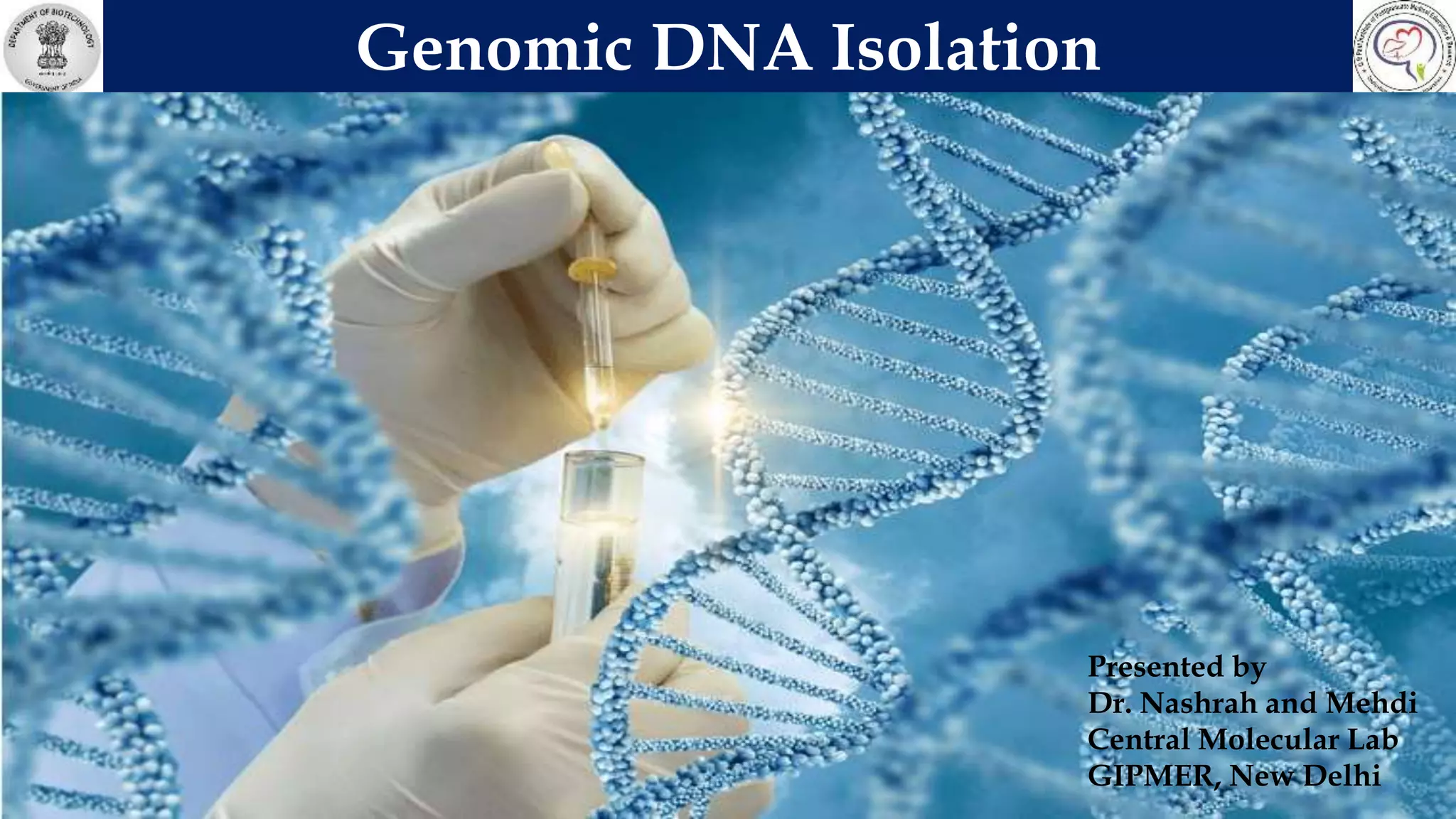
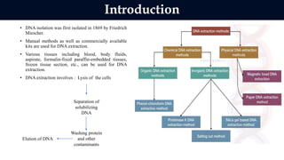
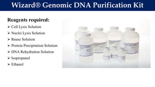
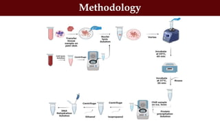
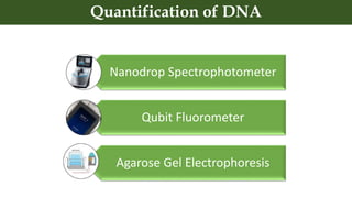
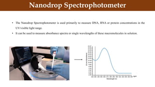
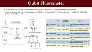
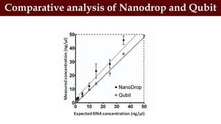
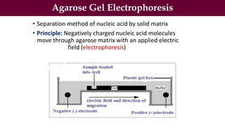
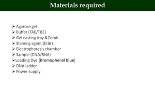
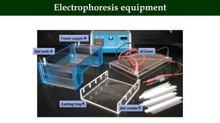
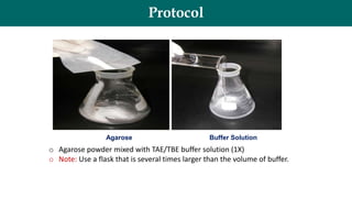
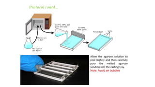
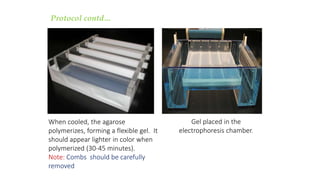
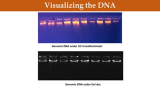
Genomic DNA Isolation
Presented by
Dr. Nashrah and Mehdi
Central Molecular Lab
GIPMER, New Delhi
• DNA isolation was first isolated in 1869 by Friedrich
Miescher.
• Manual methods as well as commercially available
kits are used for DNA extraction.
• Various tissues including blood, body fluids,
aspirate, formalin-fixed paraffin-embedded tissues,
frozen tissue section, etc., can be used for DNA
extraction.
• DNA extraction involves : Lysis of the cells
Introduction
Separation of
solubilizing
DNA
Washing protein
and other
contaminants
Elution of DNA
Reagents required:
 Cell Lysis Solution
 Nuclei Lysis Solution
 Rnase Solution
 Protein Precipitation Solution
 DNA Rehydration Solution
 Isopropanol
 Ethanol
Wizard® Genomic DNA Purification Kit
 Biological samples (Tissue/Blood)
 Microcentrifuge tubes
 Microcentrifuge tube stand
 Micropipettes
 Microtips
 Homogeniser (For tissue)
 DNA extraction kit
 Centrifuge
 Vortex mixer
 Dry bath/ water bath
Requirements for DNA Isolation
Methodology
Nanodrop Spectrophotometer
Qubit Fluorometer
Agarose Gel Electrophoresis
Quantification of DNA
Nanodrop Spectrophotometer
• The Nanodrop Spectrophotometer is used primarily to measure DNA, RNA or protein concentrations in the
UV/visible light range.
• It can be used to measure absorbance spectra or single wavelengths of these macromolecules in solution.
in continuation to previous slide…
Interpretation of the Result
 To determine the concentration of either nucleic acids or proteins in a sample by using fluorescent dye.
 Achieve accurate and precise quantification of dsDNA with Qubit dsDNA HS (High Sensitivity) and Qubit dsDNA BR
(Broad Range) Assay Kits.
Qubit Fluorometer
Comparative analysis of Nanodrop and Qubit
Agarose Gel Electrophoresis
• Separation method of nucleic acid by solid matrix
• Principle: Negatively charged nucleic acid molecules
move through agarose matrix with an applied electric
field (electrophoresis)
 Agarose gel
 Buffer (TAE/TBE)
 Gel casting tray &Comb
 Staining agent (EtBr)
 Electrophoresis chamber
 Sample (DNA/RNA)
Loading Dye (Bromophenol blue)
 DNA ladder
 Power supply
Materials required
Gel tank
Power supply
Cover
Casting tray
Gel combs
Electrophoresis equipment
Protocol
Agarose Buffer Solution
o Agarose powder mixed with TAE/TBE buffer solution (1X)
o Note: Use a flask that is several times larger than the volume of buffer.
Allow the agarose solution to
cool slightly and then carefully
pour the melted agarose
solution into the casting tray.
Note: Avoid air bubbles
Protocol contd…
Gel placed in the
electrophoresis chamber.
When cooled, the agarose
polymerizes, forming a flexible gel. It
should appear lighter in color when
polymerized (30-45 minutes).
Note: Combs should be carefully
removed
Protocol contd…
Loading the Sample
Loading of sample should be done
gently. The sample should sink into the
well.
Note: Be careful not to puncture the
gel with the pipette tip.
Sample Preparation
DNA samples mixed with
sample loading buffer.
Helps in monitoring DNA
migration
Protocol contd…
Note: Electrophoresis buffer should be enough to cover the gel
to a depth of at least 1 mm. Make sure each well is filled with
buffer.
Protocol contd…
Protocol contd…..
Cathode
(-)
DNA
(-)

Anode
(+)
 wells
 Bromophenol Blue
Bromophenol blue
will run in the same
direction as the DNA.
Applied voltage
• ↑voltage, ↑rate of migration
• But if voltage is too high, gel melts
• The best separation will apply voltage at no
more than 5V/cm of gel length.
Staining the Gel
• Ethidium bromide binds to DNA and fluoresces under UV light,
allowing the visualization of DNA on Gel.
• Ethidium bromide can be added to the gel and/or running
buffer before the gel is run or the gel can be stained after it
has run. Concentrations used is 0.5 μg/ml.
***CAUTION! Ethidium bromide is a powerful mutagen and is
moderately toxic. Gloves should be worn at all times.
Protocol contd…..
Staining the Gel
Ethidium Bromide requires an
ultraviolet light source for
visualization
• Stain for 25-30
minutes.
• Destain in water.
• Replace water several times
for efficient destain.
Protocol contd…..
Genomic DNA under UV transilluminator
Genomic DNA under Gel doc
Visualizing the DNA
Factors Affecting
 Sample type
 Purity of Nucleic acid
 DNA concentration
 Type of buffer
 pH of buffer (8.3)
 Composition of buffer
 Current and voltage
 Handling error
 Main purpose: To provide pure, unfragmented and highly concentrated DNA.
 DNA testing: DNA profiling, Genetic diagnosis, PCR, RFLP, Blotting,
Hybridization.
 Isolation of DNA is needed for genetic analysis: Scientific, Medical or forensic
purposes.
 Genetic engineering, Vaccines, Paternity test, Ancestry tracking.
Purpose of DNA Isolation
Storage of biological samples and chemicals
Sterilization of glass wares and plastic wares
Surface sterilizations
Biomedical waste management
Avoid contamination
Safety Guidelines
DNA isolation.pptx

DNA isolation.pptx

  • 1.
    Genomic DNA Isolation Presentedby Dr. Nashrah and Mehdi Central Molecular Lab GIPMER, New Delhi
  • 2.
    • DNA isolationwas first isolated in 1869 by Friedrich Miescher. • Manual methods as well as commercially available kits are used for DNA extraction. • Various tissues including blood, body fluids, aspirate, formalin-fixed paraffin-embedded tissues, frozen tissue section, etc., can be used for DNA extraction. • DNA extraction involves : Lysis of the cells Introduction Separation of solubilizing DNA Washing protein and other contaminants Elution of DNA
  • 3.
    Reagents required:  CellLysis Solution  Nuclei Lysis Solution  Rnase Solution  Protein Precipitation Solution  DNA Rehydration Solution  Isopropanol  Ethanol Wizard® Genomic DNA Purification Kit
  • 4.
     Biological samples(Tissue/Blood)  Microcentrifuge tubes  Microcentrifuge tube stand  Micropipettes  Microtips  Homogeniser (For tissue)  DNA extraction kit  Centrifuge  Vortex mixer  Dry bath/ water bath Requirements for DNA Isolation
  • 5.
  • 6.
    Nanodrop Spectrophotometer Qubit Fluorometer AgaroseGel Electrophoresis Quantification of DNA
  • 7.
    Nanodrop Spectrophotometer • TheNanodrop Spectrophotometer is used primarily to measure DNA, RNA or protein concentrations in the UV/visible light range. • It can be used to measure absorbance spectra or single wavelengths of these macromolecules in solution.
  • 8.
    in continuation toprevious slide… Interpretation of the Result
  • 9.
     To determinethe concentration of either nucleic acids or proteins in a sample by using fluorescent dye.  Achieve accurate and precise quantification of dsDNA with Qubit dsDNA HS (High Sensitivity) and Qubit dsDNA BR (Broad Range) Assay Kits. Qubit Fluorometer
  • 10.
    Comparative analysis ofNanodrop and Qubit
  • 11.
    Agarose Gel Electrophoresis •Separation method of nucleic acid by solid matrix • Principle: Negatively charged nucleic acid molecules move through agarose matrix with an applied electric field (electrophoresis)
  • 12.
     Agarose gel Buffer (TAE/TBE)  Gel casting tray &Comb  Staining agent (EtBr)  Electrophoresis chamber  Sample (DNA/RNA) Loading Dye (Bromophenol blue)  DNA ladder  Power supply Materials required
  • 13.
    Gel tank Power supply Cover Castingtray Gel combs Electrophoresis equipment
  • 14.
    Protocol Agarose Buffer Solution oAgarose powder mixed with TAE/TBE buffer solution (1X) o Note: Use a flask that is several times larger than the volume of buffer.
  • 15.
    Allow the agarosesolution to cool slightly and then carefully pour the melted agarose solution into the casting tray. Note: Avoid air bubbles Protocol contd…
  • 16.
    Gel placed inthe electrophoresis chamber. When cooled, the agarose polymerizes, forming a flexible gel. It should appear lighter in color when polymerized (30-45 minutes). Note: Combs should be carefully removed Protocol contd…
  • 17.
    Loading the Sample Loadingof sample should be done gently. The sample should sink into the well. Note: Be careful not to puncture the gel with the pipette tip. Sample Preparation DNA samples mixed with sample loading buffer. Helps in monitoring DNA migration Protocol contd…
  • 18.
    Note: Electrophoresis buffershould be enough to cover the gel to a depth of at least 1 mm. Make sure each well is filled with buffer. Protocol contd…
  • 19.
    Protocol contd….. Cathode (-) DNA (-)  Anode (+)  wells Bromophenol Blue Bromophenol blue will run in the same direction as the DNA. Applied voltage • ↑voltage, ↑rate of migration • But if voltage is too high, gel melts • The best separation will apply voltage at no more than 5V/cm of gel length.
  • 20.
    Staining the Gel •Ethidium bromide binds to DNA and fluoresces under UV light, allowing the visualization of DNA on Gel. • Ethidium bromide can be added to the gel and/or running buffer before the gel is run or the gel can be stained after it has run. Concentrations used is 0.5 μg/ml. ***CAUTION! Ethidium bromide is a powerful mutagen and is moderately toxic. Gloves should be worn at all times. Protocol contd…..
  • 21.
    Staining the Gel EthidiumBromide requires an ultraviolet light source for visualization • Stain for 25-30 minutes. • Destain in water. • Replace water several times for efficient destain. Protocol contd…..
  • 22.
    Genomic DNA underUV transilluminator Genomic DNA under Gel doc Visualizing the DNA
  • 23.
    Factors Affecting  Sampletype  Purity of Nucleic acid  DNA concentration  Type of buffer  pH of buffer (8.3)  Composition of buffer  Current and voltage  Handling error
  • 24.
     Main purpose:To provide pure, unfragmented and highly concentrated DNA.  DNA testing: DNA profiling, Genetic diagnosis, PCR, RFLP, Blotting, Hybridization.  Isolation of DNA is needed for genetic analysis: Scientific, Medical or forensic purposes.  Genetic engineering, Vaccines, Paternity test, Ancestry tracking. Purpose of DNA Isolation
  • 25.
    Storage of biologicalsamples and chemicals Sterilization of glass wares and plastic wares Surface sterilizations Biomedical waste management Avoid contamination Safety Guidelines