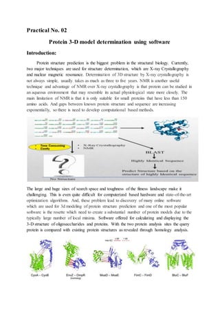
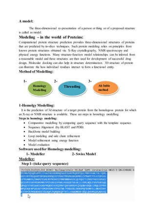
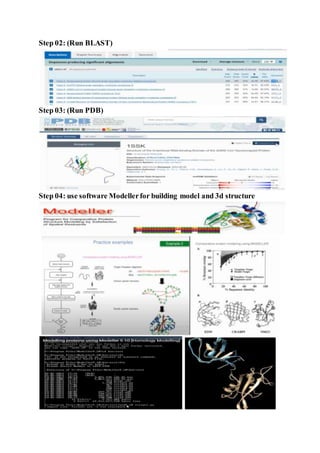
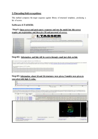
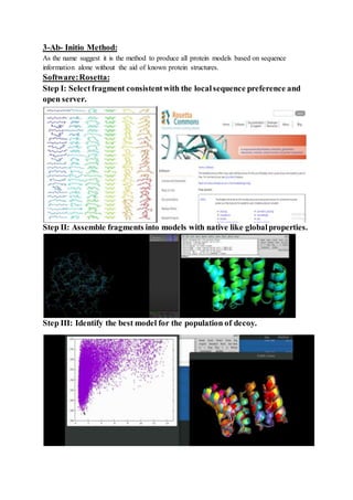
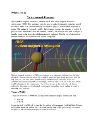
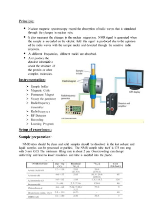
SlideShare a Scribd company logo
Practical
Introduction to proteomics
Contents:
 Nuclear Magnetic Resonance
 Protein 3D model determination using
software
Biotechnology & Bioinformatics
Government College University Faisalabad
Practical No. 02
Protein 3-D model determination using software
Introduction:
Protein structure prediction is the biggest problem in the structural biology. Currently,
two major techniques are used for structure determination, which are X-ray Crystallography
and nuclear magnetic resonance. Determination of 3D structure by X-ray crystallography is
not always simple, usually takes as much as three to five years. NMR is another useful
technique and advantage of NMR over X-ray crystallography is that protein can be studied in
an aqueous environment that may resemble its actual physiological state more closely. The
main limitation of NMR is that it is only suitable for small proteins that have less than 150
amino acids. And gaps between known protein structure and sequence are increasing
exponentially, so there is need to develop computational based methods.
The large and huge sizes of search space and toughness of the fitness landscape make it
challenging. This is even quite difficult for computerized based hardware and state-of-the-art
optimization algorithms. And, these problem lead to discovery of many online software
which are used for 3d modeling of protein structure prediction and one of the most popular
software is the rosette which need to create a substantial number of protein models due to the
typically large number of local minima. Software offered for calculating and displaying the
3-D structure of oligosaccharides and proteins. With the two protein analysis sites the query
protein is compared with existing protein structures as revealed through homology analysis.
A model:
The three-dimensional re-presentation of a person or thing or of a proposed structure
is called as model.
Modeling – in the world of Proteins:
Computational protein structure prediction provides three-dimensional structures of proteins
that are predicted by in-silico techniques. Such protein modeling relies on principles from
known protein structures obtained via: X-Ray crystallography, NMR spectroscopy and
physical energy functions. Many structure-function model relationships can be inferred from
a reasonable model and these structures are then used for development of successful drug
design. Molecular docking can also help in structure determination. 3D structure of protein
can illustrate the how individual residues interact to form a functional entity.
Method of Modelling:
1- 2- 3-
1-Homolgy Modelling:
It is the prediction of 3d structure of a target protein from the homologous protein for which
an X-ray or NMR structure is available. These are steps in homology modelling.
Steps in homology modeling:
 Comparative modelling by comparing query sequence with the template sequence.
 Sequence Alignment (by BLAST and PDB)
 Backbone model building
 Loop modeling and side chain refinement
 Model refinement using energy function
 Model evaluation
Software usedfor Homologymodelling:
1- Modeller 2- Swiss Model
Modeller:
Step 1- (take query sequence)
Homology
Modelling
Threading Ab Initio
method
Step 02: (Run BLAST)
Step 03: (Run PDB)
Step 04: use software Modellerfor building model and 3d structure
Swiss Model:
Step 01: (query sequence) Step 02: Open Swiss model software
Step 03: SWISS Model:(Putting sequence and search for templates)
Step 04: (After getting template hits run build model)
Step 05: (Model building gives result of 3d protein with different information)
2-Threading/fold recognition:
This method compares the target sequence against library of structural templates, producing a
list of scores.
Software:I-TASSER:
Step I: Open server and paste query sequence and run the model but this server
require pre-registration and then give ID and password of server.
Step 02: Information and link will be sent to through email just click on link
Step 03: Information about 2d and 2d structures were given. 5 models were given to
you select with high C-value.
3-Ab- Initio Method:
As the name suggest it is the method to produce all protein models based on sequence
information alone without the aid of known protein structures.
Software:Rosetta:
Step I: Selectfragment consistentwith the localsequence preference and
open server.
Step II: Assemble fragments into models with native like globalproperties.
Step III: Identify the best model for the population of decoy.
Practicalno. 02
Nuclearmagnetic Resonance
NMR (nuclear magnetic resonance) spectroscopy is also called Magnetic resonance
spectroscopy (MRS). This technique is mostly used to study the magnetic properties around
the atomic nuclei. It is also used to study the chemical, physical and chemical properties of
matter. This method is commonly used by the biochemist to study the structure of protein. It
provides detail information about the structure, function, and reaction state. This technique is
actually based on the absorption of electromagnetic radiations. NMR is one of most precise
method to observe the monomolecular organic compounds.
Nuclear magnetic resonance (NMR) spectroscopy is an important analytical tool for living
chemicals. Research conducted on the biological board has been greatly improved with the
help of NMR. Not only does it provide details about molecular structure, it can also
determine the content and purity of the sample. Proton (1H) NMR is one of the most widely
used NMR methods for biochemistry. The protons present in this molecule will behave
differently depending on the chemical environment surrounding them, making it easier to
determine their structure.
Types of NMR:
There are two types of NMR that can be used in chemical analysis and medical field.
1. H-NMR
2. C-NMR
In this practical H-NMR will be used for the analysis of a compound. In H-NMR on the basis
of hydrogen atoms the analysis of a compound can be done.NMR spectroscopy determines
the physical and chemical properties of atoms or molecules.
Principle:
 Nuclear magnetic spectroscopy record the absorption of radio waves that is stimulated
through the changes in nuclear spin.
 It also measures the changes in the nuclear magnetism. NMR signal is generated when
the sample is ascended on the electric field this signal is produced due to the agitation
of the radio waves with the sample nuclei and detected through the sensitive radio
receivers.
 At different frequencies, different nuclei are absorbed.
 And produce the
detailed information
about the structure of
the protein or other
complex molecules.
Instrumentation:
 Sample holder
 Magnetic Coils
 Permanent Magnet
 Sweep the generator
 Radiofrequency
transmitter
 Radiofrequency
 RF Detector
 Recording
 Learning Program
Setup of experiment:
Sample preparation:
NMR tubes should be clean and solid samples should be dissolved in the lost solvent and
liquid samples can be processed or purified. The NMR sample tube itself is 175 mm long
with 5 mm O.D. The minimum filling rate is about 2 cm. Overcrowding can disrupt
uniformity and lead to lower resolution and tube is inserted into the probe.
Temperature adjustment
Chemical changes and the formation of signal lines are highly dependent on temperature.
In addition, protein composition can change with temperature.
Lock:
In the introduction it was stated that the current energy producing magnetic field continues
indefinitely. This is almost certainly true but actually decreases slightly over time and this
means that if this is not compensated, the magnetic field at the beginning and end of the study
may vary and have a lower quality effect.
Tuning:
As a radio transmitter and / or receiver of radiofrequency radiation, the investigation must
be well-organized in order to operate efficiently.
Shimming:
The magnetic field is strong enough for an effective NMR test, and it is also important
that the magnetic field be matched to the volume of the sample. If it were not so, the nuclei of
one part of the tube would receive a different field than the nuclei of the other part of the tube
with different resonance waves as a result.
Determination of water frequency:
When making protein NMR usually contains 1 mM protein dissolved in water. The filter
of water thus is 55 M and since there are two protons in water molecule the total proton is ~
100 M, so 100,000 is higher than the heart you are interested in.
Power range drive:
The highest NMR signal is obtained when the pulse width is exactly 90 °, which means that
the magnetization vector is rotated from the z-axis to the rotating plane. In addition, most
tests require that some lumps be 180 ° exactly. So we need to measure this too. Contrary to
perceptual perception, it is not so easy to detect a high signal and therefore set up a search for
a signal (or as low as possible) signal.
Procedure
 Placed the sample in a magnetic field.
 Excite the nuclei sample into nuclear magnetic resonance with the help of radio waves
to obtain NMR signals.
 NMR signals are detected with the help of sensitive radio receivers.
 The resonance frequency of an atom in a molecule is changed with intra-molecular
magnetic field surrounding it.
 This gives details of the functional group of the individual molecule and its electronic
structure.
 NMR spectroscopy is a very precise method for the identification of monomolecular
organic compounds.
 Provide the details of reaction state, structure, chemical environment and dynamics of
a molecule.
What we canlearn from NMR spectra?
 Chemical shift: Information about the composition of atomic groups within the
molecule.
 Spin-Spin coupling constant: Information about adjacent atoms.
 Relaxation time: Information on molecular dynamics.
 Signal intensity: Quantitative information, e.g. atomic ratios within a molecule that
can be helpful in determining the molecular structure, and proportions of different
compounds in a mixture.
 Observations and results:
This test gives us the basic concepts of nuclear magnetic resonance (NMR)
spectroscopy - spin, energy levels, radiation absorption, and multiple NMR viewing
parameters, and provides experience in identifying the unknown 1H (proton) NMR
spectra. A series of well-known samples will be used to present sample preparation
methods, the performance of NMR spectrometers, and the 1 H-NMR spectra where
students will measure chemical shifts, J-couplings and spectral energy. After that, it
will be able to record three unknown objects and will use these fields to find the
structure and chemical composition.

More Related Content

What's hot

Cadd
CaddCadd
Antisense oligoneucleotides in therapy
Antisense  oligoneucleotides  in therapyAntisense  oligoneucleotides  in therapy
Antisense oligoneucleotides in therapy
Dr Sajeena Jose
 
Human Genome Project
Human Genome ProjectHuman Genome Project
Human Genome Project
PratyusshKumaarr
 
Introduction to NGS Variant Calling Analysis (UEB-UAT Bioinformatics Course -...
Introduction to NGS Variant Calling Analysis (UEB-UAT Bioinformatics Course -...Introduction to NGS Variant Calling Analysis (UEB-UAT Bioinformatics Course -...
Introduction to NGS Variant Calling Analysis (UEB-UAT Bioinformatics Course -...
VHIR Vall d’Hebron Institut de Recerca
 
Sequence assembly
Sequence assemblySequence assembly
Sequence assembly
Dr. Naveen Gaurav srivastava
 
Threading modeling methods
Threading modeling methodsThreading modeling methods
Threading modeling methods
ratanvishwas
 
Whole exome sequencing data analysis.pptx
Whole exome sequencing data analysis.pptxWhole exome sequencing data analysis.pptx
Whole exome sequencing data analysis.pptx
Haibo Liu
 
Biomarkers roohith
Biomarkers roohithBiomarkers roohith
Biomarkers roohith
roohith
 
Applications of genomics and proteomics ppt
Applications of genomics and  proteomics pptApplications of genomics and  proteomics ppt
Applications of genomics and proteomics ppt
Ibad khan
 
Ncbi basic intro_v_pitt_kent_osu
Ncbi basic intro_v_pitt_kent_osuNcbi basic intro_v_pitt_kent_osu
Ncbi basic intro_v_pitt_kent_osu
Ben Busby
 
Target identification and validation
Target identification and validationTarget identification and validation
Target identification and validation
AshishVerma571
 
Role of genomics proteomics, and bioinformatics.
Role of genomics proteomics, and bioinformatics.Role of genomics proteomics, and bioinformatics.
Role of genomics proteomics, and bioinformatics.
B V V S Hanagal Shri Kumareshwar College of Pharmacy, Bagalkote
 
Protein-protein interaction networks
Protein-protein interaction networksProtein-protein interaction networks
Protein-protein interaction networks
Bioinformatics and Computational Biosciences Branch
 
Comparitive genome mapping and model systems
Comparitive genome mapping and model systemsComparitive genome mapping and model systems
Comparitive genome mapping and model systems
Himanshi Chauhan
 
Gene silencing
Gene silencingGene silencing
Gene silencing
manojjeya
 
Target Validation
Target ValidationTarget Validation
Genomic mapping, genetic mapping
Genomic mapping, genetic mappingGenomic mapping, genetic mapping
Genomic mapping, genetic mapping
KAUSHAL SAHU
 
Structural bioinformatics.
Structural bioinformatics.Structural bioinformatics.
Structural bioinformatics.
SALIHAMUGHAL
 
Pharmacoinformatics Database basics(sree)
Pharmacoinformatics Database basics(sree)Pharmacoinformatics Database basics(sree)
Pharmacoinformatics Database basics(sree)
Sreekanth Gali
 

What's hot (20)

Cadd
CaddCadd
Cadd
 
Antisense oligoneucleotides in therapy
Antisense  oligoneucleotides  in therapyAntisense  oligoneucleotides  in therapy
Antisense oligoneucleotides in therapy
 
Human Genome Project
Human Genome ProjectHuman Genome Project
Human Genome Project
 
Introduction to NGS Variant Calling Analysis (UEB-UAT Bioinformatics Course -...
Introduction to NGS Variant Calling Analysis (UEB-UAT Bioinformatics Course -...Introduction to NGS Variant Calling Analysis (UEB-UAT Bioinformatics Course -...
Introduction to NGS Variant Calling Analysis (UEB-UAT Bioinformatics Course -...
 
Sequence assembly
Sequence assemblySequence assembly
Sequence assembly
 
Threading modeling methods
Threading modeling methodsThreading modeling methods
Threading modeling methods
 
Whole exome sequencing data analysis.pptx
Whole exome sequencing data analysis.pptxWhole exome sequencing data analysis.pptx
Whole exome sequencing data analysis.pptx
 
Biomarkers roohith
Biomarkers roohithBiomarkers roohith
Biomarkers roohith
 
Applications of genomics and proteomics ppt
Applications of genomics and  proteomics pptApplications of genomics and  proteomics ppt
Applications of genomics and proteomics ppt
 
Ncbi basic intro_v_pitt_kent_osu
Ncbi basic intro_v_pitt_kent_osuNcbi basic intro_v_pitt_kent_osu
Ncbi basic intro_v_pitt_kent_osu
 
Target identification and validation
Target identification and validationTarget identification and validation
Target identification and validation
 
Role of genomics proteomics, and bioinformatics.
Role of genomics proteomics, and bioinformatics.Role of genomics proteomics, and bioinformatics.
Role of genomics proteomics, and bioinformatics.
 
Protein-protein interaction networks
Protein-protein interaction networksProtein-protein interaction networks
Protein-protein interaction networks
 
Comparitive genome mapping and model systems
Comparitive genome mapping and model systemsComparitive genome mapping and model systems
Comparitive genome mapping and model systems
 
Bioinformatics
BioinformaticsBioinformatics
Bioinformatics
 
Gene silencing
Gene silencingGene silencing
Gene silencing
 
Target Validation
Target ValidationTarget Validation
Target Validation
 
Genomic mapping, genetic mapping
Genomic mapping, genetic mappingGenomic mapping, genetic mapping
Genomic mapping, genetic mapping
 
Structural bioinformatics.
Structural bioinformatics.Structural bioinformatics.
Structural bioinformatics.
 
Pharmacoinformatics Database basics(sree)
Pharmacoinformatics Database basics(sree)Pharmacoinformatics Database basics(sree)
Pharmacoinformatics Database basics(sree)
 

Similar to Proteomics Practical (NMR and Protein 3D software

Nuclear magnetic resonance (enzymology)
Nuclear magnetic resonance (enzymology) Nuclear magnetic resonance (enzymology)
Nuclear magnetic resonance (enzymology)
Mohsin Shad
 
Ko sponholtz NMR project
Ko sponholtz NMR projectKo sponholtz NMR project
Ko sponholtz NMR project
Kwonil Ko
 
nuclearmagneticresonance-180329132838.pdf
nuclearmagneticresonance-180329132838.pdfnuclearmagneticresonance-180329132838.pdf
nuclearmagneticresonance-180329132838.pdf
Monukumawat7
 
Nuclear magnetic resonance
Nuclear magnetic resonanceNuclear magnetic resonance
Nuclear magnetic resonance
Abhishek Indurkar
 
Nmr spectroscopy
Nmr spectroscopyNmr spectroscopy
Nmr spectroscopy
TATHAGATA CHAKRABORTY
 
NMR .pdf
NMR .pdfNMR .pdf
NMR .pdf
Nusrat Gulbarga
 
Nuclear Magnetic Resonance
Nuclear Magnetic ResonanceNuclear Magnetic Resonance
Nuclear Magnetic Resonance
govinda pathak
 
Qualitative analysis of Fruits and Vegetables using Earth’s Field Nuclear Mag...
Qualitative analysis of Fruits and Vegetables using Earth’s Field Nuclear Mag...Qualitative analysis of Fruits and Vegetables using Earth’s Field Nuclear Mag...
Qualitative analysis of Fruits and Vegetables using Earth’s Field Nuclear Mag...
IJERA Editor
 
X ray crystallography analysis
X ray crystallography analysis X ray crystallography analysis
X ray crystallography analysis
prabhakarareddy A V
 
NMRbySSS.Complete-1.pptx
NMRbySSS.Complete-1.pptxNMRbySSS.Complete-1.pptx
NMRbySSS.Complete-1.pptx
ABDULRAUF411
 
Nuclear Magnetic Resonance (NMR).pptx
Nuclear Magnetic Resonance (NMR).pptxNuclear Magnetic Resonance (NMR).pptx
Nuclear Magnetic Resonance (NMR).pptx
Ankita More
 
Determination of protein tertiary structure
Determination of protein tertiary structureDetermination of protein tertiary structure
Determination of protein tertiary structure
Bahauddin zakariya university,Multan
 
NDE by magnetic resonance.docx
NDE by magnetic resonance.docxNDE by magnetic resonance.docx
NDE by magnetic resonance.docx
Himasree Viswanadhapalli
 
NISHANT PPT TWCHNIQUES nanoparticles techniques
NISHANT PPT TWCHNIQUES nanoparticles techniquesNISHANT PPT TWCHNIQUES nanoparticles techniques
NISHANT PPT TWCHNIQUES nanoparticles techniques
Umar Karimi
 
Characterization of nanopartical
Characterization of nanoparticalCharacterization of nanopartical
Characterization of nanopartical
Amany EL-Hallaq
 
NMR Spectroscopy By Himaja Donthula
NMR Spectroscopy By Himaja DonthulaNMR Spectroscopy By Himaja Donthula
NMR Spectroscopy By Himaja Donthula
himaja donthula
 
Nanoflam product analysis
Nanoflam product analysisNanoflam product analysis
Nanoflam product analysis
JacobScholes1
 
NMR - pooja .pptx
NMR - pooja .pptxNMR - pooja .pptx
NMR - pooja .pptx
Pooja mohod04
 
Physical structure of fibers
Physical structure of fibersPhysical structure of fibers
Physical structure of fibers
Zaki Mujahid
 
2DNMRSpectroscopy_Advancedspectralanalysis (1).pdf
2DNMRSpectroscopy_Advancedspectralanalysis (1).pdf2DNMRSpectroscopy_Advancedspectralanalysis (1).pdf
2DNMRSpectroscopy_Advancedspectralanalysis (1).pdf
ssuser5c45e5
 

Similar to Proteomics Practical (NMR and Protein 3D software (20)

Nuclear magnetic resonance (enzymology)
Nuclear magnetic resonance (enzymology) Nuclear magnetic resonance (enzymology)
Nuclear magnetic resonance (enzymology)
 
Ko sponholtz NMR project
Ko sponholtz NMR projectKo sponholtz NMR project
Ko sponholtz NMR project
 
nuclearmagneticresonance-180329132838.pdf
nuclearmagneticresonance-180329132838.pdfnuclearmagneticresonance-180329132838.pdf
nuclearmagneticresonance-180329132838.pdf
 
Nuclear magnetic resonance
Nuclear magnetic resonanceNuclear magnetic resonance
Nuclear magnetic resonance
 
Nmr spectroscopy
Nmr spectroscopyNmr spectroscopy
Nmr spectroscopy
 
NMR .pdf
NMR .pdfNMR .pdf
NMR .pdf
 
Nuclear Magnetic Resonance
Nuclear Magnetic ResonanceNuclear Magnetic Resonance
Nuclear Magnetic Resonance
 
Qualitative analysis of Fruits and Vegetables using Earth’s Field Nuclear Mag...
Qualitative analysis of Fruits and Vegetables using Earth’s Field Nuclear Mag...Qualitative analysis of Fruits and Vegetables using Earth’s Field Nuclear Mag...
Qualitative analysis of Fruits and Vegetables using Earth’s Field Nuclear Mag...
 
X ray crystallography analysis
X ray crystallography analysis X ray crystallography analysis
X ray crystallography analysis
 
NMRbySSS.Complete-1.pptx
NMRbySSS.Complete-1.pptxNMRbySSS.Complete-1.pptx
NMRbySSS.Complete-1.pptx
 
Nuclear Magnetic Resonance (NMR).pptx
Nuclear Magnetic Resonance (NMR).pptxNuclear Magnetic Resonance (NMR).pptx
Nuclear Magnetic Resonance (NMR).pptx
 
Determination of protein tertiary structure
Determination of protein tertiary structureDetermination of protein tertiary structure
Determination of protein tertiary structure
 
NDE by magnetic resonance.docx
NDE by magnetic resonance.docxNDE by magnetic resonance.docx
NDE by magnetic resonance.docx
 
NISHANT PPT TWCHNIQUES nanoparticles techniques
NISHANT PPT TWCHNIQUES nanoparticles techniquesNISHANT PPT TWCHNIQUES nanoparticles techniques
NISHANT PPT TWCHNIQUES nanoparticles techniques
 
Characterization of nanopartical
Characterization of nanoparticalCharacterization of nanopartical
Characterization of nanopartical
 
NMR Spectroscopy By Himaja Donthula
NMR Spectroscopy By Himaja DonthulaNMR Spectroscopy By Himaja Donthula
NMR Spectroscopy By Himaja Donthula
 
Nanoflam product analysis
Nanoflam product analysisNanoflam product analysis
Nanoflam product analysis
 
NMR - pooja .pptx
NMR - pooja .pptxNMR - pooja .pptx
NMR - pooja .pptx
 
Physical structure of fibers
Physical structure of fibersPhysical structure of fibers
Physical structure of fibers
 
2DNMRSpectroscopy_Advancedspectralanalysis (1).pdf
2DNMRSpectroscopy_Advancedspectralanalysis (1).pdf2DNMRSpectroscopy_Advancedspectralanalysis (1).pdf
2DNMRSpectroscopy_Advancedspectralanalysis (1).pdf
 

More from iqraakbar8

Use of Microalgae for Phycoremdiation & biodiseal production
Use of Microalgae for Phycoremdiation & biodiseal productionUse of Microalgae for Phycoremdiation & biodiseal production
Use of Microalgae for Phycoremdiation & biodiseal production
iqraakbar8
 
Zero waste water treatment and biofuel production
Zero waste water treatment and biofuel productionZero waste water treatment and biofuel production
Zero waste water treatment and biofuel production
iqraakbar8
 
waste water treatment through Algae and Cyanobacteria
 waste water treatment through Algae and Cyanobacteria waste water treatment through Algae and Cyanobacteria
waste water treatment through Algae and Cyanobacteria
iqraakbar8
 
Impact of Organic & Inorganic Fertilizers on Agriculture
Impact of Organic & Inorganic Fertilizers on AgricultureImpact of Organic & Inorganic Fertilizers on Agriculture
Impact of Organic & Inorganic Fertilizers on Agriculture
iqraakbar8
 
CRISPER Cas & Food supply chain Application
CRISPER Cas & Food supply chain ApplicationCRISPER Cas & Food supply chain Application
CRISPER Cas & Food supply chain Application
iqraakbar8
 
The next generation of crispr–cas technologies and Applications
The next generation of crispr–cas technologies and ApplicationsThe next generation of crispr–cas technologies and Applications
The next generation of crispr–cas technologies and Applications
iqraakbar8
 
Magnetic particles in algae biotechnology
Magnetic particles in algae biotechnology Magnetic particles in algae biotechnology
Magnetic particles in algae biotechnology
iqraakbar8
 
Yeast two hybrid system
Yeast two hybrid systemYeast two hybrid system
Yeast two hybrid system
iqraakbar8
 
Microbial proteomics in biotechnology
Microbial proteomics in biotechnologyMicrobial proteomics in biotechnology
Microbial proteomics in biotechnology
iqraakbar8
 
(Genomics in human health)
 (Genomics in human health) (Genomics in human health)
(Genomics in human health)
iqraakbar8
 
Economic importance of cyanobacteria
Economic importance of cyanobacteriaEconomic importance of cyanobacteria
Economic importance of cyanobacteria
iqraakbar8
 
Gene transfection or Method of gene transfer
Gene transfection or Method of gene transferGene transfection or Method of gene transfer
Gene transfection or Method of gene transfer
iqraakbar8
 
Nanotechnology and drug delivery
Nanotechnology and drug deliveryNanotechnology and drug delivery
Nanotechnology and drug delivery
iqraakbar8
 
Marker assisted selection in plants
Marker assisted selection in plantsMarker assisted selection in plants
Marker assisted selection in plants
iqraakbar8
 
Genomics C elegan genome and model organism
Genomics C elegan genome and model organismGenomics C elegan genome and model organism
Genomics C elegan genome and model organism
iqraakbar8
 
Future of animal biotechnology
Future of animal biotechnologyFuture of animal biotechnology
Future of animal biotechnology
iqraakbar8
 
C3 and C4 plants Botany
C3 and C4  plants BotanyC3 and C4  plants Botany
C3 and C4 plants Botany
iqraakbar8
 
Antifreeze protein
Antifreeze proteinAntifreeze protein
Antifreeze protein
iqraakbar8
 
Electrophoretic mobility shift assay
Electrophoretic mobility shift assay Electrophoretic mobility shift assay
Electrophoretic mobility shift assay
iqraakbar8
 
Aging and animal biotechnology
Aging and animal biotechnologyAging and animal biotechnology
Aging and animal biotechnology
iqraakbar8
 

More from iqraakbar8 (20)

Use of Microalgae for Phycoremdiation & biodiseal production
Use of Microalgae for Phycoremdiation & biodiseal productionUse of Microalgae for Phycoremdiation & biodiseal production
Use of Microalgae for Phycoremdiation & biodiseal production
 
Zero waste water treatment and biofuel production
Zero waste water treatment and biofuel productionZero waste water treatment and biofuel production
Zero waste water treatment and biofuel production
 
waste water treatment through Algae and Cyanobacteria
 waste water treatment through Algae and Cyanobacteria waste water treatment through Algae and Cyanobacteria
waste water treatment through Algae and Cyanobacteria
 
Impact of Organic & Inorganic Fertilizers on Agriculture
Impact of Organic & Inorganic Fertilizers on AgricultureImpact of Organic & Inorganic Fertilizers on Agriculture
Impact of Organic & Inorganic Fertilizers on Agriculture
 
CRISPER Cas & Food supply chain Application
CRISPER Cas & Food supply chain ApplicationCRISPER Cas & Food supply chain Application
CRISPER Cas & Food supply chain Application
 
The next generation of crispr–cas technologies and Applications
The next generation of crispr–cas technologies and ApplicationsThe next generation of crispr–cas technologies and Applications
The next generation of crispr–cas technologies and Applications
 
Magnetic particles in algae biotechnology
Magnetic particles in algae biotechnology Magnetic particles in algae biotechnology
Magnetic particles in algae biotechnology
 
Yeast two hybrid system
Yeast two hybrid systemYeast two hybrid system
Yeast two hybrid system
 
Microbial proteomics in biotechnology
Microbial proteomics in biotechnologyMicrobial proteomics in biotechnology
Microbial proteomics in biotechnology
 
(Genomics in human health)
 (Genomics in human health) (Genomics in human health)
(Genomics in human health)
 
Economic importance of cyanobacteria
Economic importance of cyanobacteriaEconomic importance of cyanobacteria
Economic importance of cyanobacteria
 
Gene transfection or Method of gene transfer
Gene transfection or Method of gene transferGene transfection or Method of gene transfer
Gene transfection or Method of gene transfer
 
Nanotechnology and drug delivery
Nanotechnology and drug deliveryNanotechnology and drug delivery
Nanotechnology and drug delivery
 
Marker assisted selection in plants
Marker assisted selection in plantsMarker assisted selection in plants
Marker assisted selection in plants
 
Genomics C elegan genome and model organism
Genomics C elegan genome and model organismGenomics C elegan genome and model organism
Genomics C elegan genome and model organism
 
Future of animal biotechnology
Future of animal biotechnologyFuture of animal biotechnology
Future of animal biotechnology
 
C3 and C4 plants Botany
C3 and C4  plants BotanyC3 and C4  plants Botany
C3 and C4 plants Botany
 
Antifreeze protein
Antifreeze proteinAntifreeze protein
Antifreeze protein
 
Electrophoretic mobility shift assay
Electrophoretic mobility shift assay Electrophoretic mobility shift assay
Electrophoretic mobility shift assay
 
Aging and animal biotechnology
Aging and animal biotechnologyAging and animal biotechnology
Aging and animal biotechnology
 

Proteomics Practical (NMR and Protein 3D software

  • 1. Practical Introduction to proteomics Contents:  Nuclear Magnetic Resonance  Protein 3D model determination using software Biotechnology & Bioinformatics Government College University Faisalabad
  • 2. Practical No. 02 Protein 3-D model determination using software Introduction: Protein structure prediction is the biggest problem in the structural biology. Currently, two major techniques are used for structure determination, which are X-ray Crystallography and nuclear magnetic resonance. Determination of 3D structure by X-ray crystallography is not always simple, usually takes as much as three to five years. NMR is another useful technique and advantage of NMR over X-ray crystallography is that protein can be studied in an aqueous environment that may resemble its actual physiological state more closely. The main limitation of NMR is that it is only suitable for small proteins that have less than 150 amino acids. And gaps between known protein structure and sequence are increasing exponentially, so there is need to develop computational based methods. The large and huge sizes of search space and toughness of the fitness landscape make it challenging. This is even quite difficult for computerized based hardware and state-of-the-art optimization algorithms. And, these problem lead to discovery of many online software which are used for 3d modeling of protein structure prediction and one of the most popular software is the rosette which need to create a substantial number of protein models due to the typically large number of local minima. Software offered for calculating and displaying the 3-D structure of oligosaccharides and proteins. With the two protein analysis sites the query protein is compared with existing protein structures as revealed through homology analysis.
  • 3. A model: The three-dimensional re-presentation of a person or thing or of a proposed structure is called as model. Modeling – in the world of Proteins: Computational protein structure prediction provides three-dimensional structures of proteins that are predicted by in-silico techniques. Such protein modeling relies on principles from known protein structures obtained via: X-Ray crystallography, NMR spectroscopy and physical energy functions. Many structure-function model relationships can be inferred from a reasonable model and these structures are then used for development of successful drug design. Molecular docking can also help in structure determination. 3D structure of protein can illustrate the how individual residues interact to form a functional entity. Method of Modelling: 1- 2- 3- 1-Homolgy Modelling: It is the prediction of 3d structure of a target protein from the homologous protein for which an X-ray or NMR structure is available. These are steps in homology modelling. Steps in homology modeling:  Comparative modelling by comparing query sequence with the template sequence.  Sequence Alignment (by BLAST and PDB)  Backbone model building  Loop modeling and side chain refinement  Model refinement using energy function  Model evaluation Software usedfor Homologymodelling: 1- Modeller 2- Swiss Model Modeller: Step 1- (take query sequence) Homology Modelling Threading Ab Initio method
  • 4. Step 02: (Run BLAST) Step 03: (Run PDB) Step 04: use software Modellerfor building model and 3d structure
  • 5. Swiss Model: Step 01: (query sequence) Step 02: Open Swiss model software Step 03: SWISS Model:(Putting sequence and search for templates) Step 04: (After getting template hits run build model) Step 05: (Model building gives result of 3d protein with different information)
  • 6. 2-Threading/fold recognition: This method compares the target sequence against library of structural templates, producing a list of scores. Software:I-TASSER: Step I: Open server and paste query sequence and run the model but this server require pre-registration and then give ID and password of server. Step 02: Information and link will be sent to through email just click on link Step 03: Information about 2d and 2d structures were given. 5 models were given to you select with high C-value.
  • 7. 3-Ab- Initio Method: As the name suggest it is the method to produce all protein models based on sequence information alone without the aid of known protein structures. Software:Rosetta: Step I: Selectfragment consistentwith the localsequence preference and open server. Step II: Assemble fragments into models with native like globalproperties. Step III: Identify the best model for the population of decoy.
  • 8. Practicalno. 02 Nuclearmagnetic Resonance NMR (nuclear magnetic resonance) spectroscopy is also called Magnetic resonance spectroscopy (MRS). This technique is mostly used to study the magnetic properties around the atomic nuclei. It is also used to study the chemical, physical and chemical properties of matter. This method is commonly used by the biochemist to study the structure of protein. It provides detail information about the structure, function, and reaction state. This technique is actually based on the absorption of electromagnetic radiations. NMR is one of most precise method to observe the monomolecular organic compounds. Nuclear magnetic resonance (NMR) spectroscopy is an important analytical tool for living chemicals. Research conducted on the biological board has been greatly improved with the help of NMR. Not only does it provide details about molecular structure, it can also determine the content and purity of the sample. Proton (1H) NMR is one of the most widely used NMR methods for biochemistry. The protons present in this molecule will behave differently depending on the chemical environment surrounding them, making it easier to determine their structure. Types of NMR: There are two types of NMR that can be used in chemical analysis and medical field. 1. H-NMR 2. C-NMR In this practical H-NMR will be used for the analysis of a compound. In H-NMR on the basis of hydrogen atoms the analysis of a compound can be done.NMR spectroscopy determines the physical and chemical properties of atoms or molecules.
  • 9. Principle:  Nuclear magnetic spectroscopy record the absorption of radio waves that is stimulated through the changes in nuclear spin.  It also measures the changes in the nuclear magnetism. NMR signal is generated when the sample is ascended on the electric field this signal is produced due to the agitation of the radio waves with the sample nuclei and detected through the sensitive radio receivers.  At different frequencies, different nuclei are absorbed.  And produce the detailed information about the structure of the protein or other complex molecules. Instrumentation:  Sample holder  Magnetic Coils  Permanent Magnet  Sweep the generator  Radiofrequency transmitter  Radiofrequency  RF Detector  Recording  Learning Program Setup of experiment: Sample preparation: NMR tubes should be clean and solid samples should be dissolved in the lost solvent and liquid samples can be processed or purified. The NMR sample tube itself is 175 mm long with 5 mm O.D. The minimum filling rate is about 2 cm. Overcrowding can disrupt uniformity and lead to lower resolution and tube is inserted into the probe.
  • 10. Temperature adjustment Chemical changes and the formation of signal lines are highly dependent on temperature. In addition, protein composition can change with temperature. Lock: In the introduction it was stated that the current energy producing magnetic field continues indefinitely. This is almost certainly true but actually decreases slightly over time and this means that if this is not compensated, the magnetic field at the beginning and end of the study may vary and have a lower quality effect. Tuning: As a radio transmitter and / or receiver of radiofrequency radiation, the investigation must be well-organized in order to operate efficiently. Shimming: The magnetic field is strong enough for an effective NMR test, and it is also important that the magnetic field be matched to the volume of the sample. If it were not so, the nuclei of one part of the tube would receive a different field than the nuclei of the other part of the tube with different resonance waves as a result. Determination of water frequency: When making protein NMR usually contains 1 mM protein dissolved in water. The filter of water thus is 55 M and since there are two protons in water molecule the total proton is ~ 100 M, so 100,000 is higher than the heart you are interested in. Power range drive: The highest NMR signal is obtained when the pulse width is exactly 90 °, which means that the magnetization vector is rotated from the z-axis to the rotating plane. In addition, most
  • 11. tests require that some lumps be 180 ° exactly. So we need to measure this too. Contrary to perceptual perception, it is not so easy to detect a high signal and therefore set up a search for a signal (or as low as possible) signal. Procedure  Placed the sample in a magnetic field.  Excite the nuclei sample into nuclear magnetic resonance with the help of radio waves to obtain NMR signals.  NMR signals are detected with the help of sensitive radio receivers.  The resonance frequency of an atom in a molecule is changed with intra-molecular magnetic field surrounding it.  This gives details of the functional group of the individual molecule and its electronic structure.  NMR spectroscopy is a very precise method for the identification of monomolecular organic compounds.  Provide the details of reaction state, structure, chemical environment and dynamics of a molecule.
  • 12. What we canlearn from NMR spectra?  Chemical shift: Information about the composition of atomic groups within the molecule.  Spin-Spin coupling constant: Information about adjacent atoms.  Relaxation time: Information on molecular dynamics.  Signal intensity: Quantitative information, e.g. atomic ratios within a molecule that can be helpful in determining the molecular structure, and proportions of different compounds in a mixture.  Observations and results: This test gives us the basic concepts of nuclear magnetic resonance (NMR) spectroscopy - spin, energy levels, radiation absorption, and multiple NMR viewing parameters, and provides experience in identifying the unknown 1H (proton) NMR spectra. A series of well-known samples will be used to present sample preparation methods, the performance of NMR spectrometers, and the 1 H-NMR spectra where students will measure chemical shifts, J-couplings and spectral energy. After that, it will be able to record three unknown objects and will use these fields to find the structure and chemical composition.