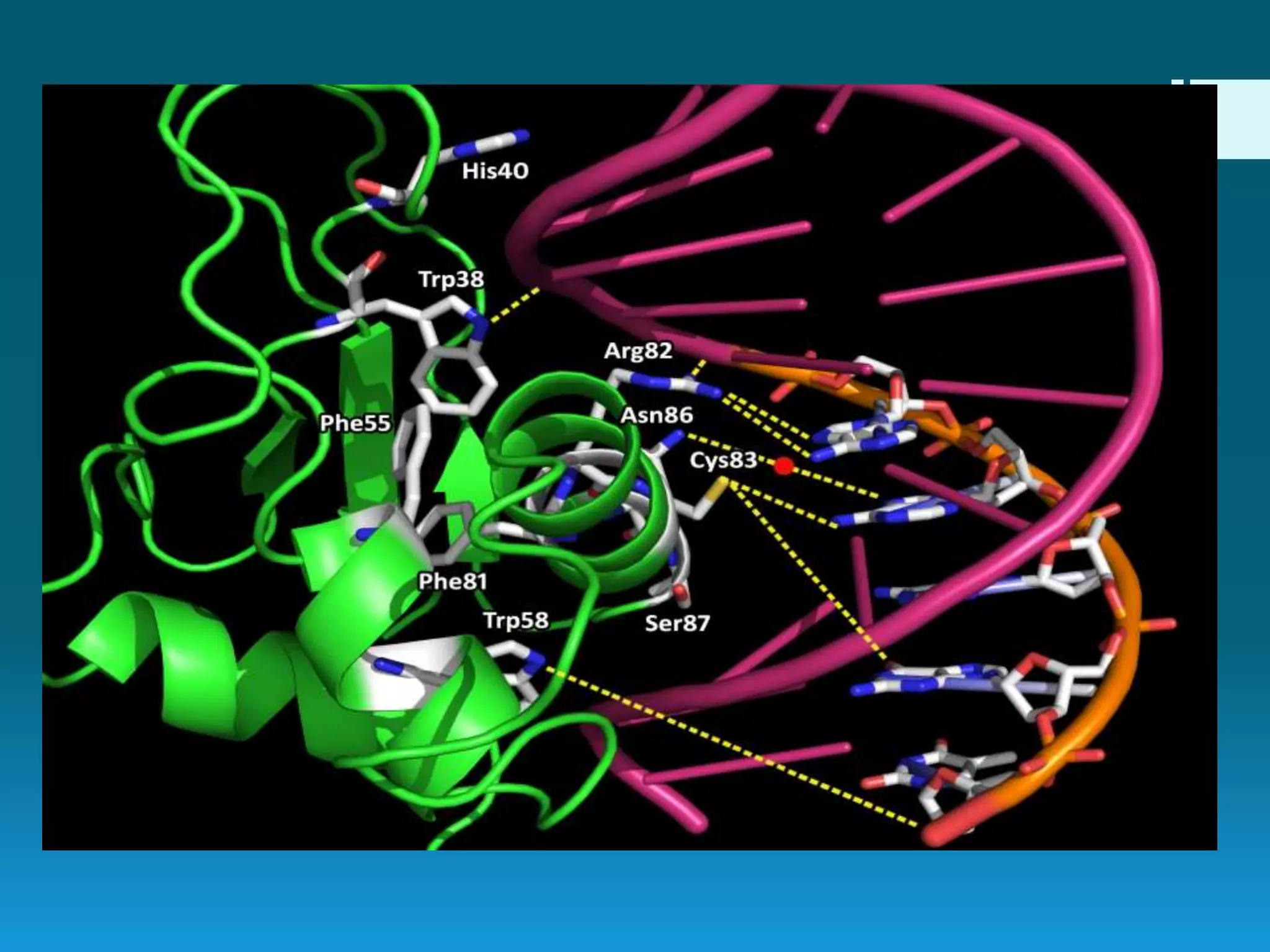
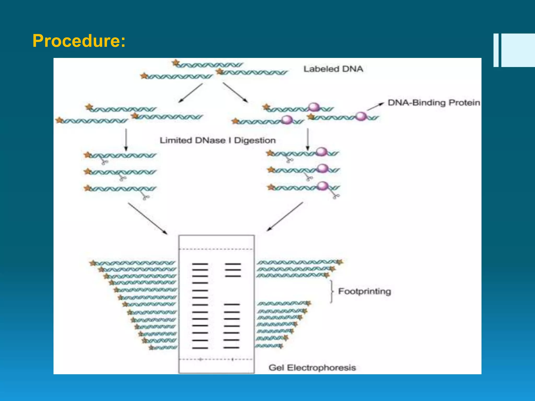
DNA footprinting
Ms Saajida Sultaana Mahusook
 DNA footprinting is an in-vitro molecular technique used to identify
protein binding regions on a DNA molecule .
 This technique is mainly used to identify the transcription factors
that bind to promoter, enhancer or silencer region of gene to
regulate its expression. The regulation of transcript ion of a gene
can be studied using this method.
 The technique is also called as DNAse I footprinting.
 Thousands of proteins (enzymes) are interacting with DNA in the
nucleus for regulating activities like replication, transcription,
translation etc.
 Transcription is a process where the DNA is converted into RNA in
a cell nucleus. Initiation of transcript ion takes place when the
enzyme RNA polymerase binds to a gene sequence known as
promoter sequence.
 DNA footprinting can be used to identify RNA polymerase
interacting DNA sequence.
History
 In 1978, David Galas and Albert Schmitz developed this technique
to study the binding specificity of the lac repressor protein.
 It was originally a modification of Maxam Gilbert chemical
sequencing technique.
Principle:
 In this technique, nucleases like DNAse I is used which will
degrade DNA molecule.
 Nucleases cannot degrade DNA if it is bounded by a protein.
 Thus that region is protected from degradation by nucleases.
 This protected DNA region is called the footprint.
Procedure:
Procedure:
1. DNA fragment (choice of interest) containing protein binding
sequence is extracted, amplified and labelled at one end of the
double helix using polymerase chain reaction technique.
2. Labelled DNA fragments with DNA binding protein; and
cleavage agent are mixed in a test tube.
3. In another test tube, labelled DNA fragments are mixed with
cleavage agents without DNA binding protein. This is used as
standard to compare the results.
4. Cleavage agent cuts the DNA fragment present in the both
test tubes but no cuts are made at the specific region
of DNA where proteins are bound. Protein has protected
the DNA binding site from cleavage agent.
5. DNA fragments are separated by polyacrylamide gel
electrophoresis and visualized using autoradiogram.
6. When compared with the standard, missing band (footprint)
indicates the protein binding specific DNA sequence.
(Comparison of both samples reveals foot prints or protein
binding sites).
Cleavage agents used in DNA footprinting are
DNase I:
 DNae I is a double strand endonuclease enzyme that cleaves the
phosphodiester bond present in the DNA. The enzyme action can be
controlled by EDTA solution.
 The enzyme acts on DNA structure and sequence specific, resulting in
an uneven ladder. This can also affect the precision of predicting
protein's specific DNA sequence.
Hydroxyl radicals:
 Hydroxyl radicals are produced from a reaction where iron salt is
reduced with hydrogen peroxide to form free hydroxyl molecule. The free
hydroxyl molecule cleaves the DNA fragment.
 Hydroxyl radicals are independent of DNA sequence for their action,
therefore form evenly distributed ladder. But the reaction rates of these
radicals are very slow therefore it requires more time to cleave
the DNA fragments.
Ultraviolet radiation:
 Ultraviolet radiations are used to excite the nucleic acid and this may
lead to damaged DNA fragments.
Application
 DNA footprinting can be used to determine the sequence
specific DNA-binding protein site.
 To study DNA-protein interaction
 Transcriptional regulations can be studied.
 To find out regions in a gene where transcription factors (proteins)
binds (that is control elements like operators, promoters etc) and
initiates transcriptions.
 Promoter, enhancer and silencer sequence of a gene can be
identified.
 To identify the functional genes present in the large genome of
human.
 To find out Hormone response elements (HREs) that is specific
sequences where hormone receptor complexes bind.
Reference
http://www.biologyexams4u.com/2014/02/dna-
footprinting-definitionprinciple.html?m=1
http://www.biotecharticles.com/Genetics-Article/DNA-
Footprinting-and-Gene-Sequencing-74.html
Thank you

DNA footprinting

  • 1.
    DNA footprinting Ms SaajidaSultaana Mahusook
  • 2.
     DNA footprintingis an in-vitro molecular technique used to identify protein binding regions on a DNA molecule .  This technique is mainly used to identify the transcription factors that bind to promoter, enhancer or silencer region of gene to regulate its expression. The regulation of transcript ion of a gene can be studied using this method.  The technique is also called as DNAse I footprinting.  Thousands of proteins (enzymes) are interacting with DNA in the nucleus for regulating activities like replication, transcription, translation etc.
  • 3.
     Transcription isa process where the DNA is converted into RNA in a cell nucleus. Initiation of transcript ion takes place when the enzyme RNA polymerase binds to a gene sequence known as promoter sequence.  DNA footprinting can be used to identify RNA polymerase interacting DNA sequence. History  In 1978, David Galas and Albert Schmitz developed this technique to study the binding specificity of the lac repressor protein.  It was originally a modification of Maxam Gilbert chemical sequencing technique.
  • 5.
    Principle:  In thistechnique, nucleases like DNAse I is used which will degrade DNA molecule.  Nucleases cannot degrade DNA if it is bounded by a protein.  Thus that region is protected from degradation by nucleases.  This protected DNA region is called the footprint.
  • 6.
  • 7.
    Procedure: 1. DNA fragment(choice of interest) containing protein binding sequence is extracted, amplified and labelled at one end of the double helix using polymerase chain reaction technique. 2. Labelled DNA fragments with DNA binding protein; and cleavage agent are mixed in a test tube. 3. In another test tube, labelled DNA fragments are mixed with cleavage agents without DNA binding protein. This is used as standard to compare the results. 4. Cleavage agent cuts the DNA fragment present in the both test tubes but no cuts are made at the specific region of DNA where proteins are bound. Protein has protected the DNA binding site from cleavage agent.
  • 8.
    5. DNA fragmentsare separated by polyacrylamide gel electrophoresis and visualized using autoradiogram. 6. When compared with the standard, missing band (footprint) indicates the protein binding specific DNA sequence. (Comparison of both samples reveals foot prints or protein binding sites).
  • 9.
    Cleavage agents usedin DNA footprinting are DNase I:  DNae I is a double strand endonuclease enzyme that cleaves the phosphodiester bond present in the DNA. The enzyme action can be controlled by EDTA solution.  The enzyme acts on DNA structure and sequence specific, resulting in an uneven ladder. This can also affect the precision of predicting protein's specific DNA sequence. Hydroxyl radicals:  Hydroxyl radicals are produced from a reaction where iron salt is reduced with hydrogen peroxide to form free hydroxyl molecule. The free hydroxyl molecule cleaves the DNA fragment.  Hydroxyl radicals are independent of DNA sequence for their action, therefore form evenly distributed ladder. But the reaction rates of these radicals are very slow therefore it requires more time to cleave the DNA fragments. Ultraviolet radiation:  Ultraviolet radiations are used to excite the nucleic acid and this may lead to damaged DNA fragments.
  • 10.
    Application  DNA footprintingcan be used to determine the sequence specific DNA-binding protein site.  To study DNA-protein interaction  Transcriptional regulations can be studied.  To find out regions in a gene where transcription factors (proteins) binds (that is control elements like operators, promoters etc) and initiates transcriptions.  Promoter, enhancer and silencer sequence of a gene can be identified.  To identify the functional genes present in the large genome of human.  To find out Hormone response elements (HREs) that is specific sequences where hormone receptor complexes bind.
  • 11.
  • 12.