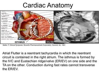
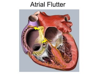
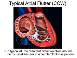
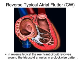
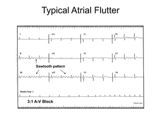
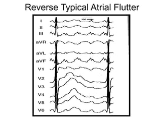
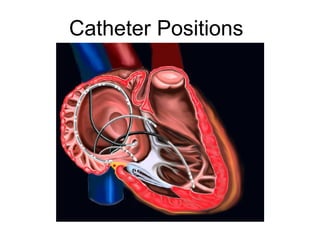
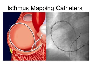
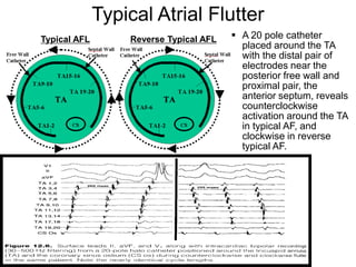
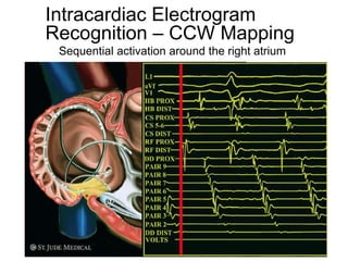
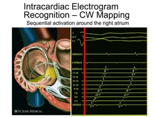
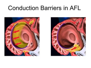
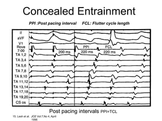
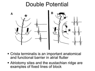
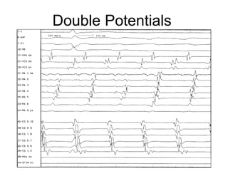
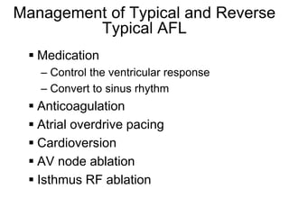
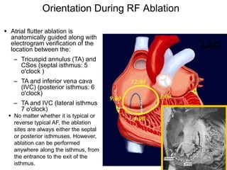
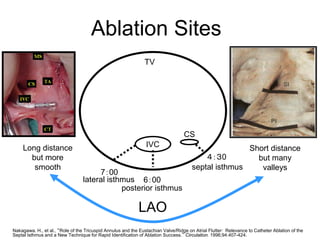
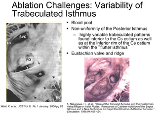
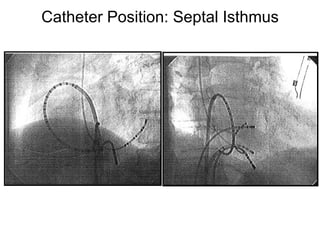
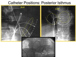
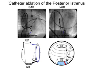
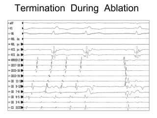
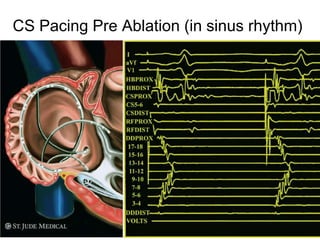
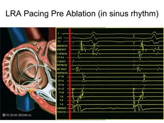
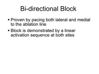
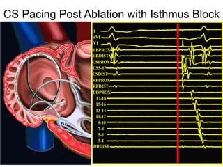
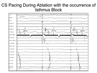
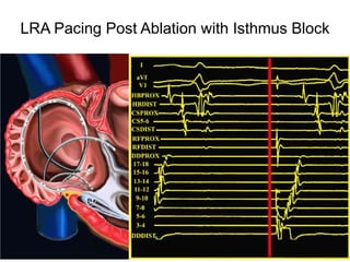
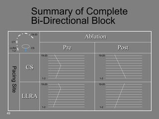
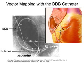
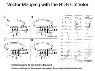
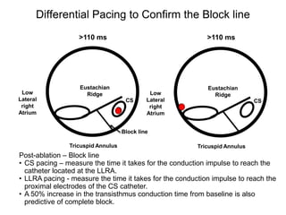
Atrial flutter is a reentrant tachycardia involving the right atrium. There are two main types - typical atrial flutter which revolves counterclockwise around the tricuspid annulus, and reverse typical atrial flutter which revolves clockwise. Catheter ablation aims to create a continuous linear lesion across the cavotricuspid isthmus to block conduction and terminate the arrhythmia. Successful ablation is confirmed by the inability to induce flutter and demonstration of bidirectional conduction block across the ablation line.
AV nodal reentrant tachycardia (AVNRT), or atrioventricular nodal reentrant tachycardia, is a type of tachycardia (fast rhythm) of the heart. It is a type of supraventricular tachycardia (SVT), meaning that it originates from a location within the heart above the bundle of His. AV nodal reentrant tachycardia is the most common regular supraventricular tachycardia. It is more common in women than men (approximately 75% of cases occur in females). The main symptom is palpitations. Treatment may be with specific physical maneuvers, medication, or, rarely, synchronized cardioversion. Frequent attacks may require radiofrequency ablation, in which the abnormally conducting tissue in the heart is destroyed.
AVNRT occurs when a reentry circuit forms within or just next to the atrioventricular node. The circuit usually involves two anatomical pathways: the fast pathway and the slow pathway, which are both in the right atrium. The slow pathway (which is usually targeted for ablation) is located inferior and slightly posterior to the AV node, often following the anterior margin of the coronary sinus. The fast pathway is usually located just superior and posterior to the AV node. These pathways are formed from tissue that behaves very much like the AV node, and some authors regard them as part of the AV node.
The fast and slow pathways should not be confused with the accessory pathways that give rise to Wolff-Parkinson-White syndrome (WPW syndrome) or atrioventricular reciprocating tachycardia (AVRT). In AVNRT, the fast and slow pathways are located within the right atrium close to or within the AV node and exhibit electrophysiologic properties similar to AV nodal tissue. Accessory pathways that give rise to WPW syndrome and AVRT are located in the atrioventricular valvular rings. They provide a direct connection between the atria and ventricles, and have electrophysiologic properties similar to ventricular myocardium.
Wolff–Parkinson–White syndrome (WPW) is one of several disorders of the conduction system of the heart that are commonly referred to as pre-excitation syndromes. WPW is caused by the presence of an abnormal accessory electrical conduction pathway between the atria and the ventricles. Electrical signals travelling down this abnormal pathway (known as the bundle of Kent) may stimulate the ventricles to contract prematurely, resulting in a unique type of supraventricular tachycardia referred to as an atrioventricular reciprocating tachycardia.The incidence of WPW is between 0.1% and 0.3% in the general population.Sudden cardiac death in people with WPW is rare (incidence of less than 0.6%), and is usually caused by the propagation of an atrial tachydysrhythmia (rapid and abnormal heart rate) to the ventricles by the abnormal accessory pathway.
Small scale generation by harnessing the wind energyeSAT Journals
Abstract Generating electricity from wind has been centered around the rotation rotating windmill in which rotating shaft coupled to the generator used to generate electricity. Other sources such as solar power hydropower, thermal power, wind power are used for generating electricity in kilo watts and megawatt but there is no such kind of application which can generate electricity on sub 100watt scale. So as to generate electricity on small scale and to provide cheap source of energy for lighting the houses in remote where there is no other sources are available. And to generate a power source that can be constructed with the help of local material and if problem occurs that can be fixed by the local people. About 2% of global electricity production comes from wind-powered generators. Their capacity has doubled in the past few years. In some countries it is more popular than solar energy because it is cheaper on a cost per watt basis. However, for powering small devices there has been minimal activity to harvest energy from the wind. This is because conventional electromagnetic wind turbines require rotating fins and gearing which adds bulk, and they become less efficient when scaled down. Key Words: Harnessing, Wind Energy, Rotating Shaft
Left ventricular non compaction is rare congenital cardiomyopathy with gaining interest due to advancement in imaging modalities for diagnosis and assessment of undulating phenotype
Michael Faraday devised the world’s first electric generator in 1821. The generator he built was a seemingly simple thing: a hand-cranked copper disk rotated between the poles of a permanent magnet.
DC GENERATOR
Are used in recharging automobile batteries.
A direct-current (DC) generator is a rotating machine that supplies an electrical output with unidirectional voltage and current.
The field is produced by direct current in field coils or by permanent magnets on the stator. The output, or armature, windings are placed in slots in the cylindrical iron rotor.
AC GENERATOR
Are used in recharging automobile batteries.
A direct-current (DC) generator is a rotating machine that supplies an electrical output with unidirectional voltage and current.
The field is produced by direct current in field coils or by permanent magnets on the stator. The output, or armature, windings are placed in slots in the cylindrical iron rotor.
MOTORS AND GENERATORS
Compare the illustrations of a dynamo and a DC motor. The action of the dynamo is just the reverse of the action of a motor,
The similarity between these two was not recognized until the mid-nineteenth century when an “accident” happened at the Vienna Exposition Hall in 1873.
The 2nd dynamo began to spark and whine and come live. The dynamo had become a generator.
The versatility of a device to act as a motor at one time and a generator at other times can be used to great advantage.
In France and Australia, trains running uphill or on level ground are fueled by electricity from power lines. When the train moves downhill, however, its motor becomes a generator. The power generated is sent to the power lines to be used by other trains. This maintains a cycle of changes from gravitational potential energy to electrical energy.
Flu Vaccine Alert in Bangalore Karnatakaaddon Scans
As flu season approaches, health officials in Bangalore, Karnataka, are urging residents to get their flu vaccinations. The seasonal flu, while common, can lead to severe health complications, particularly for vulnerable populations such as young children, the elderly, and those with underlying health conditions.
Dr. Vidisha Kumari, a leading epidemiologist in Bangalore, emphasizes the importance of getting vaccinated. "The flu vaccine is our best defense against the influenza virus. It not only protects individuals but also helps prevent the spread of the virus in our communities," he says.
This year, the flu season is expected to coincide with a potential increase in other respiratory illnesses. The Karnataka Health Department has launched an awareness campaign highlighting the significance of flu vaccinations. They have set up multiple vaccination centers across Bangalore, making it convenient for residents to receive their shots.
To encourage widespread vaccination, the government is also collaborating with local schools, workplaces, and community centers to facilitate vaccination drives. Special attention is being given to ensuring that the vaccine is accessible to all, including marginalized communities who may have limited access to healthcare.
Residents are reminded that the flu vaccine is safe and effective. Common side effects are mild and may include soreness at the injection site, mild fever, or muscle aches. These side effects are generally short-lived and far less severe than the flu itself.
Healthcare providers are also stressing the importance of continuing COVID-19 precautions. Wearing masks, practicing good hand hygiene, and maintaining social distancing are still crucial, especially in crowded places.
Protect yourself and your loved ones by getting vaccinated. Together, we can help keep Bangalore healthy and safe this flu season. For more information on vaccination centers and schedules, residents can visit the Karnataka Health Department’s official website or follow their social media pages.
Stay informed, stay safe, and get your flu shot today!
HOT NEW PRODUCT! BIG SALES FAST SHIPPING NOW FROM CHINA!! EU KU DB BK substit...GL Anaacs
Contact us if you are interested:
Email / Skype : kefaya1771@gmail.com
Threema: PXHY5PDH
New BATCH Ku !!! MUCH IN DEMAND FAST SALE EVERY BATCH HAPPY GOOD EFFECT BIG BATCH !
Contact me on Threema or skype to start big business!!
Hot-sale products:
NEW HOT EUTYLONE WHITE CRYSTAL!!
5cl-adba precursor (semi finished )
5cl-adba raw materials
ADBB precursor (semi finished )
ADBB raw materials
APVP powder
5fadb/4f-adb
Jwh018 / Jwh210
Eutylone crystal
Protonitazene (hydrochloride) CAS: 119276-01-6
Flubrotizolam CAS: 57801-95-3
Metonitazene CAS: 14680-51-4
Payment terms: Western Union,MoneyGram,Bitcoin or USDT.
Deliver Time: Usually 7-15days
Shipping method: FedEx, TNT, DHL,UPS etc.Our deliveries are 100% safe, fast, reliable and discreet.
Samples will be sent for your evaluation!If you are interested in, please contact me, let's talk details.
We specializes in exporting high quality Research chemical, medical intermediate, Pharmaceutical chemicals and so on. Products are exported to USA, Canada, France, Korea, Japan,Russia, Southeast Asia and other countries.
Ethanol (CH3CH2OH), or beverage alcohol, is a two-carbon alcohol
that is rapidly distributed in the body and brain. Ethanol alters many
neurochemical systems and has rewarding and addictive properties. It
is the oldest recreational drug and likely contributes to more morbidity,
mortality, and public health costs than all illicit drugs combined. The
5th edition of the Diagnostic and Statistical Manual of Mental Disorders
(DSM-5) integrates alcohol abuse and alcohol dependence into a single
disorder called alcohol use disorder (AUD), with mild, moderate,
and severe subclassifications (American Psychiatric Association, 2013).
In the DSM-5, all types of substance abuse and dependence have been
combined into a single substance use disorder (SUD) on a continuum
from mild to severe. A diagnosis of AUD requires that at least two of
the 11 DSM-5 behaviors be present within a 12-month period (mild
AUD: 2–3 criteria; moderate AUD: 4–5 criteria; severe AUD: 6–11 criteria).
The four main behavioral effects of AUD are impaired control over
drinking, negative social consequences, risky use, and altered physiological
effects (tolerance, withdrawal). This chapter presents an overview
of the prevalence and harmful consequences of AUD in the U.S.,
the systemic nature of the disease, neurocircuitry and stages of AUD,
comorbidities, fetal alcohol spectrum disorders, genetic risk factors, and
pharmacotherapies for AUD.
These simplified slides by Dr. Sidra Arshad present an overview of the non-respiratory functions of the respiratory tract.
Learning objectives:
1. Enlist the non-respiratory functions of the respiratory tract
2. Briefly explain how these functions are carried out
3. Discuss the significance of dead space
4. Differentiate between minute ventilation and alveolar ventilation
5. Describe the cough and sneeze reflexes
Study Resources:
1. Chapter 39, Guyton and Hall Textbook of Medical Physiology, 14th edition
2. Chapter 34, Ganong’s Review of Medical Physiology, 26th edition
3. Chapter 17, Human Physiology by Lauralee Sherwood, 9th edition
4. Non-respiratory functions of the lungs https://academic.oup.com/bjaed/article/13/3/98/278874
micro teaching on communication m.sc nursing.pdfAnurag Sharma
Microteaching is a unique model of practice teaching. It is a viable instrument for the. desired change in the teaching behavior or the behavior potential which, in specified types of real. classroom situations, tends to facilitate the achievement of specified types of objectives.
Knee anatomy and clinical tests 2024.pdfvimalpl1234
This includes all relevant anatomy and clinical tests compiled from standard textbooks, Campbell,netter etc..It is comprehensive and best suited for orthopaedicians and orthopaedic residents.
These lecture slides, by Dr Sidra Arshad, offer a quick overview of physiological basis of a normal electrocardiogram.
Learning objectives:
1. Define an electrocardiogram (ECG) and electrocardiography
2. Describe how dipoles generated by the heart produce the waveforms of the ECG
3. Describe the components of a normal electrocardiogram of a typical bipolar leads (limb II)
4. Differentiate between intervals and segments
5. Enlist some common indications for obtaining an ECG
Study Resources:
1. Chapter 11, Guyton and Hall Textbook of Medical Physiology, 14th edition
2. Chapter 9, Human Physiology - From Cells to Systems, Lauralee Sherwood, 9th edition
3. Chapter 29, Ganong’s Review of Medical Physiology, 26th edition
4. Electrocardiogram, StatPearls - https://www.ncbi.nlm.nih.gov/books/NBK549803/
5. ECG in Medical Practice by ABM Abdullah, 4th edition
6. ECG Basics, http://www.nataliescasebook.com/tag/e-c-g-basics
The prostate is an exocrine gland of the male mammalian reproductive system
It is a walnut-sized gland that forms part of the male reproductive system and is located in front of the rectum and just below the urinary bladder
Function is to store and secrete a clear, slightly alkaline fluid that constitutes 10-30% of the volume of the seminal fluid that along with the spermatozoa, constitutes semen
A healthy human prostate measures (4cm-vertical, by 3cm-horizontal, 2cm ant-post ).
It surrounds the urethra just below the urinary bladder. It has anterior, median, posterior and two lateral lobes
It’s work is regulated by androgens which are responsible for male sex characteristics
Generalised disease of the prostate due to hormonal derangement which leads to non malignant enlargement of the gland (increase in the number of epithelial cells and stromal tissue)to cause compression of the urethra leading to symptoms (LUTS
Prix Galien International 2024 Forum ProgramLevi Shapiro
June 20, 2024, Prix Galien International and Jerusalem Ethics Forum in ROME. Detailed agenda including panels:
- ADVANCES IN CARDIOLOGY: A NEW PARADIGM IS COMING
- WOMEN’S HEALTH: FERTILITY PRESERVATION
- WHAT’S NEW IN THE TREATMENT OF INFECTIOUS,
ONCOLOGICAL AND INFLAMMATORY SKIN DISEASES?
- ARTIFICIAL INTELLIGENCE AND ETHICS
- GENE THERAPY
- BEYOND BORDERS: GLOBAL INITIATIVES FOR DEMOCRATIZING LIFE SCIENCE TECHNOLOGIES AND PROMOTING ACCESS TO HEALTHCARE
- ETHICAL CHALLENGES IN LIFE SCIENCES
- Prix Galien International Awards Ceremony
Report Back from SGO 2024: What’s the Latest in Cervical Cancer?bkling
Are you curious about what’s new in cervical cancer research or unsure what the findings mean? Join Dr. Emily Ko, a gynecologic oncologist at Penn Medicine, to learn about the latest updates from the Society of Gynecologic Oncology (SGO) 2024 Annual Meeting on Women’s Cancer. Dr. Ko will discuss what the research presented at the conference means for you and answer your questions about the new developments.
Lung Cancer: Artificial Intelligence, Synergetics, Complex System Analysis, S...Oleg Kshivets
RESULTS: Overall life span (LS) was 2252.1±1742.5 days and cumulative 5-year survival (5YS) reached 73.2%, 10 years – 64.8%, 20 years – 42.5%. 513 LCP lived more than 5 years (LS=3124.6±1525.6 days), 148 LCP – more than 10 years (LS=5054.4±1504.1 days).199 LCP died because of LC (LS=562.7±374.5 days). 5YS of LCP after bi/lobectomies was significantly superior in comparison with LCP after pneumonectomies (78.1% vs.63.7%, P=0.00001 by log-rank test). AT significantly improved 5YS (66.3% vs. 34.8%) (P=0.00000 by log-rank test) only for LCP with N1-2. Cox modeling displayed that 5YS of LCP significantly depended on: phase transition (PT) early-invasive LC in terms of synergetics, PT N0—N12, cell ratio factors (ratio between cancer cells- CC and blood cells subpopulations), G1-3, histology, glucose, AT, blood cell circuit, prothrombin index, heparin tolerance, recalcification time (P=0.000-0.038). Neural networks, genetic algorithm selection and bootstrap simulation revealed relationships between 5YS and PT early-invasive LC (rank=1), PT N0—N12 (rank=2), thrombocytes/CC (3), erythrocytes/CC (4), eosinophils/CC (5), healthy cells/CC (6), lymphocytes/CC (7), segmented neutrophils/CC (8), stick neutrophils/CC (9), monocytes/CC (10); leucocytes/CC (11). Correct prediction of 5YS was 100% by neural networks computing (area under ROC curve=1.0; error=0.0).
CONCLUSIONS: 5YS of LCP after radical procedures significantly depended on: 1) PT early-invasive cancer; 2) PT N0--N12; 3) cell ratio factors; 4) blood cell circuit; 5) biochemical factors; 6) hemostasis system; 7) AT; 8) LC characteristics; 9) LC cell dynamics; 10) surgery type: lobectomy/pneumonectomy; 11) anthropometric data. Optimal diagnosis and treatment strategies for LC are: 1) screening and early detection of LC; 2) availability of experienced thoracic surgeons because of complexity of radical procedures; 3) aggressive en block surgery and adequate lymph node dissection for completeness; 4) precise prediction; 5) adjuvant chemoimmunoradiotherapy for LCP with unfavorable prognosis.
Acute scrotum is a general term referring to an emergency condition affecting the contents or the wall of the scrotum.
There are a number of conditions that present acutely, predominantly with pain and/or swelling
A careful and detailed history and examination, and in some cases, investigations allow differentiation between these diagnoses. A prompt diagnosis is essential as the patient may require urgent surgical intervention
Testicular torsion refers to twisting of the spermatic cord, causing ischaemia of the testicle.
Testicular torsion results from inadequate fixation of the testis to the tunica vaginalis producing ischemia from reduced arterial inflow and venous outflow obstruction.
The prevalence of testicular torsion in adult patients hospitalized with acute scrotal pain is approximately 25 to 50 percent
New Directions in Targeted Therapeutic Approaches for Older Adults With Mantl...i3 Health
i3 Health is pleased to make the speaker slides from this activity available for use as a non-accredited self-study or teaching resource.
This slide deck presented by Dr. Kami Maddocks, Professor-Clinical in the Division of Hematology and
Associate Division Director for Ambulatory Operations
The Ohio State University Comprehensive Cancer Center, will provide insight into new directions in targeted therapeutic approaches for older adults with mantle cell lymphoma.
STATEMENT OF NEED
Mantle cell lymphoma (MCL) is a rare, aggressive B-cell non-Hodgkin lymphoma (NHL) accounting for 5% to 7% of all lymphomas. Its prognosis ranges from indolent disease that does not require treatment for years to very aggressive disease, which is associated with poor survival (Silkenstedt et al, 2021). Typically, MCL is diagnosed at advanced stage and in older patients who cannot tolerate intensive therapy (NCCN, 2022). Although recent advances have slightly increased remission rates, recurrence and relapse remain very common, leading to a median overall survival between 3 and 6 years (LLS, 2021). Though there are several effective options, progress is still needed towards establishing an accepted frontline approach for MCL (Castellino et al, 2022). Treatment selection and management of MCL are complicated by the heterogeneity of prognosis, advanced age and comorbidities of patients, and lack of an established standard approach for treatment, making it vital that clinicians be familiar with the latest research and advances in this area. In this activity chaired by Michael Wang, MD, Professor in the Department of Lymphoma & Myeloma at MD Anderson Cancer Center, expert faculty will discuss prognostic factors informing treatment, the promising results of recent trials in new therapeutic approaches, and the implications of treatment resistance in therapeutic selection for MCL.
Target Audience
Hematology/oncology fellows, attending faculty, and other health care professionals involved in the treatment of patients with mantle cell lymphoma (MCL).
Learning Objectives
1.) Identify clinical and biological prognostic factors that can guide treatment decision making for older adults with MCL
2.) Evaluate emerging data on targeted therapeutic approaches for treatment-naive and relapsed/refractory MCL and their applicability to older adults
3.) Assess mechanisms of resistance to targeted therapies for MCL and their implications for treatment selection
ARTIFICIAL INTELLIGENCE IN HEALTHCARE.pdfAnujkumaranit
Artificial intelligence (AI) refers to the simulation of human intelligence processes by machines, especially computer systems. It encompasses tasks such as learning, reasoning, problem-solving, perception, and language understanding. AI technologies are revolutionizing various fields, from healthcare to finance, by enabling machines to perform tasks that typically require human intelligence.
2. Atrial Flutter
A macro-reentrant atrial arrhythmia that is
very regular with rates typically between
240 and 350 bpm1. There are several
recognized variations of atrial flutter.
1. Schamroth, L. The Disorders of Cardiac Rhythm. Oxford, UK, Blackwell Ltd, 1971, p 49.
2
3. Proposed Classification of Atrial Flutter
A NASPE position paper proposed an open
classification
– Typical AFL (CCW)
– Reverse Typical AFL (CW)
Saoudi, N, Cosio, F, Waldo, A, et. al. JCE Vol. 12, No. 7, pp.852-866, July, 2001
3
4. Cardiac Anatomy
TA
ER/EV
ISTHMUS
Netter, F. Clinical Symposia. Novartis Pharmaceuticals Corporation, Summit, NJ, 1997.
Atrial Flutter is a reentrant tachycardia in which the reentrant
circuit is contained in the right atrium. The isthmus is formed by
the IVC and Eustachian ridge/valve (ER/EV) on one side and the
TA on the other. Conduction during fast rates cannot transverse
the ER/EV.
4
6. Typical Atrial Flutter (CCW)
In typical AF the reentrant circuit revolves around
6 the tricuspid annulus in a counterclockwise pattern
7. Reverse Typical Atrial Flutter (CW)
In reverse typical the reentrant circuit revolves
7
around the tricuspid annulus in a clockwise pattern.
8. Electrogram Recognition
Rate
P wave morphology
12 Lead
On the surface ECG it may often be very
difficult to see the flutter waves. This may
be overcome with vagal maneuvers or
Adenosine administration.
8
10. Electrogram Recognition
Isthmus dependent Typical Atrial Flutter
(CCW)
– Atrial rhythm: regular and very stable (240-340
bpm)
– P wave:Characteristic sawtooth pattern with a
negative deflection in, II and III, and/or aVf
(inferior axis) and positive in V1 (but may be
negative or biphasic). Leads I and aVL show
low-voltage deflections
– Ventricular rate: usually 2:1 in both typical and
reverse typical aflutter (higher degrees of AV
block can occur in patients with AV nodal block
disease or increased vagal tone)
10
12. Electrogram : Reverse Typical AFL
On the surface ECG typical atrial flutter
looks similar to reverse typical flutter,
however in Reverse Typical Aflutter
(CW), the p-waves appear to be mostly
positive in the inferior leads (II, III, aVf).
P waves display an superior axis.
Wide, negative deflections in V1 (may be
most specific diagnostic sign)
May demonstrate atypical p -wave
morphologies
12
14. Catheter Positions
Catheter position varies from lab to lab
Quadripolar at the His (to define septum/HBE)
Multipolar in the CS (to define CS ostium, and
perform septal pacing)
Multipole (Duo-Decapolar™) at the RA (to
define activation anterior/lateral to CT and isthmus). This
may eliminate the HRA and CS catheters
Quadripolar at the RVA (safety pacing) optional
Exploring/Rove (mapping/RFA)
14
17. Typical Atrial Flutter
Typical AFL Reverse Typical AFL A 20 pole catheter
placed around the TA
with the distal pair of
electrodes near the
posterior free wall and
proximal pair, the
anterior septum, reveals
counterclockwise
activation around the TA
in typical AF, and
clockwise in reverse
typical AF.
17
18. Pre Ablation Methods and Strategies
Induction
– Conduction barriers
– Diagnosis
Mapping
Entrainment
Pacing maneuvers
Strategy
– Pacing maneuvers in SR
Base line measurements (Pre and post
comparison)
18
19. Atrial Flutter Induction
Induction methods for flutter include:
– Extrastimulas testing
– Atrial burst pacing
– Isoproterenol
Induction or termination using rapid atrial
pacing may also induce atrial fibrillation
(due to short cycle lengths)
19
20. Intracardiac Electrogram
Recognition – CCW Mapping
Sequential activation around the right atrium
20
21. Intracardiac Electrogram
Recognition – CW Mapping
Sequential activation around the right atrium
21
23. Concealed Entrainment
PPI :Post pacing interval FCL: Flutter cycle length
Post pacing intervals PPI=TCL
23 15. Lesh et al. JCE Vol.7,No 4, April
1996
24. Entrainment Mapping
24 Olgin et al. J of Cardiovasc Electrophysiology Vol.7,No.11,Nov 96
25. Double Potential
Crista terminalis is an important anatomical
and functional barrier in atrial flutter
Atriotomy sites and the eustachian ridge are
25 examples of fixed lines of block
27. Management of Typical and Reverse
Typical AFL
Medication
– Control the ventricular response
– Convert to sinus rhythm
Anticoagulation
Atrial overdrive pacing
Cardioversion
AV node ablation
Isthmus RF ablation
27
28. AV Node Ablation
In some situations medical therapy and
ablation attempts are unsuccessful. In
circumstances it may be necessary to
ablate the AV node and implant a
permanent pacemaker.
28
29. Goal of RF Ablation of Atrial Flutter
The goal of RF ablation is the elimination
of conduction within the critical zone of the
reentrant circuit necessary to sustain atrial
flutter.
Tachycardia may be terminated by one
lesion point along the Isthmus however this
method is associated with a high
recurrence rate
In any of the targeted ablation areas, the
key to success is a contiguous, transmural
lesion from one anatomic barrier to another
29
30. Ablation Methods and Strategies
Methods
– Point by point
– Drag (Linear lesion)
Strategy
– During SR
No acute end point
– During SR with CS pacing
Shift in activation
– During tachycardia
Termination of tachycardia
30
31. Orientation During RF Ablation
Atrial flutter ablation is
anatomically guided along with
electrogram verification of the LAO
location between the:
– Tricuspid annulus (TA) and
CSos (septal isthmus: 5
o'clock )
– TA and inferior vena cava
(IVC) (posterior isthmus: 6
o'clock)
– TA and IVC (lateral isthmus
7 o'clock)
No matter whether it is typical or
reverse typical AF, the ablation
sites are always either the septal
or posterior isthmuses. However,
ablation can be performed
anywhere along the isthmus, from
the entrance to the exit of the
31isthmus.
32. Ablation Sites
TV
CS
Long distance IVC Short distance
but more 4:30 but many
smooth septal isthmus valleys
7:00
lateral isthmus 6:00
posterior isthmus
LAO
32
Nakagawa. H., et al., “Role of the Tricuspid Annulus and the Eustachian Valve/Ridge on Atrial Flutter: Relevance to Catheter Ablation of the
Septal Isthmus and a New Technique for Rapid Identification of Ablation Success.” Circulation. 1996;94:407-424.
33. Ablation Challenges: Variability of
Trabeculated Isthmus
Blood pool
Non-uniformity of the Posterior Isthmus
– highly variable trabeculated patterns
found inferior to the Cs ostium as well
as at the inferior rim of the Cs ostium
within the “flutter isthmus”
Eustachian valve and ridge
5. Nakagawa. H., et al., “Role of the Tricuspid Annulus and the Eustachian
Waki, K. et.al. JCE Vol 11. No 1 January 2000 pg 92 Valve/Ridge on Atrial Flutter: Relevance to Catheter Ablation of the Septal
Isthmus and a New Technique for Rapid Identification of Ablation Success.” .
33 Circulation. 1996;94:407-424.
37. Catheter ablation of the Posterior Isthmus
RAO LAO
ablation catheter ablation catheter
SVC
SVC
CSo
IVC
37
IVC
38. Ablation technique
Catheter
– Normally an 8mm tip ablation catheters is used, but for
very thick or problematic isthmuses, an irrigated ablation
catheter can be used.
– Some doctors may even use a 4mm tip, but it will be a
longer procedure and recurrence may be higher
Electrogram criteria
– Initial lesion point should show big V small A.
– Electrogram should be evaluated after each point
ablation. (Point by point ablation)
– Observe for a decrease in the electrogram amplitude
and keep ablating spots with significant A waves
Use pacing maneuvers to assess the creation of
complete isthmus conduction block
38
39. Fluoroscopic Orientation During RF Ablation
Ablation of the isthmus in either the RAO or
LAO projection
LAO projection allows identification of the
position in a “clockface” relative to the location
of the TVA (point to point)
LAO projection allows visualization of the RF
catheter as it is withdrawn into the IVC
RAO projection allows discrimination of the
Anterior (TVA), initial position, to Inferior (IVC),
final position, during creation of the lesion in the
isthmus
39
40. Further Considerations during AFL Ablation
RF Power considerations
– With 4mm tip ablation catheters, 30-50 Watts will be
adequate, but 8mm tip catheters often require more than
50 Watts
Anatomical considerations
– Convective effects of blood pooling and variable,
complex anatomy may require higher power applications
– Patient discomfort in region of IVC due to stimulation of
nerve plexus
40
41. Ablation End Point
Termination of the clinical arrhythmia
– With this criteria alone there is a high
recurrence rate
Inability to re-induce atrial flutter;
Confirmation of Bi-Directional block.
– Pre and post timing
– Block indicated by a multipolar catheter
41
45. Bi-directional Block
Proven by pacing both lateral and medial
to the ablation line
Block is demonstrated by a linear
activation sequence at both sites
45
49. Summary of Complete
Bi-Directional Block
19-20
Ablation
CT
LLRA
1-2
CS Pre Post
19-20 19-20
CS
Pacing Site
1-2 1-2
19-20 19-20
LLRA
1-2 1-2
49
50. Other Methods to Confirm Bi-directional Block
Vector Mapping with the BDB Catheter
Searching for Gaps in the Blockline
Differential Pacing
50
51. Vector Mapping with the BDB Catheter
BDB
Isthmus
ABL Catheter
51 Electrogram Polarity and Cavotricuspid Isthmus Block During Ablation of Typical Atrial Flutter.Tada,H. Oral, H. et al.
Journal of Cardiovascular Electrophysiology. Volume12, No. 4, April 2001. P.394.
52. Vector Mapping with the BDB Catheter
Vector mapping to confirm the blockline
52 (Electrogram Polarity and Cavotricuspid Isthmus Block During Ablation of Typical Atrial Flutter)
53. Searching for Gaps in the Blockline
When you pace on one side of the blockline and you will note
double potentials along the line where you have made a
complete line. However, where there is a gap as you slowly
move the catheter, you will note that the double potentials
disappear meaning that you are on the Gap. You might also
find fractionated potentials. You can also look for the sites with
large electrograms meaning they have not yet been ablated
and ablate at those site.
53
54. Searching for Gaps in the Blockline
<90 >110
ms ms
Tada et al.* reported that the interval separating the two
components of a double potential was useful to distinguish
complete (>110 ms) from incomplete isthmus block (<90 ms)
in patients undergoing radiofrequency ablation of typical atrial
flutter.
* Tada H et al. J Am Coll Cardiol 2001; 38:750-5
54
55. Differential Pacing to Confirm the Bloclline
<90 ms <90 ms
Eustachian Eustachian
Low Ridge Low Ridge
Lateral CS Lateral CS
right right
Atrium Atrium
Tricuspid Annulus Tricuspid Annulus
Pre-ablation – No Blockline
• CS pacing – measure the time it takes for the conduction impulse to
reach the catheter located at the LLRA.
• LLRA pacing - measure the time it takes for the conduction impulse to
reach the proximal electrodes of the CS catheter.
55
56. Differential Pacing to Confirm the Block line
>110 ms >110 ms
Eustachian Eustachian
Low Ridge Low Ridge
Lateral CS Lateral CS
right right
Atrium Atrium
Block line
Tricuspid Annulus Tricuspid Annulus
Post-ablation – Block line
• CS pacing – measure the time it takes for the conduction impulse to reach the
catheter located at the LLRA.
• LLRA pacing - measure the time it takes for the conduction impulse to reach the
proximal electrodes of the CS catheter.
• A 50% increase in the transisthmus conduction time from baseline is also
56 predictive of complete block.