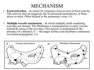
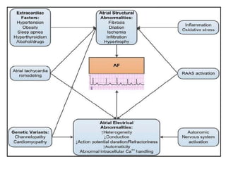
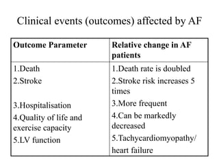
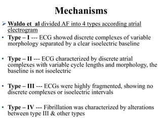
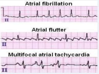
This document provides an overview of atrial fibrillation (AF) and atrial flutter. It discusses the characteristics, mechanisms, ECG features, causes and clinical outcomes of AF. It also covers the classification, mechanisms, ECG patterns and examples of atrial flutter. Key points include that AF is characterized by disorganized atrial activation and irregular ventricular rhythm, while flutter involves a reentrant circuit in the right atrium causing a regular atrial rate of 300 bpm. Complications of AF include increased risk of stroke, heart failure and cardiac death.
2. INTRODUCTION
Atrial Fibrillation (AF) is the most common sustained arrhythmia.
The incidence and prevalence of AF is increasing.
Lifetime risk over the age of 40 years is ~25%. (1)
Complications of AF include haemodynamic instability, cardiomyopathy,
cardiac failure, and embolic events such as stroke.
Characterised by disorganised atrial electrical activity and contraction.
3. Atrial Fibrillation
is characterized by disorganised, rapid and irregular atrial
activation with loss of atrial contraction and with an irregular
ventricular rate that is determined by AV nodal conduction (1)
• Paroxysmal AF
AF that terminates spontaneously or with intervention within
7 d of onset.
• Persistent AF
Continuous AF that is sustained >7 d.
• Long-standing persistent AF
Continuous AF >12 mo in duration.
• Permanent AF
When the patient and clinician make a joint decision to stop
further attempts to restore and/or maintain sinus rhythm.
4. MECHANISM
• Focal activation – In which AF originates from an area of focal activity.
This activity may be triggered, due to increased automaticity, or from
micro re-entry. Often located in the pulmonary veins. (1)
• Multiple wavelet mechanism – In which multiple small wandering
wavelets are formed. The fibrillation is maintained by re-entry circuits
formed by some of the wavelets. This process is potentiated in the
presence of a dilated LA — the larger surface area facilitates continuous
waveform propagation. (1)
5. The hallmark of AF is chaotic atrial impulses leading to
irregularly irregular ventricular contraction, usually with
incessant tachycardia
6.
7. Causes of Atrial Fibrillation
Hypertension
Heart attacks/ CAD
Valvular Heart Disease
Congenital heart defects
Hyperthyroid or other metabolic
imbalance
Exposure to stimulants such as
medications, caffeine or tobacco, or to
alcohol (holiday heart)
Sick sinus syndrome — improper
functioning of the heart's natural
pacemaker
Emphysema or other lung diseases
CABG/ Previous heart surgery
Viral infections /Pericarditis
Stress due to pneumonia, surgery or other
illnesses (Catecholamine)
Pulmonary Embolus
Pneumonia
Sleep Apnea
Pericarditis
Lone A fib (younger people)
Left ventricular Hypertrophy
Cardiomyopathy
CHF –Systolic or Diastolic
Idiopathic – mostly in younger people –
Lone Atrial Fib
Familial Predisposition.
Glucocorticoids
Electrolyte abnormalities (especially
Low Mg+2)
Atrial fibrosis from Sarcoid, collegen
vascular disease, infiltrating diseases
8. Clinical events (outcomes) affected by AF
Outcome Parameter Relative change in AF
patients
1.Death
2.Stroke
3.Hospitalisation
4.Quality of life and
exercise capacity
5.LV function
1.Death rate is doubled
2.Stroke risk increases 5
times
3.More frequent
4.Can be markedly
decreased
5.Tachycardiomyopathy/
heart failure
10. Waldo et al divided AF into 4 types according atrial
electrogram
• Type – I --- ECG showed discrete complexes of variable
morphology separated by a clear isoelectric baseline
• Type – II --- ECG characterized by discrete atrial
complexes with variable cycle lengths and morphology, the
baseline is not isoelectric
• Type – III --- ECGs were highly fragmented, showing no
discrete complexes or isoelectric intervals
• Type – IV --- Fibrillation was characterized by alterations
between type III & other types
Mechanisms
11.
12.
13. ECG FEATURES OF AF
Irregularly irregular rhythm.
No P waves.
Absence of an isoelectric baseline.
Variable ventricular rate.
QRS complexes usually < 120 ms unless pre-existing bundle branch
block, accessory pathway, or rate related aberrant conduction.
Fibrillatory waves may be present and can be either fine (amplitude <
0.5mm) or coarse (amplitude >0.5mm).
Fibrillatory waves may mimic P waves leading to misdiagnosis.
17. Example .1
Irregular ventricular response .
Coarse fibrillatory waves are visible in V1.
“Sagging” ST segment depression is visible in V6, II, III and aVF, suggestive of digoxin
effect.
21. Example 5.
AF with slow ventricular response
Irregular heart rate with no evidence of organised atrial activity. Fine
fibrillatory waves in V1.
ST depression / T wave inversion in the lateral leads could represent ischaemia
or digoxin effect.
The slow ventricular rate suggests that the patient is being treated with an AV-
nodal blocking drug (e.g. beta-blocker, verapamil/diltiazem, digoxin). Another
cause of slow AF is hypothermia.
22. Ashman’s Phenomenon – presences of aberrantly conducted beats, usually of RBBB
morphology, due a long refractory period as determined by the preceding R-R interval.
The ventricular response and thus ventricular rate in AF is dependent on several factors
including vagal tone, other pacemaker foci, AV node function, refractory period, and
medications.
Commonly AF is associated with a ventricular rate ~ 110 – 160. AF is often described as
having ‘rapid ventricular response’ once the ventricular rate is > 100 bpm.
‘Slow’AF is a term often used to describe AF with a ventricular rate < 60bpm.
Causes of ‘slow’ AF include hypothermia, digoxin toxicity, medications and sinus node
dysfunction.
23. Example 6.
Ashman aberrancy
Atrial fibrillation is diagnosed based on the irregular ventricular rate and presence of
fibrillatory waves in V1.
The third QRS complex is aberrantly conducted: it is broad with a different axis and
morphology and occurs following a long RR / short RR cycle (= Ashman aberrancy).
24. Atrial Flutter
type of supraventricular tachycardia caused by a reentry circuit within
the right atrium. (2)
The length of the re-entry circuit corresponds to the size of the RA,
resulting in a fairly predictable atrial rate of around 300 bpm (range
200-400). (2)
Ventricular rate is determined by the AV conduction ratio (“degree of
AV block”).
The commonest AV ratio is 2:1, resulting in a ventricular rate of ~150
bpm.
Higher-degree AV blocks can occur — usually due to medications or
underlying heart disease — resulting in lower rates of ventricular
conduction, e.g. 3:1 or 4:1 block.
Atrial flutter with 1:1 conduction can occur due to sympathetic
stimulation or in the presence of an accessory pathway — especially if
AV-nodal blocking agents are administered to a patient with WPW.
Atrial flutter with 1:1 conduction is associated with severe
haemodynamic instability and progression to ventricular fibrillation.
25.
26. Classification
A} Typical Atrial Flutter (Common, or Type I Atrial Flutter)
Involves the IVC & tricuspid isthmus in the reentry circuit. Can be further
classified based on the direction of the circuit:
Anticlockwise Reentry. This is the commonest form of atrial flutter (90% of
cases). Retrograde atrial conduction produces: Inverted flutter waves in leads
II,III, aVF
Positive flutter waves in V1 – may resemble upright P waves
Clockwise Reentry. This uncommon variant produces the opposite pattern:
Positive flutter waves in leads II, III, aVF
Broad, inverted flutter waves in V1
B} Atypical Atrial flutter (Uncommon, or Type II Atrial Flutter)
Does not fulfil criteria for typical atrial flutter.
Often associated with higher atrial rates and rhythm instability.
Less amenable to treatment with ablation.
27. ECG ATRIAL FLUTTER
General Features
Narrow complex tachycardia
Regular atrial activity at ~300 bpm
Flutter waves (“saw-tooth” pattern) best seen in leads II, III, aVF
— may be more easily spotted by turning the ECG upside down!
Flutter waves in V1 may resemble P waves
Loss of the isoelectric baseline
28. Fixed AV blocks
Ventricular rate is a fraction of the atrial rate, e.g.
2:1 block = 150 bpm
3:1 block = 100 bpm
4:1 block = 75 bpm
Variable AV block
The ventricular response is irregular and may mimic AF
On closer inspection, there may be a pattern of alternating 2:1, 3:1 and 4:1
conduction ratios
Atrial flutter with a 3:1 block
29. Handy tips for spotting flutter
Rapid Recognition
Narrow complex tachycardia at 150 bpm (range 130-170)? Yes ->
Suspect flutter!
Turn the ECG upside down and scrutinise the inferior leads (II, III +
aVF)
Vagal Manoeuvres + Adenosine
Atrial flutter will not usually cardiovert with these techniques (unlike
AVNRT), although typically there will be a transient period of
increased
AV block during which flutter waves may be unmasked.
30. Handy tips for spotting flutter
RR intervals
In atrial flutter with variable block the R-R intervals will be
multiples of the P-P interval — e.g. assuming an atrial rate of
300bpm (P-P interval of 200 ms), the R-R interval would be 400
ms with 2:1 block, 600 ms with 3:1 block, and 800 ms with 4:1
block.
Look for identical R-R intervals occurring sporadically along the
rhythm strip; then look to see whether there is a mathematical
relationship between the various R-R intervals on the ECG.
In contrast, atrial fibrillation will be completely irregular, with
no patterns to be discerned within the R-R intervals.
31. Example 7.
Atrial Flutter with 2:1 Block
There are inverted flutter waves in II, III + aVF at a rate of 300 bpm (one per big
square)
There are upright flutter waves in V1 simulating P waves
There is a 2:1 AV block resulting in a ventricular rate of 150 bpm
Note the occasional irregularity, with a 3:1 cycle seen in V1-3
This is the classic appearance of anticlockwise flutter.
32. Example 8.
Atrial Flutter with Variable Block
Inverted flutter waves in II, III + aVF with atrial rate ~ 300 bpm
Positive flutter waves in V1 resembling P waves
The degree of AV block varies from 2:1 to 4:1
The diagnosis of flutter with variable block could be inferred here from the R-R intervals
alone (e.g. if flutter waves were indistinct) — note how the R-R intervals during periods of
4:1 block are approximately double the R-R intervals during 2:1 block.
33. Example 9.
Atrial flutter with 4:1 block
There are inverted flutter waves in II, III + aVF at a rate of 260 bpm.
There are upright flutter waves in V1-2 (= anticlockwise circuit).
There is 4:1 block, resulting in a ventricular rate of 65 bpm.
The relatively slow ventricular response suggests treatment with an AV nodal
blocking
agent.
34. Example 10.
Atrial Flutter with Variable Block
The block varies between 2:1 and 4:1
The presence of positive flutter waves in lead II suggests a clockwise re-entry
circuit (= uncommon variant).
35. Example 11.
Atrial Flutter with High-Grade AV Block
There is anticlockwise flutter with marked AV block (varying from 5:1 up to 8:1).
The very low ventricular rate suggests treatment with AV nodal blocking drugs
(e.g.digoxin, beta-blockers). Other possibilities could include intrinsic conducting
system disease (true “AV block”) or electrolyte abnormality (e.g. hyperkalaemia).
Tip: The combination of new-onset atrial flutter with high-grade AV block is very
suspicious for digoxin toxicity.
36. Example 12.
Atrial Flutter with 1:1 Block?
There is a very rapid, regular narrow-complex tachycardia at 250-300 bpm.
Flutter waves are not clearly seen, but there is an undulation to the baseline in the
inferior leads suggestive of flutter with a 1:1 block.
Alternatively, this may just be rapid SVT (AVNRT / AVRT) with rate-related ST
depression.
With ventricular rates as rapid as this, spending any further time evaluating the ECG is
unwise! Resuscitation is the priority. This patient will almost certainly be
haemodynamically unstable, requiring emergent DC cardioversion.
37. Example 13.
Atrial Flutter with 2:1 Block
There is a narrow complex tachycardia at 150 bpm.
There are no visible P waves.
There is a sawtooth baseline in V1 with flutter waves visible at 300 bpm.
Elsewhere, flutter waves are concealed in the T waves and QRS complexes.
The heart rate of 150 bpm makes this flutter with a 2:1 block.
NB. Flutter waves are often very difficult to see when 2:1 block is present.
38. Exapmle 14.
Atrial flutter with 3:1 Block
Negative flutter waves at ~ 300bpm are best seen in the inferior leads II, III
and aVF (= anticlockwise pattern).
There is a 3:1 relationship between the flutter waves and the QRS complexes,
resulting in a ventricular rate of 100 bpm.
39. Example 15.
Suspect atrial flutter with 2:1 block whenever there is a regular narrow complex tachycardia at
150 bpm — particularly when the rate is extremely consistent.
In contrast, the rate in sinus tachycardia typically varies slightly from beat to beat, while in
AVNRT/AVRT the rate is usually faster (170-250 bpm).
To tell the difference between these rhythms, try some vagal manoeuvres or give a test dose of
adenosine — AVNRT/AVRT will often revert to sinus rhythm, whereas slowing of the ventricular
rate will unmask the underlying atrial rhythm in sinus tachycardia or atrial flutter.
40. REFERENCES
1. HARRISON’S INTERNAL MEDICINE 19th
EDITION
2. BRAUNWALD HEART DISEASE 10TH
EDITION
3. www. LIFE IN FAST LANE
4. LEO SCHAMROTH, INTRODUCTION TO
ECG
5. ACC/AHA GUIDELINES FOR ATRIAL
FIBRILLATION