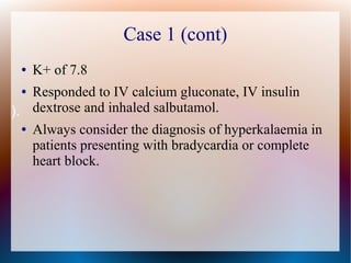
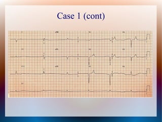
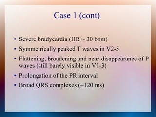
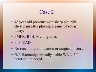
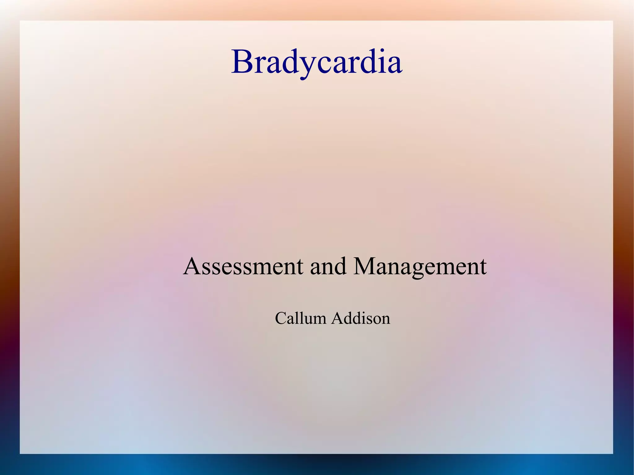
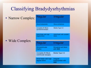
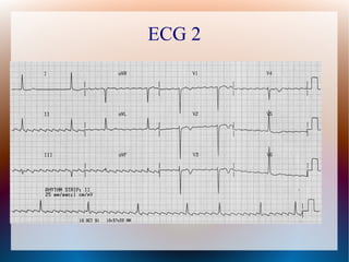
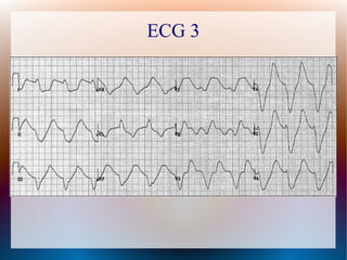
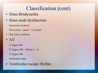
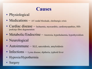
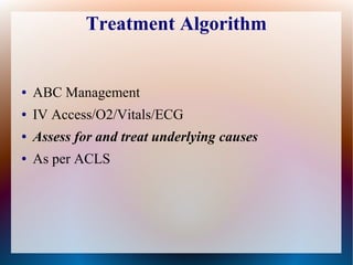
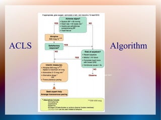
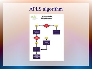
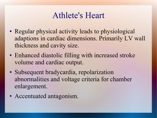
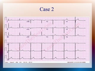
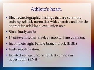
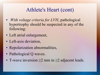
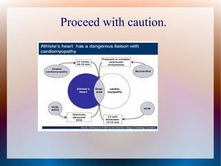
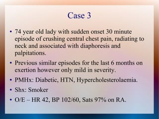
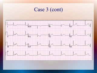
This document discusses the classification, causes, symptoms, and treatment of bradycardia. It defines different types of bradycardia based on rhythm and heart block. Common causes include medications, cardiac disease, metabolic abnormalities, and neurological or infectious etiologies. Symptoms range from dizziness to hypotension and shock. Treatment follows ACLS algorithms and may include atropine, transcutaneous pacing, or addressing underlying causes. Case examples demonstrate ECG findings and management of hyperkalemia-induced complete heart block, athlete's heart, and inferior STEMI with complete heart block.
10. Medications
● AV nodal blocking drugs
(Class II, Class IV, Digoxin, Amiodarone)
● Organophosphates
● Clonidine
● Spot Quiz – Which organophosphate was recently
used to assassinate the step-brother of a well known
political figure.
12. Ischaemia
● Up to 30% of patient with inferior STEMI will
develop second or third degree AV block.
● Associated with an increased in-hospital mortality.
● Inferior myocardial wall and the SA/AV nodes are
usually all supplied by the right coronary artery
13. Raised ICP
● Cushing response.
● Raised BP, Bradycardia, Irregular breathing.
● Bradycardia is due to the baroreceptor response to
the sympathetic efforts to increase cerebral
perfusion – or mechanical distortion of the vagus
nerve
18. Atropine
● Competitive muscarinic antagonist -
Anticholinesterase inhibitor
● Increases firing of the SA Node by blocking the
action of the vagus nerve.
● Onset of action ~ 1 minute
● Duration of action 30 – 60 minutes.
● The first drug of choice for symptomatic
bradycardia.
● Dose in the Bradycardia ACLS algorithm is 0.5mg
IV push and may repeat up to a total dose of 3mg.
19. Spot Quiz – Who Am I
● Invasive Pest
● Solanaceae family
● Contains Tropane alkaloids
20. Transcutaneous Pacing
● Non-invasive pacing is used on a temporary basis
until the patient is stabilized and either an adequate
intrinsic rhythm has returned or a transvenous
pacemaker is inserted, whether temporary or
permanent.
22. Alternative Agents
● Adrenaline (second line agent).
Non-selective A/B agonist.
2-10mcg/min
Titrated to maintain a satisfactory HR.
● Dopamine – 2-10mcg/kg/min
● Isoprenaline – 2.5mcg/min
23. Transcutaneous Pacing
● Indications
Patient with symptomatic bradycardia and a palpable pulse
who has not responded to pharmacological therapy (or no
IV access able to be established).
High-grade AV blockade (3rd
degree heart block or unstable
mobitz type II.
Cardiac arrest with ventricular standstill, but atrial activity
present.
● Recent asystole.
● Contraindication
Asystolic cardiac arrest
24. Transcutaneous Pacing
● For pacing readiness (i.e. standby mode) in the
setting of acute myocardial infarction (AMI) with
the following:
Symptomatic sinus bradycardia
Mobitz type II second-degree AV block
Third-degree AV block
New left, right or alternating bundle branch block or
bifascicular block
25. Transcutaneous Pacing - Procedure
● O2 and IV Access.
● Sedation unless contraindicated.
● Placement of pads on clean, dry, shaven skin.
● Pacing mode with rate 60-80 bpm.
● Begin pacing at 5mA amp, increase amperage in
5mA increments until visible electrical capture-
(QRS-T complexes after each pacing spike).
● Check pulse for mechanical capture.
● Increase by a further 5mA after capture.
26. Classification of Bradycardia
● Absolute or Relative
● Functional or relative bradycardia occurs when a patient may have a heart rate
within normal sinus range, but the heart rate is insufficient for the patients
condition. An example would be a patient with an heart rate of 80 bpm when they
are experiencing septic shock.
● Narrow or Wide Complex
● Regular or irregular
● Sinus vs Sick Sinus vs AV nodal vs Ventricular
27. Case 1
● 68 year old female.
● Presents with an episode of syncope on a
background of 1 week of vomiting and poor oral
intake.
● GCS 14 on arrival, HR 34, BP 80/53
● PMHx: HTN, CHF
● Medx: Spironolactone, Bisoprolol.
28. Case 1 (cont)
● K+ of 7.8
● Responded to IV calcium gluconate, IV insulin
dextrose and inhaled salbutamol.
● Always consider the diagnosis of hyperkalaemia in
patients presenting with bradycardia or complete
heart block.
).
30. Case 1 (cont)
● Severe bradycardia (HR ~ 30 bpm)
● Symmetrically peaked T waves in V2-5
● Flattening, broadening and near-disappearance of P
waves (still barely visible in V1-3)
● Prolongation of the PR interval
● Broad QRS complexes (~120 ms)
31. Case 2
● 48 year old presents with sharp pleuritic
chest pain after playing a game of squash
today.
● PMHx: BPH, Meningioma.
● Fhx: CAD.
● No recent immobilization or surgical history.
●
O/E Haemodynamically stable WNL. 3rd
heart sound heard.
32. Athlete's Heart
● Regular physical activity leads to physiological
adaptions in cardiac dimensions. Primarily LV wall
thickness and cavity size.
● Enhanced diastolic filling with increased stroke
volume and cardiac output.
● Subsequent bradycardia, repolarization
abnormalities and voltage criteria for chamber
enlargement.
● Accentuated antagonism.
34. Athlete's heart.
● Electrocardiographic findings that are common,
training-related, normalize with exercise and that do
not require additional evaluation are:
● Sinus bradycardia
● 1° atrioventricular block or mobitz 1 are common.
● Incomplete right bundle branch block (BBB)
● Early repolarization.
● Isolated voltage criteria for left ventricular
hypertrophy (LVH).
35. Athlete's Heart (cont)
● With voltage criteria for LVH, pathological
hypertrophy should be suspected in any of the
following:
● Left atrial enlargement,
● Left-axis deviation,
● Repolarization abnormalities,
● Pathological Q waves.
● T-wave inversion ≥2 mm in ≥2 adjacent leads.
37. Case 3
● 74 year old lady with sudden onset 30 minute
episode of crushing central chest pain, radiating to
neck and associated with diaphoresis and
palpitations.
● Previous similar episodes for the last 6 months on
exertion however only mild in severity.
● PMHx: Diabetic, HTN, Hypercholesterolaemia.
● Shx: Smoker
● O/E – HR 42, BP 102/60, Sats 97% on RA.
39. Case 3 (cont)
● Regular, narrow complex bradycardia.
● Ventricular rate of 43 BPM.
● Complete AV block.
● Likely junctional escape rhythm.
● Significant ST elevation in leads II, III, and AVF,
with reciprocal ST depression in leads I and AVL,
all suggestive of an inferior STEMI.
40. Case 3 (cont)
● Inferior STEMI with RV infarction was diagnosed.
● IV fluids were given.
● Aspirin, Ticagrelor and Heparin were given
● Patient taken to cath lab:
● Coronary angiography revealed an acute thrombus
with 100% occlusion of the proximal Right
Coronary Artery (proximal to the right ventricular
marginal branch), successfully stented and reduced.
41. Summary
● Remember ABC'ss
● Assess and treat underlying causes
● Reassess regularly for changes in rhythm.
● Decision to treat largely based on
haemodynamic stability and risk of asystole.
● Clear algorithms provided by APLS/ACLS
available for treatment.
43. References/Further Reading
● ACLS/APLS Australia
● Australian Resuscitation Council
● Textbook of Cardiology.org
● Family Practice Notebook
● LITFL
● Dr Smith's ECG Blog
● Dr Venkatesan.com
● Department of Agriculture and Food.