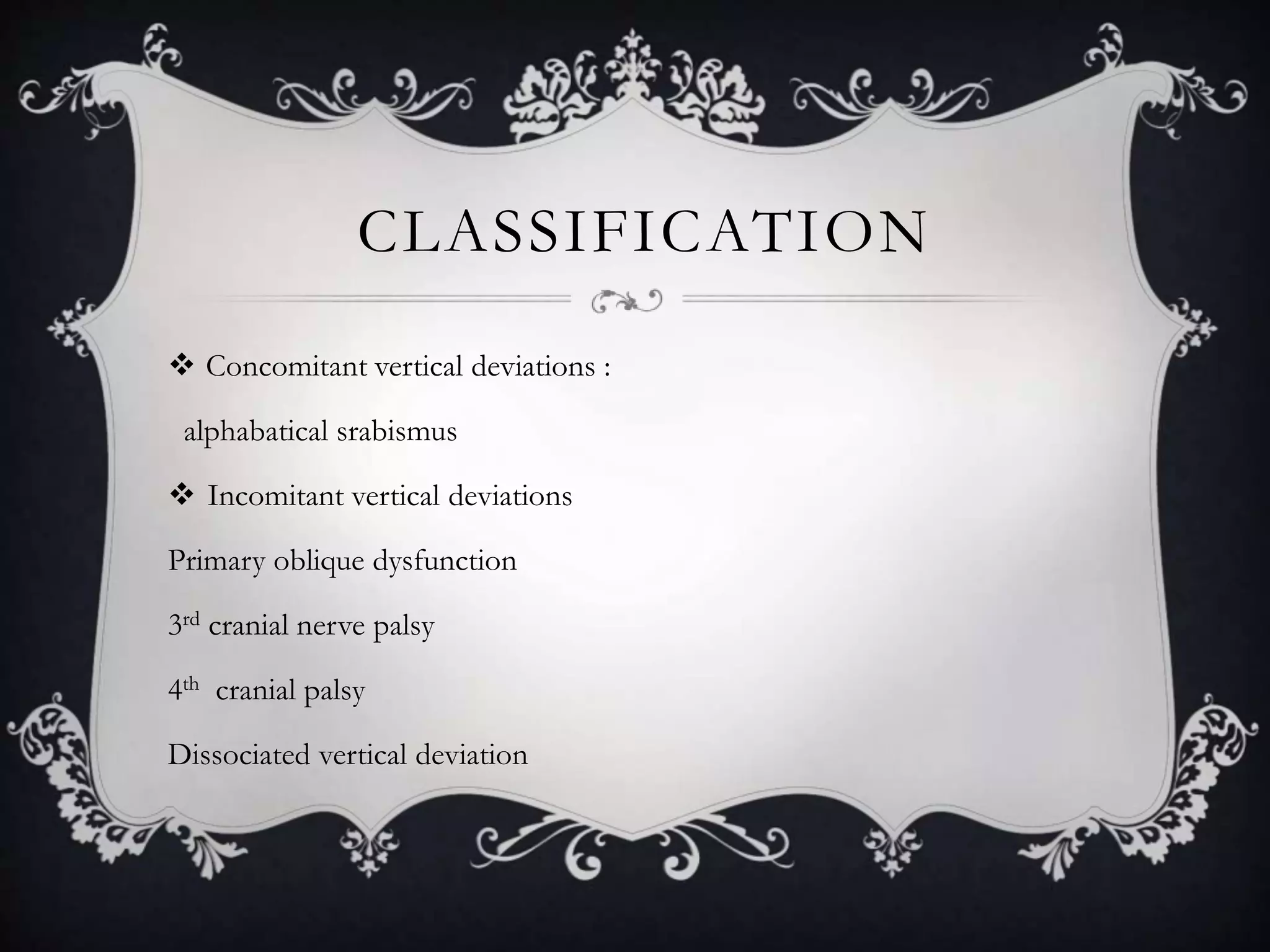
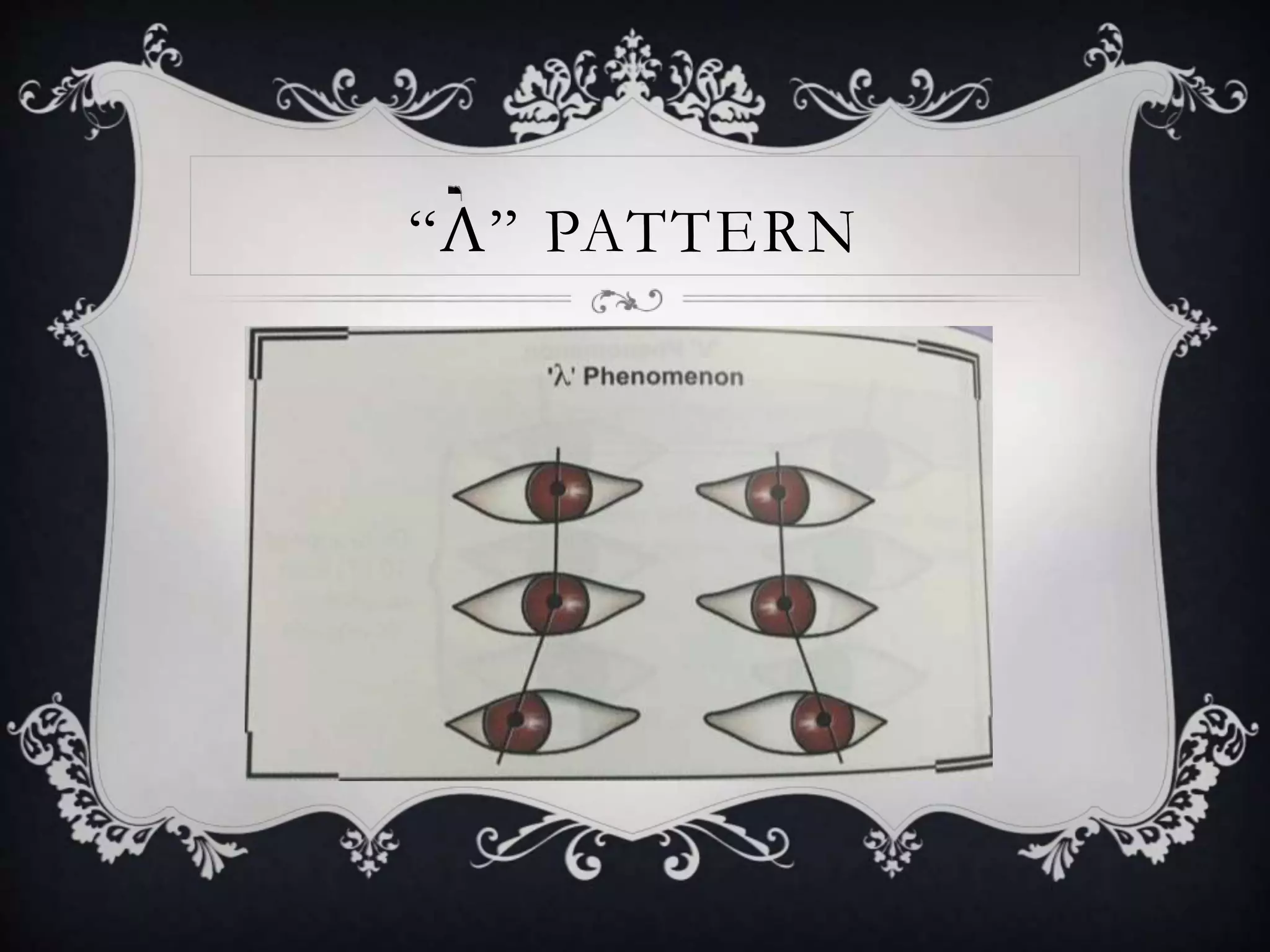
CYCLOVERTICAL
ANOMALIES
Dr NIKITA JAISWAL
IMS & SUM HOSPITAL
GLOSSARY
 DEFINITION
 CLASSIFICATION
 MANAGEMENT
CLASSIFICATION
 COMITANT HYPERDEVIATION
 DISSOCIATED VERTICAL DEVIATION
 DISSOCIATED HORIZONTAL DEVIATION
CYCLOVERTICAL MUSCLES
 RECTI—
 SUPERIOR RECTUS
 INFERIOR RECTUS
----------------------------------------------------------------------
 OBLIQUES--
 SUPERIOR OBLIQUE
 INFERIOR OBLIQUE
S
SUPERIOR RECTUS
MUSCLE
MUSCLE ORIGINATES FROM THE
ANNULUS OF ZINN .
INS .7.7 MM BEHIND THE LIMBUS
THIS IS PARALLEL TO THE ORBITAL
AXIS ND FORMS ANGLE OF 23’.
WHEN ABD. TAKES PLACE OPTICAL
& ORBITAL AXIS COINCIDE.
THESE ACT AS ELEVATORS ND
DEPRESSORS.
SUPERIOR OBLIQUE
MUSCLE
Both the obliques have oblique
course and inserted into the
posterior and temporal quadrant
sup & inferiorly.
It forms 51’ with the optical axis
in primary gaze position
ACTION SUPERIOR
RECTUS
INFERIOR
RECTUS
SUPERIOR
OBLIQUE
INFERIOR
OBLIQUE
PRIMARY ELEVATION DEPRESSION INTORSION EXTORSION
SECONDA
RY
INTORSION EXTORSION DEPRESSION ELEVATION
TERTIARY ADDUCTION ADDUCTION ABDUCTION ABDUCTION
CLASSIFICATION
 Concomitant vertical deviations :
alphabatical srabismus
 Incomitant vertical deviations
Primary oblique dysfunction
3rd cranial nerve palsy
4th cranial palsy
Dissociated vertical deviation
PATTERN STRABISMUS
DEFINITION:
Pattern strabismus is present when a horizontal deviation
changes in magnitude between upgaze & downgaze.
TYPES: V pattern
A pattern
X pattern
Y pattern
λ pattern
15%-
20%
“V” PATTERN
 The horizontal deviation is more divergent in upgaze than in downgaze.
 Presents in patient with infantile esotropia.
 It doesn’t develop when esotropia first develops but becomes apparent at
1st yr of life
 Occurs in superior oblique palsies , craniofacial malformations.
“A” PATTERN
 The second most common type of pattern
 Occurs more frequently in patients with exotropia.
 More common in infantile strabismus associated with craniofacial
malformations ,trisomy 21 & myelomeningocele.
“Y” PATTERN
 Pseudo –overaction of the IO.
 Normal in primary position & downgaze but diverge in upgaze.
 Clinical: overelevation is not seen when the eyes are moved directly
horizontally but manifested when the eyes are moved medially & slightly
elevated.
 No vertical deviations when the head tilts.
“X” PATTERN
“Λ” PATTERN
C/F & IDENTIFICATION
 It is determined by measuring alignment while the patient fixates
ON AN ACCOMMODATIVE TARGET AT DISTANCE,with
fusion prevented,in primary position & in straight upgaze &
downgaze.
 Proper refractive correction is necessary.
 A pattern- 10 PD
 V pattern-15 PD
MANAGEMENT
 PRINCIPLES:
 Weakening of the oblique muscles is performed.
 Pattern inconsistent with oblique dysfunction, vertical transposition of the
horizontal muscles is performed ,the muscles are transposed from one-half
of the width to the full width
 MR – Always towards the apex of the pattern.
 LR– moved toward the open end or empty space
VERTICAL DEVIATIONS
 CAN BE HETEROTROPIA OR HETEROPHORIA
 Vertical tropia acc to resting position of the eye.
 Hypertropia: the effected eye is higher than than the fixating eye.
 Hypotropia: if the effected eye is lower than the fixating eye.
 TRUE VERTICAL TROPIA: INNERVATIONS ARE MATCHED
IN BOTH THE EYES “RT HYPERTROPIA=LT HYPOTROPIA”
INCOMITANT V.TROPIAS
 INFERIOR OBLIQUE MUSCLE OVERACTION
 SUPERIOR OBLIQUE MUSCLE OVERACTION
 INFERIOR OBLIQUE MUSCLE PALSY
 SUPERIOR OBLIQUE MUSCLE PALSY
INF. OBLI. MUSCL E OVER ACTION
 Cause of over elevation in adduction.
 Primary: not associated with sup. Obl .muscle palsy.
 Secondary: when sup ob. Palsy or contralateral sup rectus muscle.
 C/F:-primary IO.OA develop bet 1&6 years
It occurs in acquired ESO & EXOTROPIA
B/L overaction can be asymmetric, poor vision in the eye can lead to
overaction in eye.
C/F
 Eyes in lateral gaze ,alteranate cover –the higher eye refixates with a downward
movement & lower eye refixates with an upward movement.
 Bilateral gaze: the higher & lower eyes reverses their direction of movement in
the opposite lateral gaze.
 Management : weaken the inferior oblique muscle
Recession,myotomy , dis insertion..
SUP.OBLI. MUSCL E OVE RACTION
 Cause of overdepression.
 C/F: vertical dev in primary positiom occurs with U/L or
asymmetric B/L overaction
 The muscle causes a hypotropia of the adducting eye.
 Mngt::superior oblique muscle weakening
SUP.OBLI. MUSCLE PALSY
 IT’S A 4TH NERVE PASLY .
 Can be :acquired or congenital
 Differntiate it with:
childhood photographs
facial symmetry
neuroimaging: if the signs of recovery doesn’t takes place by 3 months.
C/F
 Either normal/affected eye can be preferred for fixation.
 U/L cases: the hyperdeviation is typically incomitant.minimal amount
of hypertropia
 Sup oblique func should be tested when the patient look downwards &
inwards,loss of strength—extorsion—incycylodiplopia.
U/L B/L
pattern Little v pattern Show a v pattern
Degree of extorsion <10’ extorsion in
downgaze
=10’or >20’
incyclodiplopia uncommon common
dysfunction To the involved eye Both the eyes
MASKED B/L PASIES.-CHIN DOWN, B/L FUNDUS
EXTORSION.
INF.OBL. MUSCLE PALSY
Existence is
questioned..
COMITANT VERTICAL
TROPIAS
 Commonly exhibits-- Hypertropia or a Hypotropia. that does not change
significantly when gaze is shifted from one side to the other.
MONOCULAR ELEVATION DEFICIENCY: Involves a limitation of upward
gaze with a hypotropia ,this pattern is caused by restriction of the IR or by an
innervational deficit. Pts may have a combination of a restriction & elevator
muscle deficit.
C/F
 Hypotropia of the involved eye that increases in upgaze
 A chin-up position with fusion in downgaze/ptosis/pseudoptosis
 C/F of 3 forms of MED
 Restriction
 Elevator muscle innervational deficit
 combination
MANAGEMENT
-Indications: a)large vertical deviations in primary position.
b) a chin up head position.
If restrictions: Inferior rectus recessed using an adjustable suture
No restrictions: MR & LR can be transposed toward the superior
rectus muscless.
DISSOCIATED VERTICAL DEVIATION
DVD I S A N I N N E R VA T I O N A L D I S O R D E R F O U N D I N
5 0 % O F P T S W I T H I N F A N T I L E S T R A B I S M U S .
T H E O R Y : - - I T I S A R E S U L T O F M E C H A N I S M S T O
C O M P E N S A T E F O R L A T E N T N Y S T A G M U S , W I T H T H E
O B L I Q U E M U S C L E S P L A Y I N G T H E P R I N C I P L E R O L E .
S
C/F
 The vertical movement usually predominates
 Either eye slowly drifts upward & outward.
 DVD Is usually bilateral but frequently asyymetrical.
 Manifest dvd or latent dvd.
Measurement:a base down prism is placed infront of the upwardly deviating
eye while it is behind occluder., th eoccluder is shifted to the fixating lower
eye nd the prism power is noted when the deviated eye shows no downward
movement to refixate.
MANAGEMENT
 Surgery on the vertical muscles improves the condition
 Recessions of thcan be performed.e superior rectus muscle
Range—6 to 10 mm according to the hypertropia
Inf rectus muscle resection
Cyclovertical anomalies

Cyclovertical anomalies

  • 1.
  • 2.
  • 3.
    CLASSIFICATION  COMITANT HYPERDEVIATION DISSOCIATED VERTICAL DEVIATION  DISSOCIATED HORIZONTAL DEVIATION
  • 4.
    CYCLOVERTICAL MUSCLES  RECTI— SUPERIOR RECTUS  INFERIOR RECTUS ----------------------------------------------------------------------  OBLIQUES--  SUPERIOR OBLIQUE  INFERIOR OBLIQUE
  • 5.
  • 6.
    SUPERIOR RECTUS MUSCLE MUSCLE ORIGINATESFROM THE ANNULUS OF ZINN . INS .7.7 MM BEHIND THE LIMBUS THIS IS PARALLEL TO THE ORBITAL AXIS ND FORMS ANGLE OF 23’. WHEN ABD. TAKES PLACE OPTICAL & ORBITAL AXIS COINCIDE. THESE ACT AS ELEVATORS ND DEPRESSORS.
  • 7.
    SUPERIOR OBLIQUE MUSCLE Both theobliques have oblique course and inserted into the posterior and temporal quadrant sup & inferiorly. It forms 51’ with the optical axis in primary gaze position
  • 8.
    ACTION SUPERIOR RECTUS INFERIOR RECTUS SUPERIOR OBLIQUE INFERIOR OBLIQUE PRIMARY ELEVATIONDEPRESSION INTORSION EXTORSION SECONDA RY INTORSION EXTORSION DEPRESSION ELEVATION TERTIARY ADDUCTION ADDUCTION ABDUCTION ABDUCTION
  • 9.
    CLASSIFICATION  Concomitant verticaldeviations : alphabatical srabismus  Incomitant vertical deviations Primary oblique dysfunction 3rd cranial nerve palsy 4th cranial palsy Dissociated vertical deviation
  • 10.
  • 11.
    DEFINITION: Pattern strabismus ispresent when a horizontal deviation changes in magnitude between upgaze & downgaze. TYPES: V pattern A pattern X pattern Y pattern λ pattern 15%- 20%
  • 12.
    “V” PATTERN  Thehorizontal deviation is more divergent in upgaze than in downgaze.  Presents in patient with infantile esotropia.  It doesn’t develop when esotropia first develops but becomes apparent at 1st yr of life  Occurs in superior oblique palsies , craniofacial malformations.
  • 14.
    “A” PATTERN  Thesecond most common type of pattern  Occurs more frequently in patients with exotropia.  More common in infantile strabismus associated with craniofacial malformations ,trisomy 21 & myelomeningocele.
  • 16.
    “Y” PATTERN  Pseudo–overaction of the IO.  Normal in primary position & downgaze but diverge in upgaze.  Clinical: overelevation is not seen when the eyes are moved directly horizontally but manifested when the eyes are moved medially & slightly elevated.  No vertical deviations when the head tilts.
  • 18.
  • 19.
  • 20.
    C/F & IDENTIFICATION It is determined by measuring alignment while the patient fixates ON AN ACCOMMODATIVE TARGET AT DISTANCE,with fusion prevented,in primary position & in straight upgaze & downgaze.  Proper refractive correction is necessary.  A pattern- 10 PD  V pattern-15 PD
  • 22.
  • 23.
     PRINCIPLES:  Weakeningof the oblique muscles is performed.  Pattern inconsistent with oblique dysfunction, vertical transposition of the horizontal muscles is performed ,the muscles are transposed from one-half of the width to the full width  MR – Always towards the apex of the pattern.  LR– moved toward the open end or empty space
  • 24.
  • 25.
     CAN BEHETEROTROPIA OR HETEROPHORIA  Vertical tropia acc to resting position of the eye.  Hypertropia: the effected eye is higher than than the fixating eye.  Hypotropia: if the effected eye is lower than the fixating eye.  TRUE VERTICAL TROPIA: INNERVATIONS ARE MATCHED IN BOTH THE EYES “RT HYPERTROPIA=LT HYPOTROPIA”
  • 26.
    INCOMITANT V.TROPIAS  INFERIOROBLIQUE MUSCLE OVERACTION  SUPERIOR OBLIQUE MUSCLE OVERACTION  INFERIOR OBLIQUE MUSCLE PALSY  SUPERIOR OBLIQUE MUSCLE PALSY
  • 27.
    INF. OBLI. MUSCLE OVER ACTION  Cause of over elevation in adduction.  Primary: not associated with sup. Obl .muscle palsy.  Secondary: when sup ob. Palsy or contralateral sup rectus muscle.  C/F:-primary IO.OA develop bet 1&6 years It occurs in acquired ESO & EXOTROPIA B/L overaction can be asymmetric, poor vision in the eye can lead to overaction in eye.
  • 28.
    C/F  Eyes inlateral gaze ,alteranate cover –the higher eye refixates with a downward movement & lower eye refixates with an upward movement.  Bilateral gaze: the higher & lower eyes reverses their direction of movement in the opposite lateral gaze.  Management : weaken the inferior oblique muscle Recession,myotomy , dis insertion..
  • 29.
    SUP.OBLI. MUSCL EOVE RACTION  Cause of overdepression.  C/F: vertical dev in primary positiom occurs with U/L or asymmetric B/L overaction  The muscle causes a hypotropia of the adducting eye.  Mngt::superior oblique muscle weakening
  • 30.
    SUP.OBLI. MUSCLE PALSY IT’S A 4TH NERVE PASLY .  Can be :acquired or congenital  Differntiate it with: childhood photographs facial symmetry neuroimaging: if the signs of recovery doesn’t takes place by 3 months.
  • 31.
    C/F  Either normal/affectedeye can be preferred for fixation.  U/L cases: the hyperdeviation is typically incomitant.minimal amount of hypertropia  Sup oblique func should be tested when the patient look downwards & inwards,loss of strength—extorsion—incycylodiplopia.
  • 32.
    U/L B/L pattern Littlev pattern Show a v pattern Degree of extorsion <10’ extorsion in downgaze =10’or >20’ incyclodiplopia uncommon common dysfunction To the involved eye Both the eyes MASKED B/L PASIES.-CHIN DOWN, B/L FUNDUS EXTORSION.
  • 33.
  • 34.
    COMITANT VERTICAL TROPIAS  Commonlyexhibits-- Hypertropia or a Hypotropia. that does not change significantly when gaze is shifted from one side to the other. MONOCULAR ELEVATION DEFICIENCY: Involves a limitation of upward gaze with a hypotropia ,this pattern is caused by restriction of the IR or by an innervational deficit. Pts may have a combination of a restriction & elevator muscle deficit.
  • 35.
    C/F  Hypotropia ofthe involved eye that increases in upgaze  A chin-up position with fusion in downgaze/ptosis/pseudoptosis  C/F of 3 forms of MED  Restriction  Elevator muscle innervational deficit  combination
  • 36.
    MANAGEMENT -Indications: a)large verticaldeviations in primary position. b) a chin up head position. If restrictions: Inferior rectus recessed using an adjustable suture No restrictions: MR & LR can be transposed toward the superior rectus muscless.
  • 37.
  • 38.
    DVD I SA N I N N E R VA T I O N A L D I S O R D E R F O U N D I N 5 0 % O F P T S W I T H I N F A N T I L E S T R A B I S M U S . T H E O R Y : - - I T I S A R E S U L T O F M E C H A N I S M S T O C O M P E N S A T E F O R L A T E N T N Y S T A G M U S , W I T H T H E O B L I Q U E M U S C L E S P L A Y I N G T H E P R I N C I P L E R O L E . S
  • 39.
    C/F  The verticalmovement usually predominates  Either eye slowly drifts upward & outward.  DVD Is usually bilateral but frequently asyymetrical.  Manifest dvd or latent dvd. Measurement:a base down prism is placed infront of the upwardly deviating eye while it is behind occluder., th eoccluder is shifted to the fixating lower eye nd the prism power is noted when the deviated eye shows no downward movement to refixate.
  • 40.
    MANAGEMENT  Surgery onthe vertical muscles improves the condition  Recessions of thcan be performed.e superior rectus muscle Range—6 to 10 mm according to the hypertropia Inf rectus muscle resection