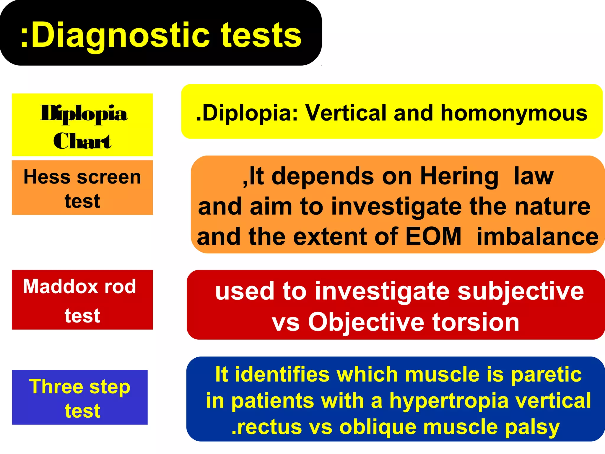
Superior Oblique Palsy
Submitted for partial fulfillment
of the Master Degree in
ophthalmology
By
Mohammad Kamel Mohammad Noor El-Mahdy
M.B.B.Ch. - Al-Azhar University
Superior Oblique Palsy
Supervised by
Prof. Dr. Attiat Mostafa
El-Sayed Mostafa
Prof. of ophthalmology
Faculty of Medicine
Al-Azhar University
Prof. Dr. Abubakr Mohammad Farid AbulNaga
Prof. of Ophthalmology
faculty of medicine
Al-Azhar University
Dr. Ahmad El-Sayed Hodieb
Lecturer of Ophthalmology
faculty of medicine
Al-Azhar University
Aim of the Essay
The aim of this essay is to
review of literature of superior
oblique muscle palsy.
Introduction•Bielchowsky was first to describe SOP as the
leading cause of vertical double vision.
•It has no predilection for males or females
•It is the single most common form of paralytic
strabismus diagnosed in routine practice (von
Noorden et al., 1986).
Review of literature
Anatomy of SO muscle:
• Origin: It arises from the periosteum close to the annulus of Zinn
(the apex of the orbit) above and medial to the optic foramen till
reach the troclea.
• Insertion: Pass under the superior rectus muscle to insert on sclera
along the temporal border of the superior rectus muscle behined the
equator.
• Innervation: The trochlear nerve.
Physiology of SO muscle:
1) Action of superior oblique muscle:
 In 1ry position intorte, in 2ry position depress
and in 3ry position abduct the eye.
The maximum action of the superior oblique muscle as
a depressor is in adduction:
In adduction, with adduction of 54°, the angle between the
median plane of the eye and the muscle plane is reduced.
The maximum incyclotortion occurs in abduction:
In abduction, with 36° of abduction, the angle between the
median plane of the globe and the muscle plane increases.
2) Control of superior oblique muscle movement:
 Remember that:
 Ipsilateral Inferior oblique muscle ( D. antagonist)
 Contralateral Superior rectus muscle ( Ind. Antagonist)
 Contralateral Inferior rectus muscle (Yoke m.)
 The muscle governed by The laws of ocular
motility:
 Dander's law: concerned with axis of positions.
 Listing's law: concerned with Cylotortion.
 Hering's law: concerned with Binoccular vision
(innervation of Yoke ms).
Management of Superior oblique palsy
Causes can be classified as
Congenital palsy: present
at birth may be isolated or
associated with congenital
anomalies.
Aquired palsy: a common
cause is head trauma.
Clinical pictureA) Symptoms
Diplopia:
Vertical and homonymous.
notable when reading or, walking down stairs.
Compensatory head posture:
The head tilt to the opposite side and
the face turn to the opposite side
with the chin depressed.
Rt. SOP (mostafa,2004):
• Chin depression.
• Head tilt to left.
• Face turn to left.
B) Signs: 1-Ocular motility testing
Lt Superior oblique palsy
Rt Lt
2-Macular Torsion
Macularextorsion seen
by
fluorescein fundus
camera, fovea seen
below that line
(Mostafa, 2004).
Normal macula at
level of horizontal
line drown
between upper2/3
and lower1/3 of
optic disc
Torsion as seen by fluorescein
Diagnostic tests:
Diplopia: Vertical and homonymous.
It depends on Hering law,
and aim to investigate the nature
and the extent of EOM imbalance
used to investigate subjective
vs Objective torsion
It identifies which muscle is paretic
in patients with a hypertropia vertical
rectus vs oblique muscle palsy.
Diplopia
Chart
Hess screen
test
Maddox rod
test
Three step
test
Diplopia
Chart
Hess screen test
Maddox rod test Three step test
red- green goggles and Lt. SOP
Rt. superior oblique palsy, Rt.
secondary IOOA and Lt. IR
overaction
Bielschowsky Park's head tilt
test:
(A) (B) (C)
Rt. Superioroblique palsy: (Mostafa, 2004)
(A)Head tilt to Lt.
(B)Rt. hypertropia on forced head tilt to Rt.
(C)Upshoot on adduction due to Rt.
secondary IOOA.
Treatment• Strategies require identifying where the
hypertropia is greatest.
• Surgical methods of treatment are as follows
(Özkan, 2010):
 Superior oblique strengthening procedures.
 Inferior oblique weakening procedures.
 Superior rectus recession in the affected eye.
 Inferior rectus recession in the contralateral
eye.
1- Superior oblique strengthening
procedures
A- Superior Oblique Tuck
 The triad of indications for superior
oblique tendon tuck is:
1) Large angled vertical deviation,
2) Prominent abnormal head posture and,
3) Superior oblique tendon laxity
Superior oblique tendon tuck
SR
MR
LR
IR
SR
LR
RM
IR
IOIO
Dr. G.Vicente
SO
Superior oblique tendon tuck
SR
LR
RM
IR
IOIO
Dr. G.Vicente
SO SR
MR
LR
IR
Superior oblique tendon tuck
SR
LR
RM
IR
IOIO
Dr. G.Vicente
SO SR
MR
LR
IR
Superior oblique tendon tuck
SR
LR
RM
IR
IOIO
Dr. G.Vicente
SO SR
MR
LR
IR
Superior oblique tendon tuck
SR
LR
RM
IR
IOIO
Dr. G.Vicente
SO SR
MR
LR
IR
Superior oblique tendon tuck
SR
LR
RM
IR
IOIO
Dr. G.Vicente
SO SR
MR
LR
IR
B-Harada Ito surgery:
• Indications:
(1) Patients whose primary complaint is
torsional diplopia.
This is most often in adult patients with
bilateral, post traumatic superior oblique
muscle palsy.
(2) Patients with little or no vertical
deviation in primary gaze position.
(3) In the treatment of ocular torticollis with
tilt -dependent nystagmus.
Harada-Ito
Anterior displacement of ½ SO tendon
Dr. G.Vicente
Harada-Ito
Anterior displacement of ½ SO tendon
Dr. G.Vicente
Harada-Ito
Anterior displacement of ½ SO tendon
Dr. G.Vicente
Harada-Ito
Anterior displacement of ½ SO tendon
Dr. G.Vicente
2-Inferior oblique weakening procedures.
The patient's right eye viewed from below; (a) natural position of the inferior
oblique muscle (b) recession; (c) anterior transposition; (d) anterior nasal
transposition; (e and f) nasal myectomy.
1-Inferior oblique muscle recession:
LR
MR
IR
SR
Is a suitable procedure
for most congenital
SO palsies with a
moderate-to-large
vertical deviation in
adduction, resulting in
a lower incidence of
consecutive Brown's
pattern.
IO
Rt. Superior Oblique Palsy (mostafa, 2004(
AHP “preoperative” After Rt. IO recession
After Lt. IO recessionLt. Inferior oblique overaction
Lt. SO palsy (mostafa, 2004)
2- Anterior Transposition (AT)
• It weakens the classic functions of the IO
(eliminate IOOA) .
• converts the muscle to an “antielevator”(reserve
the action of IO).
3- Myectomy or myotomy inferotemporally
A complete myotomy is considered by some surgeons
to be as effective as myectomy or recession of the
inferior oblique muscle.
3-Superior Rectus Muscle Recession
• Indication:
In a vertical deviation exceeding 15 prism
diopters.
• In cases with agenesis of the superior
oblique tendon, superior rectus recession
is the procedure of choice with inferior
oblique weakening.
3-Inferior Rectus Muscle Recession in
the contralateral eye
• Indication:
Acquired superior oblique palsy
surgery to improve torsion and vertical
alignment.
• A minimum recession of the inferior rectus
is 2.5 mm.
• A maximum recession of the inferior rectus
under most circumstances is 5 mm.
Inferior rectus muscle recession
(contralateral eye(
SR
MR
LR
IR
SR
LR
RM
IR
IOIO
Dr. G.Vicente
Recess IO
Recess IR (contralateral(
Affected eyeLtRt
Thank You

Superior oblique palsy

  • 2.
    Superior Oblique Palsy Submittedfor partial fulfillment of the Master Degree in ophthalmology By Mohammad Kamel Mohammad Noor El-Mahdy M.B.B.Ch. - Al-Azhar University
  • 3.
    Superior Oblique Palsy Supervisedby Prof. Dr. Attiat Mostafa El-Sayed Mostafa Prof. of ophthalmology Faculty of Medicine Al-Azhar University Prof. Dr. Abubakr Mohammad Farid AbulNaga Prof. of Ophthalmology faculty of medicine Al-Azhar University Dr. Ahmad El-Sayed Hodieb Lecturer of Ophthalmology faculty of medicine Al-Azhar University
  • 4.
    Aim of theEssay The aim of this essay is to review of literature of superior oblique muscle palsy.
  • 5.
    Introduction•Bielchowsky was firstto describe SOP as the leading cause of vertical double vision. •It has no predilection for males or females •It is the single most common form of paralytic strabismus diagnosed in routine practice (von Noorden et al., 1986).
  • 6.
  • 7.
    Anatomy of SOmuscle: • Origin: It arises from the periosteum close to the annulus of Zinn (the apex of the orbit) above and medial to the optic foramen till reach the troclea. • Insertion: Pass under the superior rectus muscle to insert on sclera along the temporal border of the superior rectus muscle behined the equator. • Innervation: The trochlear nerve.
  • 8.
    Physiology of SOmuscle: 1) Action of superior oblique muscle:  In 1ry position intorte, in 2ry position depress and in 3ry position abduct the eye. The maximum action of the superior oblique muscle as a depressor is in adduction: In adduction, with adduction of 54°, the angle between the median plane of the eye and the muscle plane is reduced. The maximum incyclotortion occurs in abduction: In abduction, with 36° of abduction, the angle between the median plane of the globe and the muscle plane increases.
  • 9.
    2) Control ofsuperior oblique muscle movement:  Remember that:  Ipsilateral Inferior oblique muscle ( D. antagonist)  Contralateral Superior rectus muscle ( Ind. Antagonist)  Contralateral Inferior rectus muscle (Yoke m.)  The muscle governed by The laws of ocular motility:  Dander's law: concerned with axis of positions.  Listing's law: concerned with Cylotortion.  Hering's law: concerned with Binoccular vision (innervation of Yoke ms).
  • 10.
  • 11.
    Causes can beclassified as Congenital palsy: present at birth may be isolated or associated with congenital anomalies. Aquired palsy: a common cause is head trauma.
  • 12.
    Clinical pictureA) Symptoms Diplopia: Verticaland homonymous. notable when reading or, walking down stairs. Compensatory head posture: The head tilt to the opposite side and the face turn to the opposite side with the chin depressed. Rt. SOP (mostafa,2004): • Chin depression. • Head tilt to left. • Face turn to left.
  • 13.
    B) Signs: 1-Ocularmotility testing Lt Superior oblique palsy Rt Lt
  • 14.
    2-Macular Torsion Macularextorsion seen by fluoresceinfundus camera, fovea seen below that line (Mostafa, 2004). Normal macula at level of horizontal line drown between upper2/3 and lower1/3 of optic disc Torsion as seen by fluorescein
  • 15.
    Diagnostic tests: Diplopia: Verticaland homonymous. It depends on Hering law, and aim to investigate the nature and the extent of EOM imbalance used to investigate subjective vs Objective torsion It identifies which muscle is paretic in patients with a hypertropia vertical rectus vs oblique muscle palsy. Diplopia Chart Hess screen test Maddox rod test Three step test
  • 16.
    Diplopia Chart Hess screen test Maddoxrod test Three step test red- green goggles and Lt. SOP Rt. superior oblique palsy, Rt. secondary IOOA and Lt. IR overaction
  • 17.
    Bielschowsky Park's head tilt test: (A)(B) (C) Rt. Superioroblique palsy: (Mostafa, 2004) (A)Head tilt to Lt. (B)Rt. hypertropia on forced head tilt to Rt. (C)Upshoot on adduction due to Rt. secondary IOOA.
  • 18.
    Treatment• Strategies requireidentifying where the hypertropia is greatest. • Surgical methods of treatment are as follows (Özkan, 2010):  Superior oblique strengthening procedures.  Inferior oblique weakening procedures.  Superior rectus recession in the affected eye.  Inferior rectus recession in the contralateral eye.
  • 19.
    1- Superior obliquestrengthening procedures A- Superior Oblique Tuck  The triad of indications for superior oblique tendon tuck is: 1) Large angled vertical deviation, 2) Prominent abnormal head posture and, 3) Superior oblique tendon laxity
  • 20.
    Superior oblique tendontuck SR MR LR IR SR LR RM IR IOIO Dr. G.Vicente SO
  • 21.
    Superior oblique tendontuck SR LR RM IR IOIO Dr. G.Vicente SO SR MR LR IR
  • 22.
    Superior oblique tendontuck SR LR RM IR IOIO Dr. G.Vicente SO SR MR LR IR
  • 23.
    Superior oblique tendontuck SR LR RM IR IOIO Dr. G.Vicente SO SR MR LR IR
  • 24.
    Superior oblique tendontuck SR LR RM IR IOIO Dr. G.Vicente SO SR MR LR IR
  • 25.
    Superior oblique tendontuck SR LR RM IR IOIO Dr. G.Vicente SO SR MR LR IR
  • 26.
    B-Harada Ito surgery: •Indications: (1) Patients whose primary complaint is torsional diplopia. This is most often in adult patients with bilateral, post traumatic superior oblique muscle palsy. (2) Patients with little or no vertical deviation in primary gaze position. (3) In the treatment of ocular torticollis with tilt -dependent nystagmus.
  • 27.
    Harada-Ito Anterior displacement of½ SO tendon Dr. G.Vicente
  • 28.
    Harada-Ito Anterior displacement of½ SO tendon Dr. G.Vicente
  • 29.
    Harada-Ito Anterior displacement of½ SO tendon Dr. G.Vicente
  • 30.
    Harada-Ito Anterior displacement of½ SO tendon Dr. G.Vicente
  • 31.
    2-Inferior oblique weakeningprocedures. The patient's right eye viewed from below; (a) natural position of the inferior oblique muscle (b) recession; (c) anterior transposition; (d) anterior nasal transposition; (e and f) nasal myectomy.
  • 32.
    1-Inferior oblique musclerecession: LR MR IR SR Is a suitable procedure for most congenital SO palsies with a moderate-to-large vertical deviation in adduction, resulting in a lower incidence of consecutive Brown's pattern. IO
  • 33.
    Rt. Superior ObliquePalsy (mostafa, 2004( AHP “preoperative” After Rt. IO recession
  • 34.
    After Lt. IOrecessionLt. Inferior oblique overaction Lt. SO palsy (mostafa, 2004)
  • 35.
    2- Anterior Transposition(AT) • It weakens the classic functions of the IO (eliminate IOOA) . • converts the muscle to an “antielevator”(reserve the action of IO). 3- Myectomy or myotomy inferotemporally A complete myotomy is considered by some surgeons to be as effective as myectomy or recession of the inferior oblique muscle.
  • 36.
    3-Superior Rectus MuscleRecession • Indication: In a vertical deviation exceeding 15 prism diopters. • In cases with agenesis of the superior oblique tendon, superior rectus recession is the procedure of choice with inferior oblique weakening.
  • 38.
    3-Inferior Rectus MuscleRecession in the contralateral eye • Indication: Acquired superior oblique palsy surgery to improve torsion and vertical alignment. • A minimum recession of the inferior rectus is 2.5 mm. • A maximum recession of the inferior rectus under most circumstances is 5 mm.
  • 39.
    Inferior rectus musclerecession (contralateral eye( SR MR LR IR SR LR RM IR IOIO Dr. G.Vicente Recess IO Recess IR (contralateral( Affected eyeLtRt
  • 41.