4. The term ocular surface squamous neoplasia
(OSSN) was first described in 1995 by Lee and
Hirst to denote a spectrum of neoplasm
originate from squamous epithelium ranging
from simple dysplasia to invasive squamous
cell carcinoma(SCC), involving the
conjunctiva, the limbus, and the cornea.
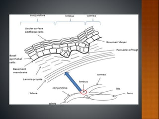
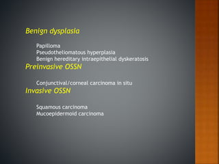
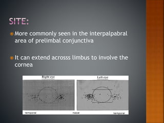
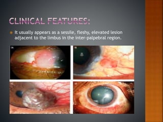
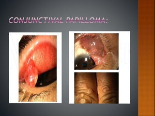
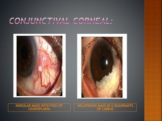
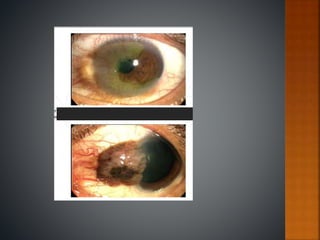
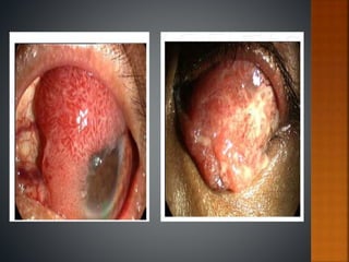
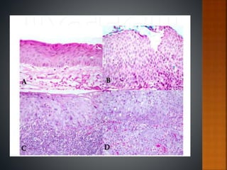
5. Ocular surface squamous neoplasia (OSSN)
It comprises of:Conjunctival intraepithelial neoplastic
lesions (CIN)
Invasive squamous cell carcinoma (SCC) of conjunctiva and
cornea.
10. linear relationship between exposure to
ultraviolet radiation and development of OSSN
Male sex, temporal lesions being more common
and more aggressive than nasal lesions .
UVA (320–400 nm), UVB (280–320 nm), and UVC
(200–280 nm). Exposure to UVB light-- formation of
pyrimidine dimers in DNA and also causes damage to
the nucleotide excision repair pathway and injured
DNA undergoes one cycle of proliferation, this
stimulus of proliferation could be provided by the UV
rays themselves or could be provided by virus such as
HPV or some chemical stimulus.
11. HPV is a nonencapsulated, icosahedral,
non-lipid containing DNA virus that
replicates in the nuclei of infect cell.
High-risk HPVs (e.g., types 16 and 18)
have been implicated in the genesis of
several cancers, particularly squamous
cell carcinoma of the cervix, anogenital
region, oropharyngeal region and ocular
surface.
12. HIV infection is strongly associated with the
occurrence of ossn
Mean age ranges from 32-37 years
Thus this is an established risk factor for
squamous cell neoplasia.
13. It usually appears as a sessile, fleshy, elevated lesion
adjacent to the limbus in the inter-palpebral region.
14.
15. NODULAR MASS WITH FOCI OF
LEUKOPLAKIA
GELATINOUS MASS IN 2 QUADRANTS
OF LIMBUS
16.
17.
18. Histopathological evaluation of the lesion
following incisional & excisional biopsy.
Impression cytology
Anterior segment OCT.
19.
20.
21. Impression cytology (IC) is a technique for
collecting the superficial layers of the ocular
surface by applying collecting devices.
Commonly used are cellulose acetate filter
paper
with a pore size ranging from 0.025 – 0.45
micron or other materials (nitrocellulose
filters,
Biopore membranes, or polyether sulfone
filters
24. Complete excision for most localised lesions.
Alcohol assisted kerato-epitheliectomy.
Double-freeze cryotherapy to the resected margins and
base of the lesion.
Confirmed by the histopathological examination
The residual defect after excision can be covered by
amniotic membrane or buccal mucous membrane.
Negative surgical margin: the most import. Recurrence
indicator
5% to 33% after negative margins to as high as 56%
25. Topical applications 5 fluorouracil(5FU) &
mitomycin C (MMC)
Primary t/t is limited to localised lesion.
Both of these can be used as adjuvant therapy
for recurrent lesions
Due to toxicity they are less preferred.
26.
27.
28. Topical interferon (IFNα2b) 1 million IU/ml) 4
times a day has been used effectively in
treatment of primary OSSN.
OSSN measuring <8 mm.
choice for wider and extensive OSSN
involving >4 clock hours of the limbus.
Flu-like symptoms and ocular surface
irritation are the side effects.
29. The overall prognosis in OSSN is good.
Modern T/T are effective with local recurrence
rates reported to be 5% and regional lymph node
metastasis at <2%.
Aggressive variants like muco-epidermoid and
spindle cell carcinoma and OSSN in
immunocompromised patients have a worse
prognosis.