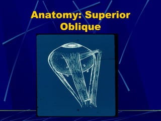
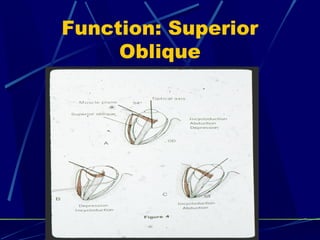
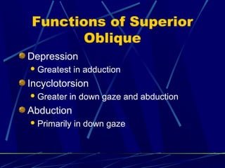
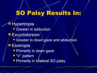
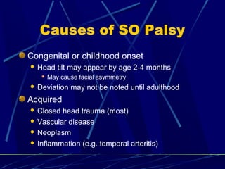
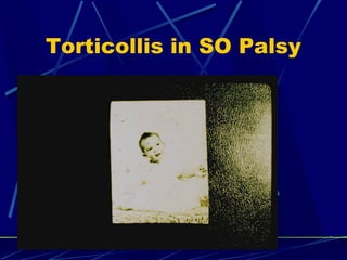
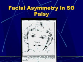
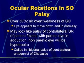
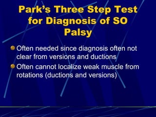
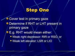
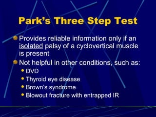
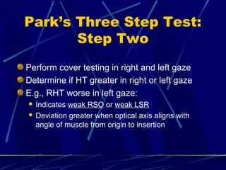
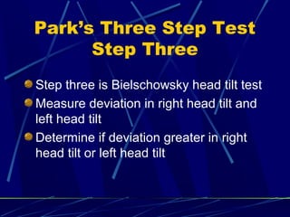
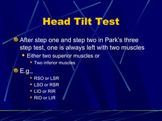
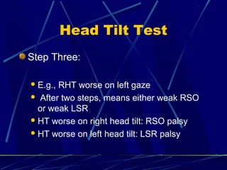
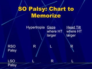
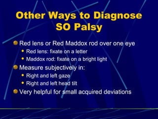
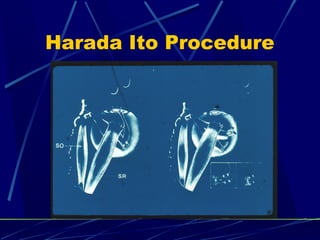
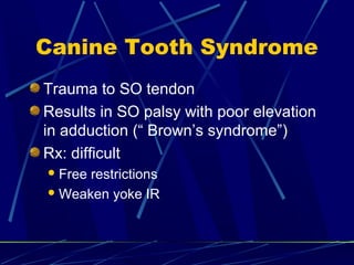
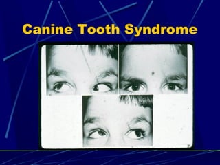
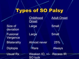
This document discusses the diagnosis and management of superior oblique palsy. It begins by describing the anatomy and function of the superior oblique muscle. Superior oblique palsy can result in hypertropia, excyclotorsion, and esotropia that are greater in certain gazes. Causes may be congenital or acquired from trauma or vascular issues. Diagnosis involves evaluating eye movements, diplopia, and head tilt. Non-surgical treatment includes patching or prisms while surgery involves weakening the antagonist inferior oblique muscle or tucking the superior oblique tendon. The goal of treatment is to expand the field of single vision while minimizing complications.
Duane syndrome, also called Duane retraction syndrome (DRS), is a congenital and non-progressive type of strabismus due to abnormal development of the 6th cranial nerve.
It is characterized by difficulty rotating one or both eyes outward (abduction) or inward (adduction).
On the other hands Duane Retraction Syndrome is a congenital strabismus syndrome occurring in isolated or syndromic forms. It presents with a variety of clinical features including diplopia, anisometropia, and amblyopia.
This presentation is a detailed description of how a patient should be examined in an oprthoptic clinic. it lists down all the investigations sequentially. the order of investigations mentioned is the best way to investigate a squint case.
It describes about the procedure of Hess charting. it serves as a great tool to understand the concepts involved. Suitable for optometry course. This is not a routine procedure but an important procedure which is used in diagnosis.
Presenters :
Jenil Shelsiya
Sisira PS
Gopika Jyothirmayan
Special Thanks To Sushma Shrestha
and Mentor Deepak Rai (Optometrist).
If any query,Viewers are requested to refer to book for detailed explanation or can ask me question directly in the comment box. Answers will be given from Presenter's side.
PARALYTIC SQUINT
KANISHK DEEP SHARMA
ROLL NO. 50
Uncoordinated eye movement
Angle of squint varies
Motor imbalance
ETIOLOGY
Lesion of nerve
Lesion of muscles
Lesions due to
Injury
Inflammation-syphilis, disseminated sclerosis
Vascular diseases-hemorrhage, aneurysm,arteriosclerosis
Neoplasms-brain tumor
Toxins-alcohol, lead, carbon monoxide
Degeneration-chronic nuclear ophthalmoplegia
Myasthenia gravis
SYMPTOMS
DIPLOPIA
In field of action of paralyzed muscle
Long duration- suppression of false image
Vertigo & nausea
Action required towards paralysed muscle
False projection
Binocular diplopia
Secondry deviation
Defective ocular motility
Complementary head postures
Attempt to lessen diplopia
Head tilt to avoid torsion
SEQUALAE
Weakness of paretic muscle
Overacting contralateral synergistic muscle
Inhibitory palsy of contralateral antagonist
TESTS
Record of visual acuity
Ocular motility
Perimetry
Inspection of compensatory head postures
Diplopia charting
Dark room procedure
Armstrong's glasses
4ft distance, fine linear light
Primary & other positions of gaze measured
Hess charting
Explains muscle paralysis & pathological sequlae
Field of binocular fixation
Forced duction test
MANAGEMENT
Treatment of cause
Conservative measures
Vit B complex, systemic steroids
Diplopia treatment
Occluder on affected eye
SURGERIES
Muscle weakening procedure
Recession, marginal myotomy, myectomy
Muscle strengthening procedures
Resection, tucking advancement
Changing direction of muscle action
THANK YOU
The presentation I have made and uploaded provides you with an in-depth insight into the patterns the strabismus may take following anomalies of extraocular muscles, deformities of the orbital structures,innnervational disturbances.
The author does not assume responsibility or legal liability for any errors in the text or for the misuse or misapplication of material in this work.
No copyright infringement, or plagiarism intended.
Amrit Pokharel
Antibiotic Stewardship by Anushri Srivastava.pptxAnushriSrivastav
Stewardship is the act of taking good care of something.
Antimicrobial stewardship is a coordinated program that promotes the appropriate use of antimicrobials (including antibiotics), improves patient outcomes, reduces microbial resistance, and decreases the spread of infections caused by multidrug-resistant organisms.
WHO launched the Global Antimicrobial Resistance and Use Surveillance System (GLASS) in 2015 to fill knowledge gaps and inform strategies at all levels.
ACCORDING TO apic.org,
Antimicrobial stewardship is a coordinated program that promotes the appropriate use of antimicrobials (including antibiotics), improves patient outcomes, reduces microbial resistance, and decreases the spread of infections caused by multidrug-resistant organisms.
ACCORDING TO pewtrusts.org,
Antibiotic stewardship refers to efforts in doctors’ offices, hospitals, long term care facilities, and other health care settings to ensure that antibiotics are used only when necessary and appropriate
According to WHO,
Antimicrobial stewardship is a systematic approach to educate and support health care professionals to follow evidence-based guidelines for prescribing and administering antimicrobials
In 1996, John McGowan and Dale Gerding first applied the term antimicrobial stewardship, where they suggested a causal association between antimicrobial agent use and resistance. They also focused on the urgency of large-scale controlled trials of antimicrobial-use regulation employing sophisticated epidemiologic methods, molecular typing, and precise resistance mechanism analysis.
Antimicrobial Stewardship(AMS) refers to the optimal selection, dosing, and duration of antimicrobial treatment resulting in the best clinical outcome with minimal side effects to the patients and minimal impact on subsequent resistance.
According to the 2019 report, in the US, more than 2.8 million antibiotic-resistant infections occur each year, and more than 35000 people die. In addition to this, it also mentioned that 223,900 cases of Clostridoides difficile occurred in 2017, of which 12800 people died. The report did not include viruses or parasites
VISION
Being proactive
Supporting optimal animal and human health
Exploring ways to reduce overall use of antimicrobials
Using the drugs that prevent and treat disease by killing microscopic organisms in a responsible way
GOAL
to prevent the generation and spread of antimicrobial resistance (AMR). Doing so will preserve the effectiveness of these drugs in animals and humans for years to come.
being to preserve human and animal health and the effectiveness of antimicrobial medications.
to implement a multidisciplinary approach in assembling a stewardship team to include an infectious disease physician, a clinical pharmacist with infectious diseases training, infection preventionist, and a close collaboration with the staff in the clinical microbiology laboratory
to prevent antimicrobial overuse, misuse and abuse.
to minimize the developme
Telehealth Psychology Building Trust with Clients.pptxThe Harvest Clinic
Telehealth psychology is a digital approach that offers psychological services and mental health care to clients remotely, using technologies like video conferencing, phone calls, text messaging, and mobile apps for communication.
Struggling with intense fears that disrupt your life? At Renew Life Hypnosis, we offer specialized hypnosis to overcome fear. Phobias are exaggerated fears, often stemming from past traumas or learned behaviors. Hypnotherapy addresses these deep-seated fears by accessing the subconscious mind, helping you change your reactions to phobic triggers. Our expert therapists guide you into a state of deep relaxation, allowing you to transform your responses and reduce anxiety. Experience increased confidence and freedom from phobias with our personalized approach. Ready to live a fear-free life? Visit us at Renew Life Hypnosis..
QA Paediatric dentistry department, Hospital Melaka 2020Azreen Aj
QA study - To improve the 6th monthly recall rate post-comprehensive dental treatment under general anaesthesia in paediatric dentistry department, Hospital Melaka
CHAPTER 1 SEMESTER V PREVENTIVE-PEDIATRICS.pdfSachin Sharma
This content provides an overview of preventive pediatrics. It defines preventive pediatrics as preventing disease and promoting children's physical, mental, and social well-being to achieve positive health. It discusses antenatal, postnatal, and social preventive pediatrics. It also covers various child health programs like immunization, breastfeeding, ICDS, and the roles of organizations like WHO, UNICEF, and nurses in preventive pediatrics.
Navigating Challenges: Mental Health, Legislation, and the Prison System in B...Guillermo Rivera
This conference will delve into the intricate intersections between mental health, legal frameworks, and the prison system in Bolivia. It aims to provide a comprehensive overview of the current challenges faced by mental health professionals working within the legislative and correctional landscapes. Topics of discussion will include the prevalence and impact of mental health issues among the incarcerated population, the effectiveness of existing mental health policies and legislation, and potential reforms to enhance the mental health support system within prisons.
Leading the Way in Nephrology: Dr. David Greene's Work with Stem Cells for Ki...Dr. David Greene Arizona
As we watch Dr. Greene's continued efforts and research in Arizona, it's clear that stem cell therapy holds a promising key to unlocking new doors in the treatment of kidney disease. With each study and trial, we step closer to a world where kidney disease is no longer a life sentence but a treatable condition, thanks to pioneers like Dr. David Greene.
Global launch of the Healthy Ageing and Prevention Index 2nd wave – alongside...ILC- UK
The Healthy Ageing and Prevention Index is an online tool created by ILC that ranks countries on six metrics including, life span, health span, work span, income, environmental performance, and happiness. The Index helps us understand how well countries have adapted to longevity and inform decision makers on what must be done to maximise the economic benefits that comes with living well for longer.
Alongside the 77th World Health Assembly in Geneva on 28 May 2024, we launched the second version of our Index, allowing us to track progress and give new insights into what needs to be done to keep populations healthier for longer.
The speakers included:
Professor Orazio Schillaci, Minister of Health, Italy
Dr Hans Groth, Chairman of the Board, World Demographic & Ageing Forum
Professor Ilona Kickbusch, Founder and Chair, Global Health Centre, Geneva Graduate Institute and co-chair, World Health Summit Council
Dr Natasha Azzopardi Muscat, Director, Country Health Policies and Systems Division, World Health Organisation EURO
Dr Marta Lomazzi, Executive Manager, World Federation of Public Health Associations
Dr Shyam Bishen, Head, Centre for Health and Healthcare and Member of the Executive Committee, World Economic Forum
Dr Karin Tegmark Wisell, Director General, Public Health Agency of Sweden
Welcome to Secret Tantric, London’s finest VIP Massage agency. Since we first opened our doors, we have provided the ultimate erotic massage experience to innumerable clients, each one searching for the very best sensual massage in London. We come by this reputation honestly with a dynamic team of the city’s most beautiful masseuses.
6. SO Palsy Results In:
Hypertropia
Greater in adduction
Excyclotorsion
Greater in down gaze and abduction
Esotropia
Primarily in down gaze
“V” pattern
Primarily in bilateral SO palsy
7. Causes of SO Palsy
Congenital or childhood onset
Head tilt may appear by age 2-4 months
May cause facial asymmetry
Deviation may not be noted until adulthood
Acquired
Closed head trauma (most)
Vascular disease
Neoplasm
Inflammation (e.g. temporal arteritis)
8. Systemic Workup of SO
Palsy
Usually unrewarding if SO palsy
isolated (i.e., no other new ocular or
neurologic signs or symptoms)
Definitely not needed if signs of
childhood onset present
9. Evolution of New SO
Palsy
Initially:
Vertical deviation greatest in field of SO
(i.e., gaze down and in)
Later
Contracture of antagonist and changes in other
vertical muscles often occurs
Vertical deviation often greater in other fields of
gaze
12. Torsional Diplopia in SO
Palsy
Eye itself is extorted
Visible on fundus exam
Superior pole of image seen by patient
appears intorted
If normal eye is occluded and patient
asked to hold object straight, he will
hold it in an extorted position
15. Diplopia in SO Palsy
Diplopia may be vertical, torsional
and/or horizontal
Occasionally, torsional diplopia occurs
with little or no vertical deviation
Bilateral SO palsy: large amount of
torsional diplopia, no vertical deviation
in primary if bilateral palsy symmetric
17. Torticollis in SO Palsy
Classical:
Head tilt to normal side
Face turn to normal side
Chin down
Not stereotyped: variations exist
Head tilt may be “paradoxical”: i.e. to side
of paretic eye
18. Torticollis In SO Palsy
Torticollis may be large and noted at
age 2-4 months of age
Head tilt may be small and not noted by
patient
Old pictures (or spouse) helpful
22. Ocular Rotations in SO
Palsy
Over 50%: no overt weakness of SO
Eye appears to move down and in normally
May look like palsy of contralateral SR
(if patient fixates with paretic eye in
adduction, non paretic eye will be
hypotropic)
Called inhibitional palsy of contralateral
antagonist of Chavasse
23. Park’s Three Step Test
for Diagnosis of SO
Palsy
Often needed since diagnosis often not
clear from versions and ductions
Often cannot localize weak muscle from
rotations (ductions and versions)
24. Step One
Cover test in primary gaze
Determine if RHT or LHT present in
primary gaze
E.g. RHT would mean either:
Weak right depressor: RIR or RSO, or
Weak left elevator: LSR or LIO
25. Park’s Three Step Test
Provides reliable information only if an
isolated palsy of a cyclovertical muscle
is present
Not helpful in other conditions, such as:
DVD
Thyroid eye disease
Brown’s syndrome
Blowout fracture with entrapped IR
26. Park’s Three Step Test:
Step Two
Perform cover testing in right and left gaze
Determine if HT greater in right or left gaze
E.g., RHT worse in left gaze:
Indicates weak RSO or weak LSR
Deviation greater when optical axis aligns with
angle of muscle from origin to insertion
27. Park’s Three Step Test
Step Three
Step three is Bielschowsky head tilt test
Measure deviation in right head tilt and
left head tilt
Determine if deviation greater in right
head tilt or left head tilt
28. Head Tilt Test
If a superior muscle is weak, HT greater
on tilt toward involved muscle
If an inferior muscle is weak, HT greater
on head tilt opposite involved side
32. Head Tilt Test
After step one and step two in Park’s three
step test, one is always left with two muscles
Either two superior muscles or
Two inferior muscles
E.g.,
RSO or LSR
LSO or RSR
LIO or RIR
RIO or LIR
33. Head Tilt Test
Step Three:
E.g., RHT worse on left gaze
After two steps, means either weak RSO
or weak LSR
HT worse on right head tilt: RSO palsy
HT worse on left head tilt: LSR palsy
34. SO Palsy: Chart to
Memorize
Hypertropia Gaze
where HT
larger
Head Tilt
where HT
larger
RSO
Palsy
R L R
LSO
Palsy
L R L
35. Other Ways to Diagnose
SO Palsy
Red lens or Red Maddox rod over one eye
Red lens: fixate on a letter
Maddox rod: fixate on a bright light
Measure subjectively in:
Right and left gaze
Right and left head tilt
Very helpful for small acquired deviations
36. Bilateral SO Palsy
V pattern with esotropia in downgaze
common
Excylotorsion over 10-15 degrees
HT changes from right to left gaze
E.g., RHT on left gaze, LHT on right gaze
Type of HT changes on head tilt
E.g., RHT on right head tilt, LHT on left
head tilt
37. Bilateral SO Palsy
Vertical deviation often asymmetric
If symmetric, little or no vertical in primary gaze
Often first diagnosed after surgery for
apparent unilateral SO palsy (called “masked
bilateral SO palsy”)
Double Maddox Rod helpful to diagnose pre-
operatively
38. Measurement of Torsion
Double Maddox rod placed in trial frame
One red, one white (or two red Maddox rods)
Patient views a single white light source
Patient sees a red line and a white line
Rotate one lens to make the two lines parallel
(subjectively)
Use vertical prism if needed to separate lines
40. Treatment of SO Palsy
Surgical Treatment
Non-Surgical Treatment
41. Non Surgical Treatment
of SO Palsy
Wait six months if new palsy occurs
Many improve spontaneously
Patch for diplopia
Let adult choose which eye to patch, usually non
paretic eye
Prisms
May be helpful in adults with diplopia and with
smaller less incomitant deviations
Amblyopia
Treat if present
May occur in either eye
43. Which Muscle to
Operate On
Measure vertical deviation in all fields
Pay particular attention to:
Primary gaze
Right and left gaze
Oblique gazes opposite palsy
45. Surgery for SO Palsy
Surgery planned primarily based on the
deviation and where the deviation is
largest
Head Tilt primarily useful for diagnosis
Presence of bilateral SO palsy will
change treatment plan
46. Surgical Treatment of
SO Palsy
Some patients, mostly childhood onset
types, have laxity in the SO tendon
Found with forced duction of SO under
general anesthesia
Patients with laxity of SO tendon need
SO surgery (generally SO tuck) to
equalize forced duction with normal SO
47. Surgical Treatment of
SO Palsy
Superior Oblique tuck performed on
patient without laxity in the tendon likely
to cause a “Brown syndrome”, or
inability to look upward in adduction
SO tuck still often indicated in:
SO palsy worse in straight down gaze
Bilateral SO Palsy
49. Surgical Treatment of
SO Palsy
Patients without laxity of SO, primary
surgical procedure is weakening
(generally recession) of IO, the direct
antagonist to the weak SO
Unilateral cases
IO weakening done in 50-80% of cases
Can be done with adjustable suture
50. Surgical Treatment of
SO Palsy
If deviation greatest in field of SO and
no tendon laxity present, choices are:
Weaken opposite inferior rectus
Tuck SO
Which muscle depends on difference of
deviation in primary and lateral
gaze,and down and down and lateral
gaze
51. Surgical Treatment of
SO Palsy
Recess ipsilateral superior rectus if:
Positive forced duction on attempted
depression of paretic eye
Five diopter or more vertical in abduction of
paretic eye
52. How Many Muscles to
Operate on
Determine deviation in field of greatest
deviation
If that deviation is under 15-20 diopters,
operate on one muscle
If that deviation is over 15-20 diopters,
operate on two muscles
Three muscles: usually results in
overcorrection
53. IO Weakening
Amount of correction varies with
amount of overaction of IO
Can correct 10-15 diopters in primary
gaze
Can be done as adjustable suture in
adults
54. Treatment of Bilateral
SO Palsy
Mostly torsional with little vertical
deviation: Harada Ito procedure
With large HT in side gaze: tuck SO OU
Usually bilateral IO overaction not seen
55. SO Tuck: Dangers
Can easily overcorrect and create
restriction if SO tendon is not lax
In the past, large percentage of SO
tucks had to be “taken down”
Use forced ductions at surgery as guide
to amount of tuck
56. Other Surgical
Complications
IO weakening
Can cause “ adherence syndrome” if fat pad
penetrated: will look like IO overaction on other
side (restriction of elevation in abduction of
operated eye)
IR Recession
Can easily cause lower eyelid retraction
Can prevent eyelid retraction with recession of lower lid
retractors
57. Amount of Surgery
IO weakening
Recess to just posterior and lateral to IR
insertion
Can do asymmetric IO recession OU
SO tuck
Determine at surgery
Usually 6-14 mm, sometimes more
58. Amount of Surgery
IR Recession
Usually 3-5 mm: use adjustable suture in
adults
SR Recession
Usually 3-5 mm: use adjustable sutures in
adults
59. Work Up of SO Palsy
History
Trauma
Diplopia
Torticollis
Other neurological signs or symptoms
60. Work Up of SO Palsy
Observe torticollis
Measurement in all cardinal fields and
head tilt right and left
Double Maddox Rod to measure torsion
Observe fundus for torsion
61. SO Palsy with Torsion
and No Or Minimal HT
Tuck or advance anterior portion of SO
tendon
Advancement of anterior SO called
Harada Ito procedure
63. Canine Tooth Syndrome
Trauma to SO tendon
Results in SO palsy with poor elevation
in adduction (“ Brown’s syndrome”)
Rx: difficult
Free restrictions
Weaken yoke IR
65. Types of SO Palsy
Childhood
Onset
Adult Onset
Size of
deviation
Large Small
Fusional
Vergence
Large Small
Bilaterality Almost never 25%
Diplopia Rare Always
Usual Rx Weaken IO, +/-
SO tuck
Recess IR
66. Course of SO Palsy
May present early in childhood with
torticollis or strabismus
May present later ( often age 30-50)
with symptoms from strabismus or
torticollis
67. Field of Single Vision
Very important to patient
Often ignored by physician
Measure pre-op and post-op ( can use
Goldman perimeter) or estimate
Warn patients that deviation will
probably be present in some fields post-
op