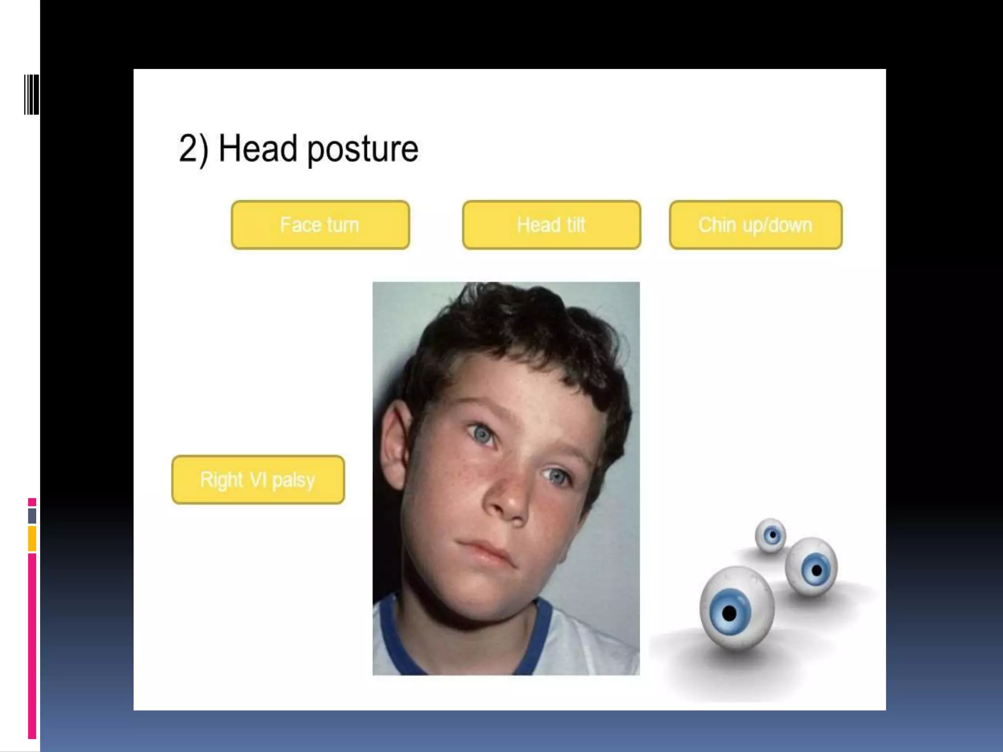
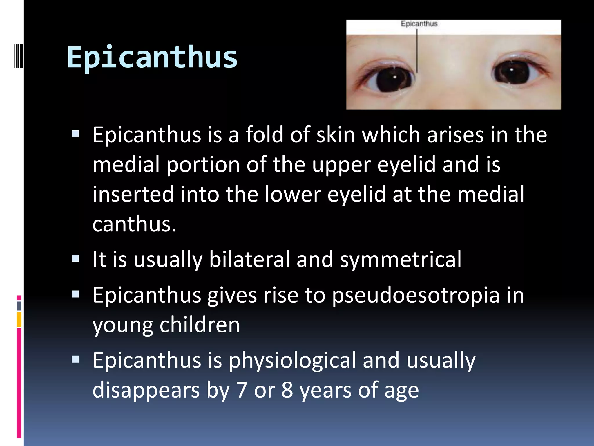
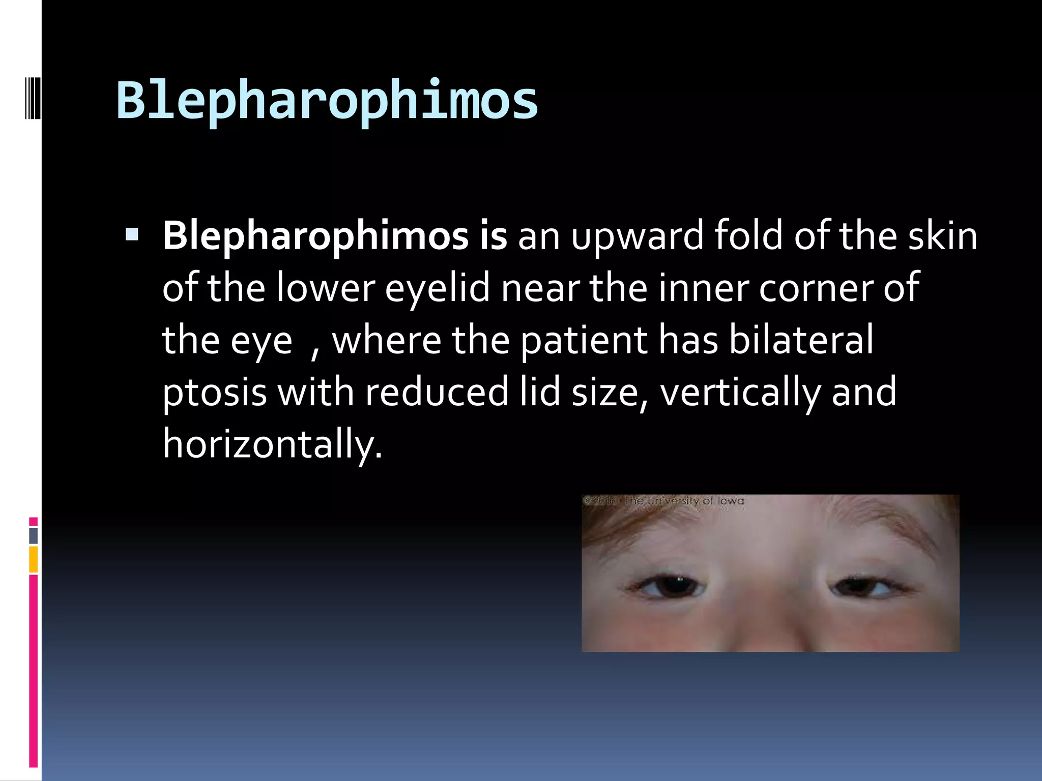
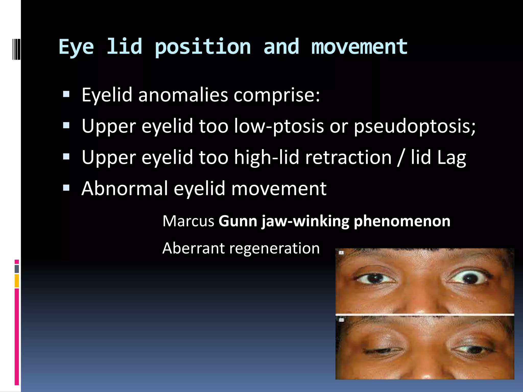
This document outlines the order and components of a general orthoptic examination. The examination aims to diagnose ocular motor disorders and detect other eye or non-eye issues. It involves assessing appearance, measuring visual acuity, observing eyelids and eye position, and measuring eye movements and deviations. It also includes refraction with cycloplegia, investigating binocular function, and potential laboratory/radiology tests. Specific examination points cover general appearance, head posture, eyelid assessment, ocular deviation and movement testing, and evaluating binocular visual functions.
2. Introduction
Examination of the patient has two main aims.
Diagnosing the nature and degree of the
ocular motor disorder.
Detecting other ocular and non-ocular
disorders and assessing their relationship to
the motility problem.
3. Examination Order
Assessment of the patient’s appearance,
including his facial features and head posture.
Measurement of the uncorrected and
corrected visual acuity
Observation of the position and function of
the eyelids.
Observation and measurement of the position
of the globe.
4. Examination
Assessment and measurement of the ocular
deviation and ocular movement
Refraction with cycloplegia
Investigation of binocular function
Laboratory and radiological investigations
5. Examination Order
General Appearance :
The following condition are associated with
high incidence of strabismus and eye
movement problems
Hydrocephalus
Microcephalus
Albinism
Downs syndrome etc
6.
7. Examination Order
Head posture?
An abnormal head posture has three possible
components
Face turn to the right or left side
Head tilt to the right or left shoulder
Chin elevation or depression
10. Examination Order
Visual Acuity
Applying proper test and method of
assessment
To find out any decrease in vision
11. Examination Order
The eye lid
The eyelid position is symmetrical on both sides.
The height of each palpebral fissure is 9-11 mm
The resting position of the upper eyelid margin is
1-2 mm below the superior limbus.
Movement of the upper eyelid from down-gaze
to up-gaze measures 15-1 8 mm.
This relationship is normally maintained in the
different positions of gaze.
12. Examination Order
Eyelids
Palpebral fissures
Variants from the normal palpebral fissure
shape or size can result in pseudostrabismus
or can be characteristic of certain ocular
motility disorders.
13. Fissure changes during ocular movement
An increase or decrease in the height of the
palpebral fissure can be characteristic of some
incomitant conditions,
for example:
Narrowing of the fissure on adduction and
widening on abduction are diagnostic features
of Duane’s retraction syndrome.
The fissure commonly widens on adduction in
Brown’s syndrome.
14.
15.
16. Epicanthus
Epicanthus is a fold of skin which arises in the
medial portion of the upper eyelid and is
inserted into the lower eyelid at the medial
canthus.
It is usually bilateral and symmetrical
Epicanthus gives rise to pseudoesotropia in
young children
Epicanthus is physiological and usually
disappears by 7 or 8 years of age
17. Blepharophimos
Blepharophimos is an upward fold of the skin
of the lower eyelid near the inner corner of
the eye , where the patient has bilateral
ptosis with reduced lid size, vertically and
horizontally.
18. Eye lid position and movement
Eyelid anomalies comprise:
Upper eyelid too low-ptosis or pseudoptosis;
Upper eyelid too high-lid retraction / lid Lag
Abnormal eyelid movement
Marcus Gunn jaw-winking phenomenon
Aberrant regeneration
19.
20.
21. ocular deviation and ocular
movement
ocular deviation
Preliminary inspection should include
comparison of the corneal reflections in the two
eyes as the patient fixates a light source at 33 cm
A cover test should be used at near and distance
to detect the deviation and differentiate manifest
and latent strabismus
The prism cover test is the preferred routine
method of measurement in suitable cases
22. ocular deviation and ocular movement
Ocular movement
Versions and ductions should be assessed by
asking the patient to follow a moving target,
usually a spotlight, from the primary position to
the limit of each of the other eight positions of
gaze
The movement of the two eyes should be
compared and underaction and overaction
noted.
An alternate cover test should be used to confirm
the findings
23. Cycloplegic Refraction
Cycloplegia is essential for accurate refraction
of children, otherwise full correction of the
refractive error may not be achieved