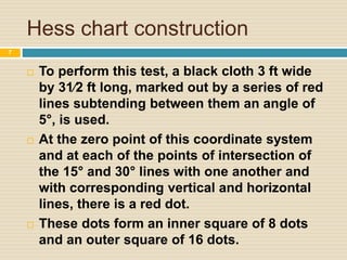
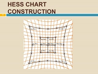
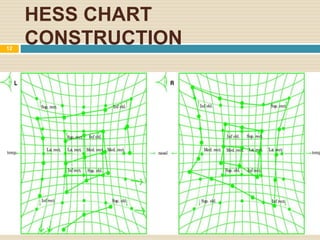
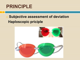
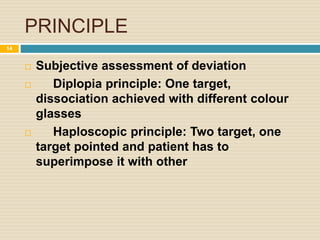
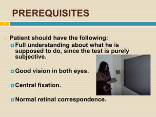
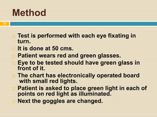
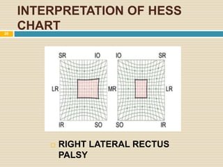
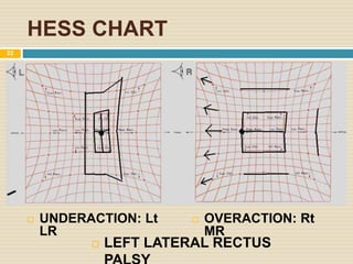
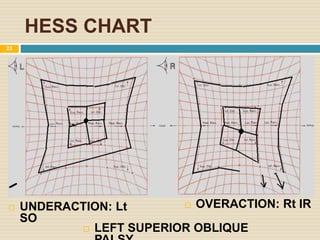
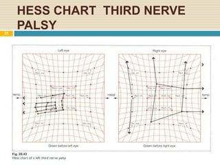
HESS CHARTING
NEURO OPHTHALMOLOGY
1
EOM and their Movements
2
EOM and their Movements
3
 1. Synergists. It refers to the muscles having the
same primary action in the same eye; e.g., superior
rectus and inferior oblique of the same eye act as
synergistic elevators.
 2.Antagonists. These are the muscles having
opposite actions in the same eye. For example,
medial and lateral recti, superior and inferior recti and
superior and inferior obliques are antagonists to each
other in the same eye.
 3. Yoke muscles (contralateral synergists). It
refers to the pair of muscles (one from each eye)
which contract simultaneously during version
movements. For example, right lateral rectus and left
medial rectus act as yoke muscles for dextroversion
movements. Other pairs of yoke muscles are: right
MR and left LR, right LR and left MR
4
 Hering’s law of equal innervation. According
to it an equal and simultaneous innervation flows
from the brain to a pair of muscles which contract
simultaneously (yoke muscles) in different
binocular Movements
 Sherrington’s law of reciprocal innervation.
According to it, during ocular motility increased
flow of innervation to the contracting muscle is
accompanied by decreased flow of innervation to
the relaxing antagonist muscle. For example,
during dextroversion, an increased innervation
flow to the right LR and left MR is accompanied by
decreased flow to the right MR and left LR
muscles.
5
Basic concepts
 Agonist
 Antagonist
 Synergist
 Yoke muscle
Herring’s Law
Sherrington’s law
6
Hess chart construction
 To perform this test, a black cloth 3 ft wide
by 31⁄2 ft long, marked out by a series of red
lines subtending between them an angle of
5°, is used.
 At the zero point of this coordinate system
and at each of the points of intersection of
the 15° and 30° lines with one another and
with corresponding vertical and horizontal
lines, there is a red dot.
 These dots form an inner square of 8 dots
and an outer square of 16 dots.
7
HESS CHART
CONSTRUCTION8
 An indicator is provided consisting of three
short green cords knotted to form the letter Y.
 The end of the vertical green cord is fastened
to a movable black rod 50 cm long.
 The ends of the other two cords are kept taut
by black threads that pass through loops at
upper corners of screen to small weights at
corresponding upper corners of the screen.
 This arrangement enables the patient to move
the indicator freely and smoothly over the
whole surface of the screen in all directions.
9
HESS CHART
CONSTRUCTION10
HESS CHART
CONSTRUCTION11
HESS CHART
CONSTRUCTION12
PRINCIPLE
 Subjective assessment of deviation
Haploscopic priciple
13
PRINCIPLE
 Subjective assessment of deviation
 Diplopia principle: One target,
dissociation achieved with different colour
glasses
 Haploscopic principle: Two target, one
target pointed and patient has to
superimpose it with other
14
PREREQUISITES
 Patient should have the following:
 Full understanding about what he is
supposed to do, since the test is purely
subjective.
 Good vision in both eyes.
 Central fixation.
 Normal retinal correspondence.
15
METHOD
16
METHOD
17
Method
 Test is performed with each eye fixating in
turn.
 It is done at 50 cms.
 Patient wears red and green glasses.
 Eye to be tested should have green glass in
front of it.
 The chart has electronically operated board
with small red lights.
 Patient is asked to place green light in each of
points on red light as illuminated.
 Next the goggles are changed.
18
Interpretation
 Compression of space between the two plotted
fixation points indicates underaction of a muscle
acting in that direction.
 Expansion indicates overaction.
 Smaller field belongs to eye with paretic muscle.
 Unaffected eye shows larger field expressing the
overaction of the contralateral synergist.
 Fields of similar shape and size seen in comitant
deviation, while dissimilar shape and size indicate
19
INTERPRETATION OF HESS
CHART
 RIGHT LATERAL RECTUS
PALSY
20
 RIGHT LATERAL RECTUS
UNDERACTION: Rt
LR
OVERACTION: Lt
MR
21
HESS CHART
 LEFT LATERAL RECTUS
 OVERACTION: Rt
MR
 UNDERACTION: Lt
LR
22
HESS CHART
 LEFT SUPERIOR OBLIQUE
 OVERACTION: Rt IR UNDERACTION: Lt
SO
23
HESS CHART
 RIGHT INFERIOR OBLIQUE
 UNDERACTION: Rt
IO
 OVERACTION: Lt
SR
24
HESS CHART THIRD NERVE
PALSY25
HESS CHART FOURTH NERVE
PALSY
26
HESS CHART SIXTH NERVE
PALSY27
28

Hess chart

  • 1.
  • 2.
    EOM and theirMovements 2
  • 3.
    EOM and theirMovements 3
  • 4.
     1. Synergists.It refers to the muscles having the same primary action in the same eye; e.g., superior rectus and inferior oblique of the same eye act as synergistic elevators.  2.Antagonists. These are the muscles having opposite actions in the same eye. For example, medial and lateral recti, superior and inferior recti and superior and inferior obliques are antagonists to each other in the same eye.  3. Yoke muscles (contralateral synergists). It refers to the pair of muscles (one from each eye) which contract simultaneously during version movements. For example, right lateral rectus and left medial rectus act as yoke muscles for dextroversion movements. Other pairs of yoke muscles are: right MR and left LR, right LR and left MR 4
  • 5.
     Hering’s lawof equal innervation. According to it an equal and simultaneous innervation flows from the brain to a pair of muscles which contract simultaneously (yoke muscles) in different binocular Movements  Sherrington’s law of reciprocal innervation. According to it, during ocular motility increased flow of innervation to the contracting muscle is accompanied by decreased flow of innervation to the relaxing antagonist muscle. For example, during dextroversion, an increased innervation flow to the right LR and left MR is accompanied by decreased flow to the right MR and left LR muscles. 5
  • 6.
    Basic concepts  Agonist Antagonist  Synergist  Yoke muscle Herring’s Law Sherrington’s law 6
  • 7.
    Hess chart construction To perform this test, a black cloth 3 ft wide by 31⁄2 ft long, marked out by a series of red lines subtending between them an angle of 5°, is used.  At the zero point of this coordinate system and at each of the points of intersection of the 15° and 30° lines with one another and with corresponding vertical and horizontal lines, there is a red dot.  These dots form an inner square of 8 dots and an outer square of 16 dots. 7
  • 8.
  • 9.
     An indicatoris provided consisting of three short green cords knotted to form the letter Y.  The end of the vertical green cord is fastened to a movable black rod 50 cm long.  The ends of the other two cords are kept taut by black threads that pass through loops at upper corners of screen to small weights at corresponding upper corners of the screen.  This arrangement enables the patient to move the indicator freely and smoothly over the whole surface of the screen in all directions. 9
  • 10.
  • 11.
  • 12.
  • 13.
    PRINCIPLE  Subjective assessmentof deviation Haploscopic priciple 13
  • 14.
    PRINCIPLE  Subjective assessmentof deviation  Diplopia principle: One target, dissociation achieved with different colour glasses  Haploscopic principle: Two target, one target pointed and patient has to superimpose it with other 14
  • 15.
    PREREQUISITES  Patient shouldhave the following:  Full understanding about what he is supposed to do, since the test is purely subjective.  Good vision in both eyes.  Central fixation.  Normal retinal correspondence. 15
  • 16.
  • 17.
  • 18.
    Method  Test isperformed with each eye fixating in turn.  It is done at 50 cms.  Patient wears red and green glasses.  Eye to be tested should have green glass in front of it.  The chart has electronically operated board with small red lights.  Patient is asked to place green light in each of points on red light as illuminated.  Next the goggles are changed. 18
  • 19.
    Interpretation  Compression ofspace between the two plotted fixation points indicates underaction of a muscle acting in that direction.  Expansion indicates overaction.  Smaller field belongs to eye with paretic muscle.  Unaffected eye shows larger field expressing the overaction of the contralateral synergist.  Fields of similar shape and size seen in comitant deviation, while dissimilar shape and size indicate 19
  • 20.
    INTERPRETATION OF HESS CHART RIGHT LATERAL RECTUS PALSY 20
  • 21.
     RIGHT LATERALRECTUS UNDERACTION: Rt LR OVERACTION: Lt MR 21
  • 22.
    HESS CHART  LEFTLATERAL RECTUS  OVERACTION: Rt MR  UNDERACTION: Lt LR 22
  • 23.
    HESS CHART  LEFTSUPERIOR OBLIQUE  OVERACTION: Rt IR UNDERACTION: Lt SO 23
  • 24.
    HESS CHART  RIGHTINFERIOR OBLIQUE  UNDERACTION: Rt IO  OVERACTION: Lt SR 24
  • 25.
    HESS CHART THIRDNERVE PALSY25
  • 26.
    HESS CHART FOURTHNERVE PALSY 26
  • 27.
    HESS CHART SIXTHNERVE PALSY27
  • 28.

Editor's Notes

  • #26 Limited adduction, elevation, and depression occurs in third nerve palsy. This is because of third nerve oculomotor nerve supplying inferior rectus, superior rectus, medial rectus, inferior oblique
  • #27 Fourth nerve trochlear supplies superior oblique muscle which is involved in depression and abduction
  • #28 Paralysis of lateral rectus muscle incomitant eso deviation involving abduction.