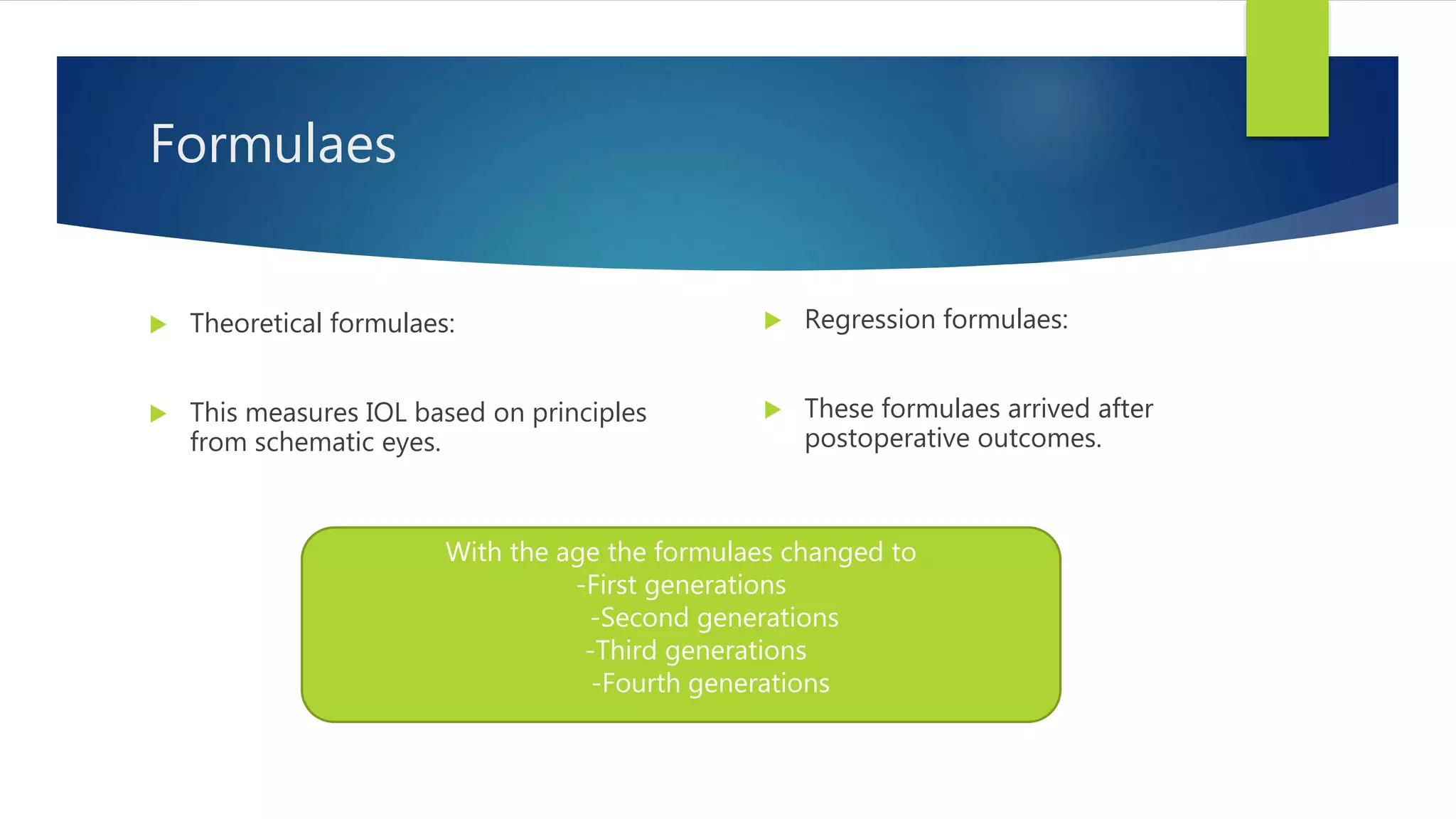
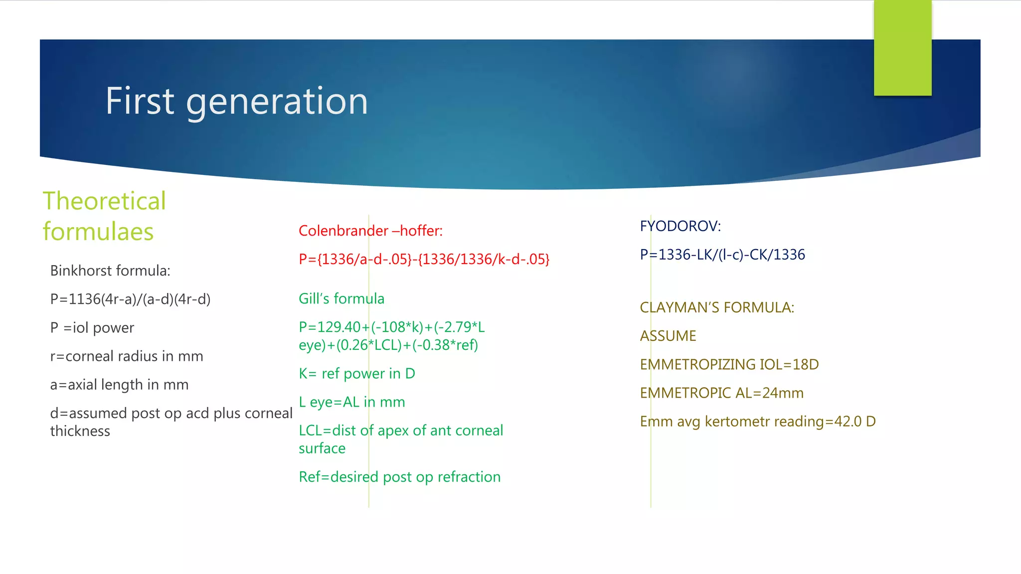
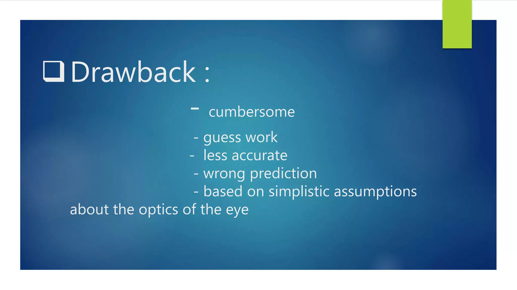
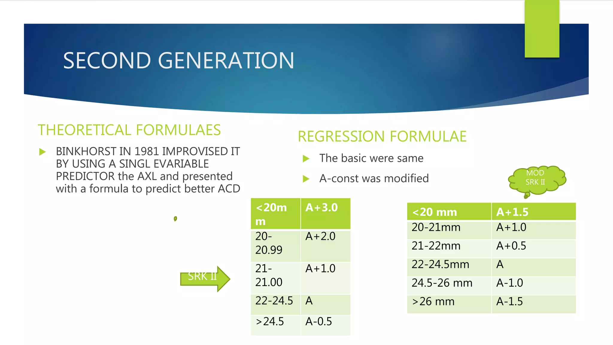
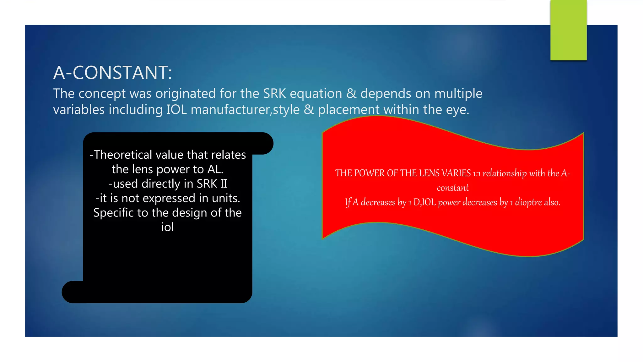
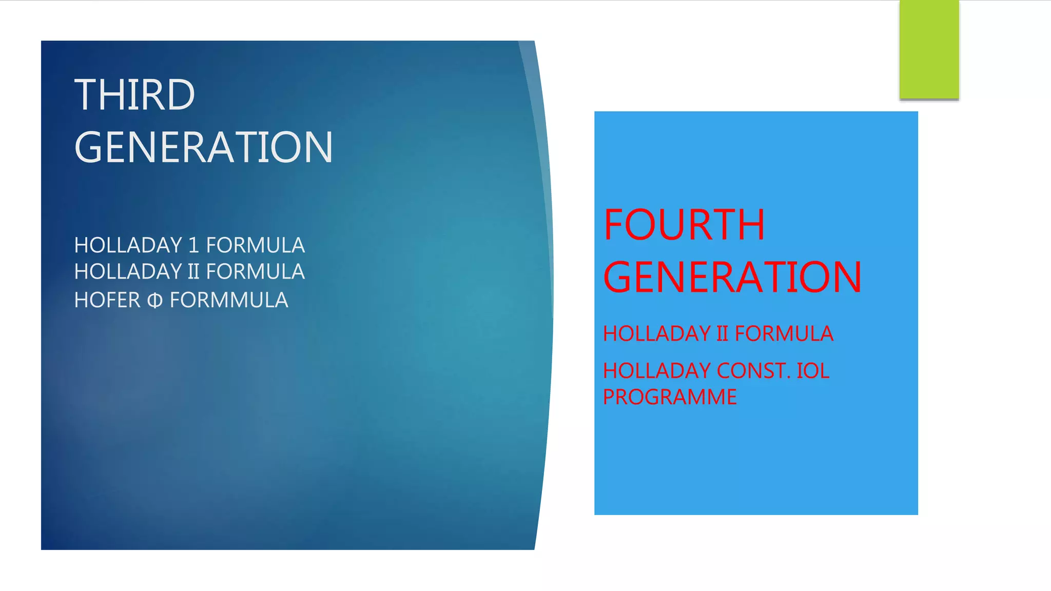
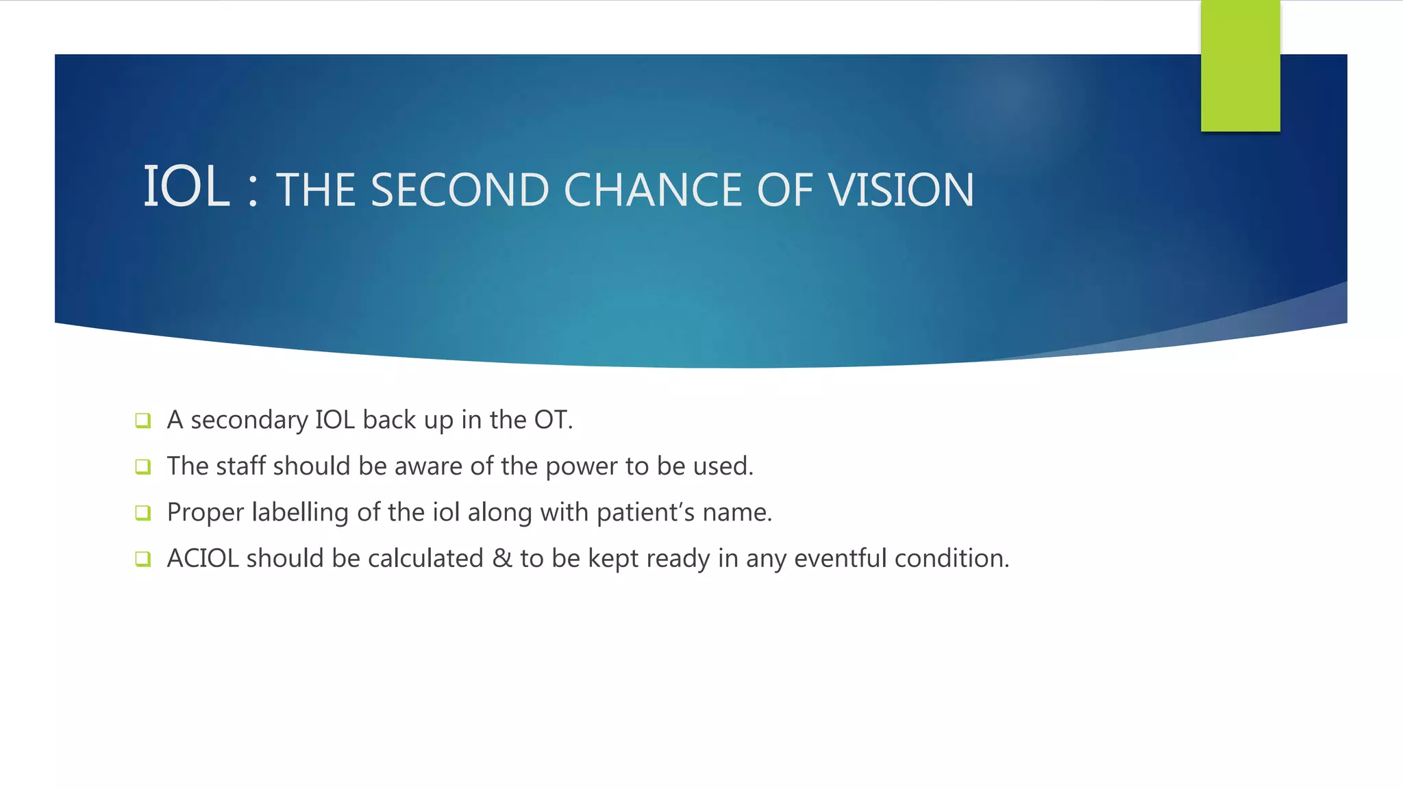
BIOMETRY
BY
DR.NIKITA JAISWAL
1ST YEAR RESIDENT
IMS & SUM HOSPITAL
Biometry :The analysis of
biological data using
mathematical &
statistical methods.
Glossary
 Measurement of axial length
 Keratometry{ measurement of k reading}
 Iol power calculation
A-SCAN
 Axial length-it is the distance between the anterior surface of the cornea and the
fovea.
 A-scan is used to measure axial length.
 It measures the time required for a sound pulse to travel from the cornea to retina.
 Normal axial length of an eye is –----22-25mm
 1 mm error of IOL = 2.88 D 0r 3.0 D of IOL POWER
Ultrasonography
 Crystal oscillates—high frequency sound wave penetrates into eye—sound wave
encounters a media interface—part of the sound wave is reflected back to the
probe
 USG doesn’t measure the distance rather it measures the TIME required for a sound
pulse to travel from cornea to retina.
cornea 1620m/s
Anterior
chamber
1532m/s
Lens thickness 1641m/s
Vitreous cavity 1532m/s
1555m/s
Types of a scan {methods of
measurement}
1- ultrasonic measurement –this comprises of –applanationn method
immersion technique
2- optical measurement –this uses partial coherence laser ,the iol master measures
time required for infrared light to travel to the retina & this technique doesn’t
requires contact with the globe. {iol master}
Applanation
Things required:
A scan machine
Probe
Anaesthetic drops
Chair
Patient
Procedure:
 Anaesthetize the eye
 Touch the probe
 “DO NOT PRESS OVER THE CORNEA”
 NOTE THE READING
 a=initial spike(probe tip& cornea)
 b=ant lens capsule
 c=post lens capsule
 d=retina
 e=sclera
 f=orbital fat
Immersion A scan biometry
 The ultrasonic beam is coupled to the eye through fluid.
 Because of no corneal compression the results display true axial length.
 Procedure:pt lies supine
looking up at the ceiling
scleral shell is placed between the eyelids centered over the
cornea.
Scleral shell
Filled with 40-60
mixture of goniosol &
dacriose & the probe
tip into the solution
Align the ultrasound
with macula by asking
the pt to look to the
fixation light of the
probe.
Reading
a-probe tip
b- cornea- double peaked
echo will show both ant &
post surfaces
c-anterior lens capsule
d-posterior lens capsule
e- retina this echo needs to
havre sharp 90’ take off from
baseline
f-sclera
g-orbital fat
Essentials :
 Calibration
 Gain or sensitivity
 Sound velocity
Calibration: it is done with the help of model eye.
Required time to time
Instructions are specific and the model eye is provided
with the manufacturers
Gain/sensitivity:
 Gain = electronic amplification of the sound waves received by the transducer …{
is called as a decibel(db)}
• Normal is 70% in most of the
biometers
Normal
• When the echo height is inadequate.
• High myopia,dense cataracts,ocular opacities
Increase
• When artifacts are seen
• Eg: silicone oil, pseudophakic eye
decrease
Features of a good scan:
 One dimensional image in which spikes of variable hts are seen:
 Cornea: single tall peak
 Aqueous chamber: does not produce any echo
 Ant & post lens capsule : produce tall echoes
 Vitreous: no to few echoes
 Retina: tall,sharp echoes with staircase at the origin
 Orbital fat produces : medium to low echoes
Keratometry : It is used to measure the corneal
curvature.
In the optical area i.e 2-3 mm
History :
Helmholtz—javal & schiotz---Bausch & laumb{reichert}
{1854} {1881} {1932}
}}}]}}
HELMHOLTZ: This keratometer consist of two plates which
displaces the image through half of its length & the total displacement
gives the size of image.
Javal schiotz
Javal schiotz- This is on the principle of “variable object size
constant image size”
The object
Objective lens & doubling prism
The eyepiece lens
The object: 2 mires A & B mounted on an arc
A+B= object thus of variable size.
“Stepped mire
rectangular mire”
--divided horizontally through the center.
Image of two mires=object.
Bausch & laumb
keratometer (1932)
“constant object size
variable image size”
B & L:- THE OBJECT {CIRCULAR MIRE}
The imag eof the circular mires appears on the patients cornea
appears diminished & serves as object.
The objective lens : from the image of the mire (new object)
DIAPHRAGM & DOUBLING PRISMS: -A 4 APERTURE DIAPHRAGM
-A 2 DOUBLING PRISMS{ONE WITH
BASE UP AND ONE WITH BASE OUT}
moved independently
parallel to the central axis
UNIQUE– Image doubling mechanism is unique
image is produced side by side or at 90’
thus also k/as “one position keratometer”
Procedure:
Instrument:calibration is done with the a steel ball along with the
machine with a known radius of curvature mires formed correctly machine is
calibrated.
PATIENT: chin on chin rest
head on head rest h
occlude occludes the non examining eye
patients pupil & projective knob at the same level
Focusing of mire:center of cornea
patients view
examiners view
Reading:
perfectly spherical
mires.
Oval mires
horizontal-with the rule astig
Vertical –against with the rule
Keratoconus:irregular
Jumping of mires
Wavy mires
Uses:
--It helps to assess the radius of curvature of cornea.
--To monitor the shape of cornea in keratoglobus & keratoconus.
--The K readings have been taken to measure the iol power with axial
length by SRK formula.
Limitations:
refractive status of very small central area of cornea is
measured{3-4 mm}
It loses its accuracy when measuring very flat and steep
cornea.
Small corneal irregularities would preclude the use due
to irregular astigmatism.
Sources of error:
Improper calibration
Position of the patient at fault
Any corneal pathology
Examiners fault
Any lid position abnormality
tearing.
Recent advances :
automated keratometer
IOL calculation
Formulaes
 Theoretical formulaes:
 This measures IOL based on principles
from schematic eyes.
 Regression formulaes:
 These formulaes arrived after
postoperative outcomes.
With the age the formulaes changed to
-First generations
-Second generations
-Third generations
-Fourth generations
First generation
Theoretical
formulaes
Binkhorst formula:
P=1136(4r-a)/(a-d)(4r-d)
P =iol power
r=corneal radius in mm
a=axial length in mm
d=assumed post op acd plus corneal
thickness
Colenbrander –hoffer:
P={1336/a-d-.05}-{1336/1336/k-d-.05}
Gill’s formula
P=129.40+(-108*k)+(-2.79*L
eye)+(0.26*LCL)+(-0.38*ref)
K= ref power in D
L eye=AL in mm
LCL=dist of apex of ant corneal
surface
Ref=desired post op refraction
FYODOROV:
P=1336-LK/(l-c)-CK/1336
CLAYMAN’S FORMULA:
ASSUME
EMMETROPIZING IOL=18D
EMMETROPIC AL=24mm
Emm avg kertometr reading=42.0 D
Drawback :
- cumbersome
- guess work
- less accurate
- wrong prediction
- based on simplistic assumptions
about the optics of the eye
Regression formula
 SRK 1 (sanders ,retzlaff & kraff)
 a breakthrough in calculating iol: they analysed the post op results and found that the
theoretical formulaes can be odified
 They replaced ACD with A constant which was unique for diff types of iols
 P=A-2.5L-0.9K
 P= IOL POWER
 A=A CONSTANT
 L=AXIAL LENGTH
 K=AVERAGE KERATOMETRY IN DIOPTRES
SECOND GENERATION
THEORETICAL FORMULAES
 BINKHORST IN 1981 IMPROVISED IT
BY USING A SINGL EVARIABLE
PREDICTOR the AXL and presented
with a formula to predict better ACD
REGRESSION FORMULAE
 The basic were same
 A-const was modified
<20m
m
A+3.0
20-
20.99
A+2.0
21-
21.00
A+1.0
22-24.5 A
>24.5 A-0.5
<20 mm A+1.5
20-21mm A+1.0
21-22mm A+0.5
22-24.5mm A
24.5-26 mm A-1.0
>26 mm A-1.5
MOD
SRK II
SRK II
A-CONSTANT:
The concept was originated for the SRK equation & depends on multiple
variables including IOL manufacturer,style & placement within the eye.
-Theoretical value that relates
the lens power to AL.
-used directly in SRK II
-it is not expressed in units.
Specific to the design of the
iol
THE POWER OF THE LENS VARIES 1:1 relationship with the A-
constant
If A decreases by 1 D,IOL power decreases by 1 dioptre also.
THIRD
GENERATION
HOLLADAY 1 FORMULA
HOLLADAY II FORMULA
HOFER Φ FORMMULA
FOURTH
GENERATION
HOLLADAY II FORMULA
HOLLADAY CONST. IOL
PROGRAMME
FORMULAES AXIAL LENGTH
SRK1 22.0-24.5MM
HOFFER Q <24.5MM
SRK/ T >26.0MM
HOLLADAY I NORMAL as well as AL 24.5-26.0mm
4th generation More universal application
A recent study in 2011 showed
this formulaes on 8000 eyes
IOL : THE SECOND CHANCE OF VISION
 A secondary IOL back up in the OT.
 The staff should be aware of the power to be used.
 Proper labelling of the iol along with patient’s name.
 ACIOL should be calculated & to be kept ready in any eventful condition.
Biometry

Biometry

  • 1.
    BIOMETRY BY DR.NIKITA JAISWAL 1ST YEARRESIDENT IMS & SUM HOSPITAL
  • 2.
    Biometry :The analysisof biological data using mathematical & statistical methods.
  • 3.
    Glossary  Measurement ofaxial length  Keratometry{ measurement of k reading}  Iol power calculation
  • 4.
    A-SCAN  Axial length-itis the distance between the anterior surface of the cornea and the fovea.  A-scan is used to measure axial length.  It measures the time required for a sound pulse to travel from the cornea to retina.  Normal axial length of an eye is –----22-25mm  1 mm error of IOL = 2.88 D 0r 3.0 D of IOL POWER
  • 5.
    Ultrasonography  Crystal oscillates—highfrequency sound wave penetrates into eye—sound wave encounters a media interface—part of the sound wave is reflected back to the probe  USG doesn’t measure the distance rather it measures the TIME required for a sound pulse to travel from cornea to retina. cornea 1620m/s Anterior chamber 1532m/s Lens thickness 1641m/s Vitreous cavity 1532m/s 1555m/s
  • 6.
    Types of ascan {methods of measurement} 1- ultrasonic measurement –this comprises of –applanationn method immersion technique 2- optical measurement –this uses partial coherence laser ,the iol master measures time required for infrared light to travel to the retina & this technique doesn’t requires contact with the globe. {iol master}
  • 7.
    Applanation Things required: A scanmachine Probe Anaesthetic drops Chair Patient
  • 8.
    Procedure:  Anaesthetize theeye  Touch the probe  “DO NOT PRESS OVER THE CORNEA”  NOTE THE READING  a=initial spike(probe tip& cornea)  b=ant lens capsule  c=post lens capsule  d=retina  e=sclera  f=orbital fat
  • 9.
    Immersion A scanbiometry  The ultrasonic beam is coupled to the eye through fluid.  Because of no corneal compression the results display true axial length.  Procedure:pt lies supine looking up at the ceiling scleral shell is placed between the eyelids centered over the cornea.
  • 10.
    Scleral shell Filled with40-60 mixture of goniosol & dacriose & the probe tip into the solution Align the ultrasound with macula by asking the pt to look to the fixation light of the probe.
  • 11.
    Reading a-probe tip b- cornea-double peaked echo will show both ant & post surfaces c-anterior lens capsule d-posterior lens capsule e- retina this echo needs to havre sharp 90’ take off from baseline f-sclera g-orbital fat
  • 12.
    Essentials :  Calibration Gain or sensitivity  Sound velocity
  • 13.
    Calibration: it isdone with the help of model eye. Required time to time Instructions are specific and the model eye is provided with the manufacturers
  • 14.
    Gain/sensitivity:  Gain =electronic amplification of the sound waves received by the transducer …{ is called as a decibel(db)} • Normal is 70% in most of the biometers Normal • When the echo height is inadequate. • High myopia,dense cataracts,ocular opacities Increase • When artifacts are seen • Eg: silicone oil, pseudophakic eye decrease
  • 15.
    Features of agood scan:  One dimensional image in which spikes of variable hts are seen:  Cornea: single tall peak  Aqueous chamber: does not produce any echo  Ant & post lens capsule : produce tall echoes  Vitreous: no to few echoes  Retina: tall,sharp echoes with staircase at the origin  Orbital fat produces : medium to low echoes
  • 16.
    Keratometry : Itis used to measure the corneal curvature. In the optical area i.e 2-3 mm
  • 17.
    History : Helmholtz—javal &schiotz---Bausch & laumb{reichert} {1854} {1881} {1932} }}}]}}
  • 18.
    HELMHOLTZ: This keratometerconsist of two plates which displaces the image through half of its length & the total displacement gives the size of image.
  • 19.
  • 20.
    Javal schiotz- Thisis on the principle of “variable object size constant image size” The object Objective lens & doubling prism The eyepiece lens
  • 22.
    The object: 2mires A & B mounted on an arc A+B= object thus of variable size. “Stepped mire rectangular mire” --divided horizontally through the center. Image of two mires=object.
  • 24.
    Bausch & laumb keratometer(1932) “constant object size variable image size”
  • 25.
    B & L:-THE OBJECT {CIRCULAR MIRE} The imag eof the circular mires appears on the patients cornea appears diminished & serves as object. The objective lens : from the image of the mire (new object)
  • 26.
    DIAPHRAGM & DOUBLINGPRISMS: -A 4 APERTURE DIAPHRAGM -A 2 DOUBLING PRISMS{ONE WITH BASE UP AND ONE WITH BASE OUT} moved independently parallel to the central axis UNIQUE– Image doubling mechanism is unique image is produced side by side or at 90’ thus also k/as “one position keratometer”
  • 28.
    Procedure: Instrument:calibration is donewith the a steel ball along with the machine with a known radius of curvature mires formed correctly machine is calibrated. PATIENT: chin on chin rest head on head rest h occlude occludes the non examining eye patients pupil & projective knob at the same level
  • 29.
    Focusing of mire:centerof cornea patients view examiners view
  • 30.
    Reading: perfectly spherical mires. Oval mires horizontal-withthe rule astig Vertical –against with the rule Keratoconus:irregular Jumping of mires Wavy mires
  • 32.
    Uses: --It helps toassess the radius of curvature of cornea. --To monitor the shape of cornea in keratoglobus & keratoconus. --The K readings have been taken to measure the iol power with axial length by SRK formula.
  • 33.
    Limitations: refractive status ofvery small central area of cornea is measured{3-4 mm} It loses its accuracy when measuring very flat and steep cornea. Small corneal irregularities would preclude the use due to irregular astigmatism.
  • 34.
    Sources of error: Impropercalibration Position of the patient at fault Any corneal pathology Examiners fault Any lid position abnormality tearing.
  • 35.
  • 36.
  • 37.
    Formulaes  Theoretical formulaes: This measures IOL based on principles from schematic eyes.  Regression formulaes:  These formulaes arrived after postoperative outcomes. With the age the formulaes changed to -First generations -Second generations -Third generations -Fourth generations
  • 38.
    First generation Theoretical formulaes Binkhorst formula: P=1136(4r-a)/(a-d)(4r-d) P=iol power r=corneal radius in mm a=axial length in mm d=assumed post op acd plus corneal thickness Colenbrander –hoffer: P={1336/a-d-.05}-{1336/1336/k-d-.05} Gill’s formula P=129.40+(-108*k)+(-2.79*L eye)+(0.26*LCL)+(-0.38*ref) K= ref power in D L eye=AL in mm LCL=dist of apex of ant corneal surface Ref=desired post op refraction FYODOROV: P=1336-LK/(l-c)-CK/1336 CLAYMAN’S FORMULA: ASSUME EMMETROPIZING IOL=18D EMMETROPIC AL=24mm Emm avg kertometr reading=42.0 D
  • 39.
    Drawback : - cumbersome -guess work - less accurate - wrong prediction - based on simplistic assumptions about the optics of the eye
  • 40.
    Regression formula  SRK1 (sanders ,retzlaff & kraff)  a breakthrough in calculating iol: they analysed the post op results and found that the theoretical formulaes can be odified  They replaced ACD with A constant which was unique for diff types of iols  P=A-2.5L-0.9K  P= IOL POWER  A=A CONSTANT  L=AXIAL LENGTH  K=AVERAGE KERATOMETRY IN DIOPTRES
  • 41.
    SECOND GENERATION THEORETICAL FORMULAES BINKHORST IN 1981 IMPROVISED IT BY USING A SINGL EVARIABLE PREDICTOR the AXL and presented with a formula to predict better ACD REGRESSION FORMULAE  The basic were same  A-const was modified <20m m A+3.0 20- 20.99 A+2.0 21- 21.00 A+1.0 22-24.5 A >24.5 A-0.5 <20 mm A+1.5 20-21mm A+1.0 21-22mm A+0.5 22-24.5mm A 24.5-26 mm A-1.0 >26 mm A-1.5 MOD SRK II SRK II
  • 42.
    A-CONSTANT: The concept wasoriginated for the SRK equation & depends on multiple variables including IOL manufacturer,style & placement within the eye. -Theoretical value that relates the lens power to AL. -used directly in SRK II -it is not expressed in units. Specific to the design of the iol THE POWER OF THE LENS VARIES 1:1 relationship with the A- constant If A decreases by 1 D,IOL power decreases by 1 dioptre also.
  • 43.
    THIRD GENERATION HOLLADAY 1 FORMULA HOLLADAYII FORMULA HOFER Φ FORMMULA FOURTH GENERATION HOLLADAY II FORMULA HOLLADAY CONST. IOL PROGRAMME
  • 44.
    FORMULAES AXIAL LENGTH SRK122.0-24.5MM HOFFER Q <24.5MM SRK/ T >26.0MM HOLLADAY I NORMAL as well as AL 24.5-26.0mm 4th generation More universal application A recent study in 2011 showed this formulaes on 8000 eyes
  • 45.
    IOL : THESECOND CHANCE OF VISION  A secondary IOL back up in the OT.  The staff should be aware of the power to be used.  Proper labelling of the iol along with patient’s name.  ACIOL should be calculated & to be kept ready in any eventful condition.