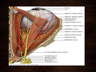
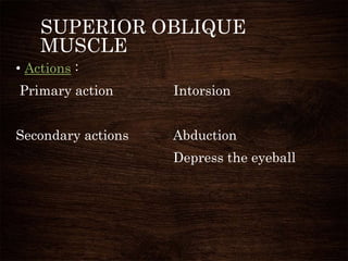
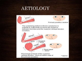
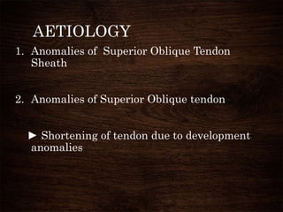
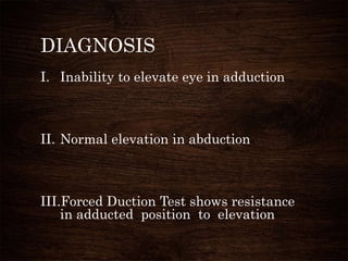
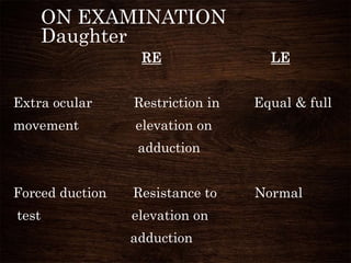
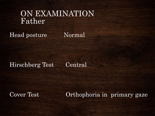
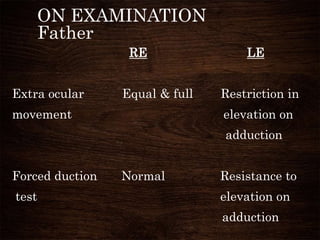
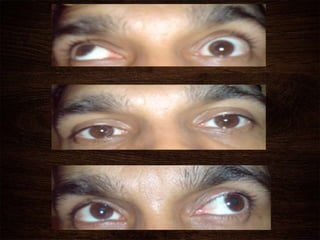
HEREDITARY
BROWN
SYNDROME
DR. JOSEPH KURIAN
PG STUDENT , ACME
PARIYARAM
DEFINITION
• Brown syndrome is a disorder of elevation
involving mechanical restriction of extra
ocular movement.
• Brown was the first to describe this
anomaly
• He suspected a congenitally short superior
oblique tendon sheath to be the cause
hence
“superior oblique tendon sheath syndrome”
INCIDENCE
• Incidence is low –
6 in 2583 (Croswell & Haldi )
• Familial occurrence is rare
2%(Birgit Lorenz, Michael C. Brodsky)
• Usually unilateral but 10% is bilateral
• Syndrome can be : Congenital
Acquired
Constant
Intermittent
SUPERIOR OBLIQUE
MUSCLE
• Origin : Annulus of Zinn at the orbital apex,
medial to optic canal
• Insertion :Outer posterior quadrant of the
eyeball (insertion line 11mm)
• Artery : Lateral muscular branch of the
ophthalmic artery
• Nerve : Trochlear nerve
SUPERIOR OBLIQUE
MUSCLE
• Actions :
Primary action Intorsion
Secondary actions Abduction
Depress the eyeball
AETIOLOGY
AETIOLOGY
1. Anomalies of Superior Oblique Tendon
Sheath
2. Anomalies of Superior Oblique tendon
► Shortening of tendon due to development
anomalies
AETIOLOGY
3. Anomalies of Inferior oblique & adjacent
structures
► Dense attachments causing restriction of
action
4. Impaired slippage of tendon through Trochlea
► Anomalies of Trochlea
►Retro bulbar thickening of tendon
AETIOLOGY
5. Acquired causes
► Post operative after superior oblique
tucking surgery, scleral buckling & Molteno
implant
► Trauma to orbital roof
► Inflammatory conditions like Rheumatoid
arthritis
CLINICAL FEATURES
CONSISTENT
• Absence of elevation in adduction
• Normal elevation in abduction
• Forced duction test shows restriction in
adduction
CLINICAL FEATURES
VARIABLE
• Mild limitation of elevation in primary gaze
• Down shoot in adduction
• Widening of palpebral fissure
• Hypotropia in primary position
• Compensatory head posture>Head Tilt ,Chin lift
DIAGNOSIS
I. Inability to elevate eye in adduction
II. Normal elevation in abduction
III.Forced Duction Test shows resistance
in adducted position to elevation
DIFFERENTIAL
DIAGNOSIS
i. Paralysis of Inferior oblique muscle
ii. Congenital fibrosis of inferior rectus
muscle
iii. Grave’s ophthalmopathy
CASE HISTORY
8yr old girl presented to our ophthal OPD .
Her mother had noticed squinting in the RE
in certain gaze positions. No complaints of
diplopia .
Family History: Father gives history of squint
in LE. He is a diagnosed case of Browns
syndrome in LE.
ON EXAMINATION
Daughter
Head posture Normal
Hirschberg Test Central
Cover Test Orthophoria in primary gaze
ON EXAMINATION
Daughter
RE LE
Extra ocular Restriction in Equal & full
movement elevation on
adduction
Forced duction Resistance to Normal
test elevation on
adduction
ON EXAMINATION
Father
Head posture Normal
Hirschberg Test Central
Cover Test Orthophoria in primary gaze
ON EXAMINATION
Father
RE LE
Extra ocular Equal & full Restriction in
movement elevation on
adduction
Forced duction Normal Resistance to
test elevation on
adduction
TREATMENT
INDICATIONS:
i. Involved eye hypotropic in primary gaze
position
ii. Significant anomaly in head posture
Head tilt towards affected side
Chin lift
TREATMENT
PROCEDURES
 Complete Tenectomy of Superior Oblique muscle.
* Forced duction test becomes negative
* In case of symptoms of superior oblique
paralysis consider recession of contra lateral IR
muscle or ipsilateral IO muscle.
TREATMENT
Silicon superior oblique tendon expanders to
lengthen tendon
* Preferred procedure
* Advantage: No symptoms of SO palsy
REFERENCES
• BINOCULAR VISION & OCULAR
MOTILITY 5th edition by Gunter K von
Noorden
• DELHI JOURNAL OF
OPHTHALMOLOGY vol 22
• KANSKI BOWLING 7th edition
THANK YOU

Hereditary brown syndrome

  • 1.
  • 2.
    DEFINITION • Brown syndromeis a disorder of elevation involving mechanical restriction of extra ocular movement. • Brown was the first to describe this anomaly • He suspected a congenitally short superior oblique tendon sheath to be the cause hence “superior oblique tendon sheath syndrome”
  • 3.
    INCIDENCE • Incidence islow – 6 in 2583 (Croswell & Haldi ) • Familial occurrence is rare 2%(Birgit Lorenz, Michael C. Brodsky) • Usually unilateral but 10% is bilateral
  • 4.
    • Syndrome canbe : Congenital Acquired Constant Intermittent
  • 5.
    SUPERIOR OBLIQUE MUSCLE • Origin: Annulus of Zinn at the orbital apex, medial to optic canal • Insertion :Outer posterior quadrant of the eyeball (insertion line 11mm) • Artery : Lateral muscular branch of the ophthalmic artery • Nerve : Trochlear nerve
  • 8.
    SUPERIOR OBLIQUE MUSCLE • Actions: Primary action Intorsion Secondary actions Abduction Depress the eyeball
  • 9.
  • 10.
    AETIOLOGY 1. Anomalies ofSuperior Oblique Tendon Sheath 2. Anomalies of Superior Oblique tendon ► Shortening of tendon due to development anomalies
  • 11.
    AETIOLOGY 3. Anomalies ofInferior oblique & adjacent structures ► Dense attachments causing restriction of action 4. Impaired slippage of tendon through Trochlea ► Anomalies of Trochlea ►Retro bulbar thickening of tendon
  • 12.
    AETIOLOGY 5. Acquired causes ►Post operative after superior oblique tucking surgery, scleral buckling & Molteno implant ► Trauma to orbital roof ► Inflammatory conditions like Rheumatoid arthritis
  • 13.
    CLINICAL FEATURES CONSISTENT • Absenceof elevation in adduction • Normal elevation in abduction • Forced duction test shows restriction in adduction
  • 14.
    CLINICAL FEATURES VARIABLE • Mildlimitation of elevation in primary gaze • Down shoot in adduction • Widening of palpebral fissure • Hypotropia in primary position • Compensatory head posture>Head Tilt ,Chin lift
  • 15.
    DIAGNOSIS I. Inability toelevate eye in adduction II. Normal elevation in abduction III.Forced Duction Test shows resistance in adducted position to elevation
  • 16.
    DIFFERENTIAL DIAGNOSIS i. Paralysis ofInferior oblique muscle ii. Congenital fibrosis of inferior rectus muscle iii. Grave’s ophthalmopathy
  • 17.
    CASE HISTORY 8yr oldgirl presented to our ophthal OPD . Her mother had noticed squinting in the RE in certain gaze positions. No complaints of diplopia . Family History: Father gives history of squint in LE. He is a diagnosed case of Browns syndrome in LE.
  • 18.
    ON EXAMINATION Daughter Head postureNormal Hirschberg Test Central Cover Test Orthophoria in primary gaze
  • 19.
    ON EXAMINATION Daughter RE LE Extraocular Restriction in Equal & full movement elevation on adduction Forced duction Resistance to Normal test elevation on adduction
  • 21.
    ON EXAMINATION Father Head postureNormal Hirschberg Test Central Cover Test Orthophoria in primary gaze
  • 22.
    ON EXAMINATION Father RE LE Extraocular Equal & full Restriction in movement elevation on adduction Forced duction Normal Resistance to test elevation on adduction
  • 24.
    TREATMENT INDICATIONS: i. Involved eyehypotropic in primary gaze position ii. Significant anomaly in head posture Head tilt towards affected side Chin lift
  • 25.
    TREATMENT PROCEDURES  Complete Tenectomyof Superior Oblique muscle. * Forced duction test becomes negative * In case of symptoms of superior oblique paralysis consider recession of contra lateral IR muscle or ipsilateral IO muscle.
  • 26.
    TREATMENT Silicon superior obliquetendon expanders to lengthen tendon * Preferred procedure * Advantage: No symptoms of SO palsy
  • 27.
    REFERENCES • BINOCULAR VISION& OCULAR MOTILITY 5th edition by Gunter K von Noorden • DELHI JOURNAL OF OPHTHALMOLOGY vol 22 • KANSKI BOWLING 7th edition
  • 28.