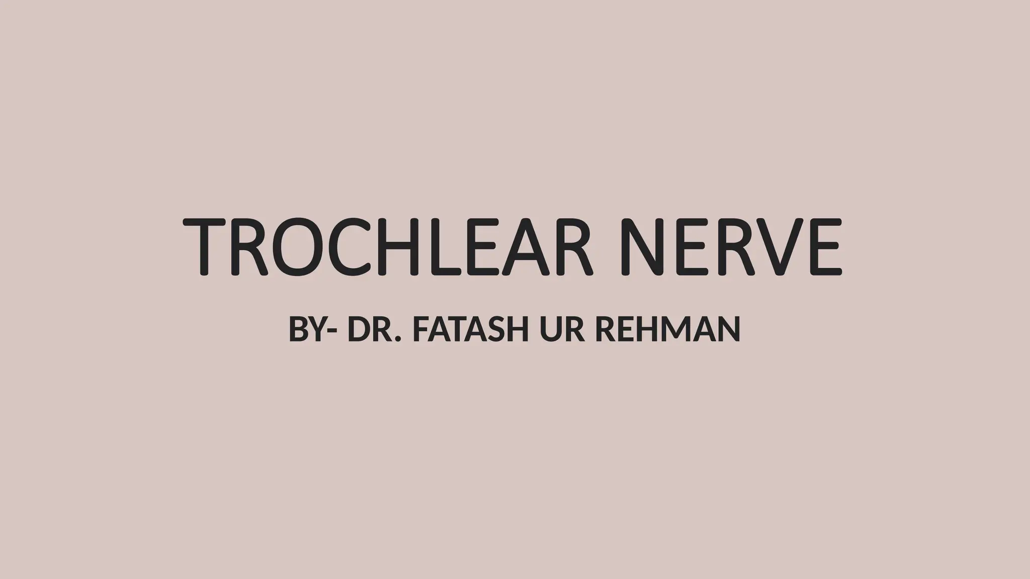
introduction
• 4TH
cranial nerve
•Motor in function, supplies only
superior oblique
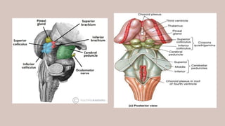
• Only cranial nerve that arises from
the dorsal aspect of the brain
• Only cranial nerve to cross
completely to the other side (arises
from the contralateral nucleus)
• Longest and thinnest of all cranial
nerves
3.
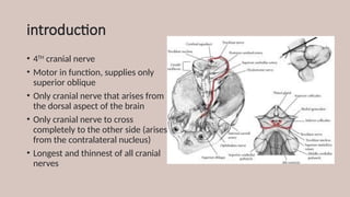
FUNCTIONAL COMPONENTS
• SOMATICEFFERENT- movement of the eyeball through superior
oblique muscle
• GENERAL SOMATIC AFFERENT- proprioceptive impulses from superior
oblique relayed in mesencephalic nucleus of trigeminal nerve
4.
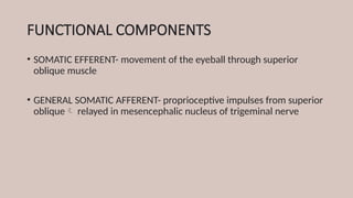
NUCLEUS
• Midbrain- Ventromedialpart of
central gray matter at the level
of inferior colliculus
• Caudal to and continuous with
3rd
nerve nucleus complex
• Belongs to somatic efferent
column of nuclei and closely
related to medial longitudinal
bundle
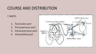
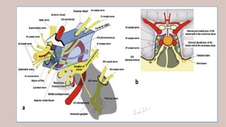
COURSE AND DISTRIBUTION
•PARTS
1. Fascicular part
2. Precavernous part
3. Intracavernous part
4. Intraorbital part
8.
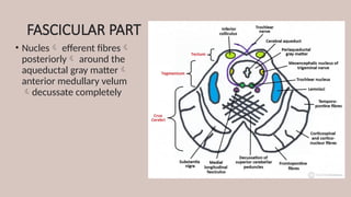
FASCICULAR PART
• Nuclesefferent fibres
posteriorly around the
aqueductal gray matter
anterior medullary velum
decussate completely
9.
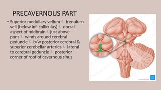
PRECAVERNOUS PART
• Superiormedullary vellum frenulum
veli (below inf. colliculus) dorsal
aspect of midbrainjust above
pons winds around cerebral
peduncle b/w posterior cerebral &
superior cerebellar arteries lateral
to cerebral peduncle posterior
corner of roof of cavernous sinus
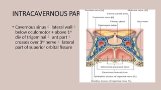
11.
INTRACAVERNOUS PART
• Cavernoussinus lateral wall
below oculomotor + above 1st
div of trigeminal ant part
crosses over 3rd
nerve lateral
part of superior orbital fissure
13.
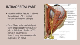
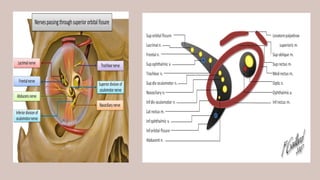
INTRAORBITAL PART
• Superiororbital fissure above
the origin of LPS orbital
surface of superior oblique
• Extra fibres in intraorbital part
carry proprioceptive impulses
join ophthalmic division of 5th
nerve in cavernouos
sinusrelay in mesencephalic
nucleus of 5th
nerve
15.
SUPERIOR OBLIQUE MUSCLE
•PRIMARY POSITION OF GAZE
• Primary action- INTORSION (along A-P axis)
• Secondary action- DEPRESSION (along horizontal axis)
• Tertiary action- ABDUCTION ( along vertical axis)
• GLOBE 51° ADDUCTED
• Axis of muscle rotation coincides with optical axis
• DEPRESSION only
• GLOBE 39° ABDUCTED
• Optical axis and line of pull of muscle mae an angle of
90°
• INTORSION only
16.
4th
NERVE PARALYSIS
1. CONGENITAL-40%
2. TRAUMA- 34%
• Usually bilateral
• Impact on anterior medullary velum at decussation
3. IDIOPATHIC- 20%
4. VASCULAR AND NEUROGENIC- 3-5%
• in older individuals microvasculopathy secondary to diabetes
atherosclerosis or hypertension
• Aneyrusms and tumor
• Ocular myasthenia- isolated unilateral
• SO palsy is most common form of paralytic squint
17.
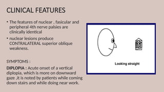
CLINICAL FEATURES
• Thefeatures of nuclear , fasicular and
peripheral 4th nerve palsies are
clinically identical
• nuclear lesions produce
CONTRALATERAL superior oblique
weakness.
SYMPTOMS :
DIPLOPIA : Acute onset of a vertical
diplopia, which is more on downward
gaze ,it is noted by patients while coming
down stairs and while doing near work.
18.
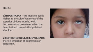
SIGNS :
1)HYPERTROPIA –the involved eye is
higher as a result of weakness of the
superior oblique muscle, which
becomes more prominent when the
head is tilted towards the ipsilateral
shoulder
2)RESTRICTED OCULAR MOVEMENTS:
there is limitation of depression on
adduction.
19.
3)ABNORMAL HEAD POSTURE:
toavoid diplopia ,head takes a
posture towards the action of the
superior oblique muscle, face is
slightly turned to the opposite side,
chin is depressed, and head is tilted
towards the opposite side.
HEAD IS TILTED TO THE RIGHT.
FACE IS TURNED TO THE RIGHT.
CHIN IS DEPRESSED
20.
UNILATERAL PALSY BILATERALPALSY
EXOTROPIA IN
DOWNWARD GAZE
little V-pattern exotrpia
TORSION (double
Maddox rod test)
Excyclodeviation <10° Excyclodeviation > 10°
DUCTIONS Normal or diminished diminished
HEAD TILT TEST Positive for involved eye
Hypertropia increases on
tilting head towards
ipsilateral shoulder
Tilting to either side
increases hypertropia
21.
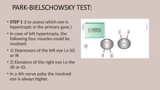
PARK-BIELSCHOWSKY TEST:
• STEP1 :( to assess which eye is
hypertropic in the primary gaze.)
• In case of left hypertropia, the
following four muscles could be
involved:
• 1) Depressors of the left eye i.e SO
or IR
• 2) Elevators of the right eye i.e the
SR or IO.
• In a 4th nerve palsy the involved
eye is always higher.
22.
• STEP 2: (which lateral direction has
worse hypertropia )
• If the left hypertropia increases on
right gaze implicates a left superior
oblique or right superior rectus
involvement.
• Increase in the left gaze implicates that
either the right inferior oblique or left
inferior rectus are involved.
• In 4th nerve palsy the deviation IS
WORSE ON OPPOSITE GAZE . (WOOG)
23.
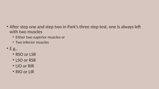
• After stepone and step two in Park’s three step test, one is always left
with two muscles
• Either two superior muscles or
• Two inferior muscles
• E.g.,
• RSO or LSR
• LSO or RSR
• LIO or RIR
• RIO or LIR
24.
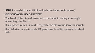
• STEP 3:( in which head tilt direction is the hypertropia worse )
• BIELSCHOWSKY HEAD TILT TEST
• The head tilt test is performed with the patient fixating at a straight
ahead target at 3 mts.
• If a superior muscle is weak, HT greater on tilt toward involved muscle
• If an inferior muscle is weak, HT greater on head tilt opposite involved
side
25.
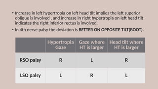
• Increase inleft hypertropia on left head tilt implies the left superior
oblique is involved , and increase in right hypertropia on left head tilt
indicates the right inferior rectus is involved.
• In 4th nerve palsy the deviation is BETTER ON OPPOSITE TILT(BOOT).
Hypertropia
Gaze
Gaze where
HT is larger
Head tilt where
HT is larger
RSO palsy R L R
LSO palsy L R L
26.
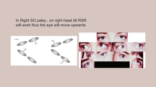
In Right SOpalsy , on right head tilt RSR
will work thus the eye will move upwards
28.
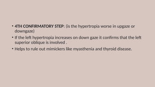
• 4TH CONFIRMATORYSTEP: (is the hypertropia worse in upgaze or
downgaze)
• If the left hypertropia increases on down gaze it confirms that the left
superior oblique is involved .
• Helps to rule out mimickers like myasthenia and thyroid disease.
29.
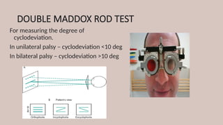
DOUBLE MADDOX RODTEST
For measuring the degree of
cyclodeviation.
In unilateral palsy – cyclodeviation <10 deg
In bilateral palsy – cyclodeviation >10 deg
31.
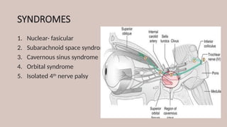
SYNDROMES
1. Nuclear- fasicular
2.Subarachnoid space syndrome
3. Cavernous sinus syndrome
4. Orbital syndrome
5. Isolated 4th
nerve palsy
32.
1. -NUCLEAR- FASICULAR
•Difficult to distinguish nuclear and fasicular lesions due to short course of
fascicles within midbrain
1. Haemorrhage
2. Infarction
3. Demyelination
4. Trauma
• Fascicular lesions may get contralateral Horner’s syndrome; and trauma
(especially near anterior medullary velum) may cause bilateral CN IV
palsies
• 3. CAVERNOUSSINUS SYNDROME
• Associated with 3rd
, 5th
, 6th
nerve palsies and ocular sympathetic
paralysis
35.
3. ORBITAL SYNDROME
•Trauma
• Infalmmation
• Tumors
• Seen in association with other cranial nerve palsies: 3rd
, 5th
, 6th
• Associated with
• Proptosis
• Chemosis
• Conjunctival congestion
36.
ISOLATED SUPERIOR OBLIQUEPALSY
• Most common etiologies are congenital and traumatic
1. CONGENITAL-
• large vertical fusion amplitude (10-15 prism diopters)
• FAT- family album tomography scan
2. ACQUIRED-
• In ischaemic conditions- diabetes, herpes zoster
37.
Differential Diagnosis ofVertical Binocular
Diplopia
• Superior Oblique Palsy
• Thyroid Ophthalmopathy
• Myasthenia Gravis
• Brown Syndrome
• Orbital fracture with entrapment
• Cyclovertical paresis or overaction
• Skew Deviation/Ocular Tilt
• Dissociated Vertical Deviation
38.
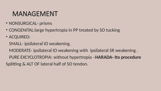
MANAGEMENT
• NONSURGICAL- prisms
•CONGENITAL:large hypertropia in PP treated by SO tucking
• ACQUIRED:
SMALL- ipsilateral IO weakening.
MODERATE- ipsilateral IO weakening with ipsilateral SR weakening .
PURE EXCYCLOTROPIA: without hypertropia –HARADA- Ito procedure
Splitting & ALT OF lateral half of SO tendon.