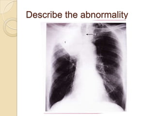
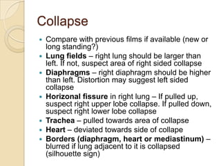
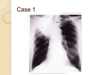
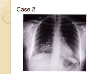
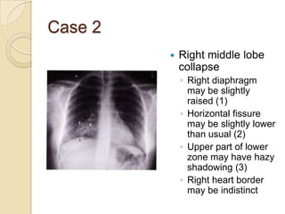
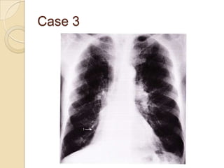
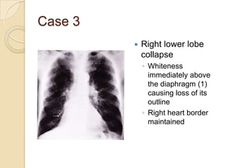
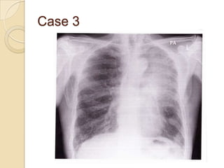
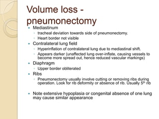
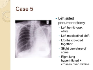
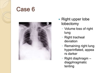
This document describes how to identify different types of abnormalities on a chest radiograph, including lung collapse, pneumonectomy, and mediastinal masses. It provides examples of specific abnormalities like right upper lobe collapse, left lower lobe collapse, and left-sided pneumonectomy. For each abnormality, it identifies key radiographic findings such as changes in lung volumes, diaphragm or heart positioning, tracheal deviation, and silhouette signs. The goal is to recognize changes that indicate different pathologies based on comparison to normal anatomy.


![Imaging in opaqe hemithorax [autosaved]](https://cdn.slidesharecdn.com/ss_thumbnails/imaginginopaqehemithoraxautosaved-161030071708-thumbnail.jpg?width=640&height=640&fit=bounds)


