Introduction
• • Lungconsolidation and collapse are
common radiological findings.
• • Understanding their features is crucial for
diagnosis and management.
• • This session will cover:
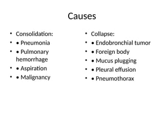
• – Definitions
• – Causes
• – Radiological features
• – Case examples with X-rays
3.
Definition: Consolidation
• •Consolidation refers to alveolar air replaced
by:
• – Fluid (e.g., pneumonia)
• – Pus (e.g., abscess)
• – Blood (e.g., pulmonary hemorrhage)
• – Cells (e.g., malignancy)
• • Radiologically appears as increased lung
opacity.
4.
Definition: Collapse (Atelectasis)
•• Collapse (atelectasis) is loss of lung volume
due to airway obstruction or compression.
• • Types:
• – Obstructive (e.g., tumor, mucus plug)
• – Compressive (e.g., pleural effusion,
pneumothorax)
• • Radiologically seen as displacement of
structures and increased density.
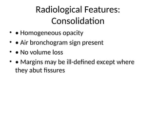
Radiological Features:
Consolidation
• •Homogeneous opacity
• • Air bronchogram sign present
• • No volume loss
• • Margins may be ill-defined except where
they abut fissures
7.
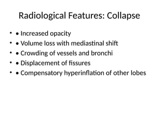
Radiological Features: Collapse
•• Increased opacity
• • Volume loss with mediastinal shift
• • Crowding of vessels and bronchi
• • Displacement of fissures
• • Compensatory hyperinflation of other lobes
8.
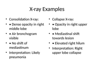
X-ray Examples
• ConsolidationX-ray:
• • Dense opacity in right
middle lobe
• • Air bronchogram
visible
• • No shift of
mediastinum
• Interpretation: Likely
pneumonia
• Collapse X-ray:
• • Opacity in right upper
lobe
• • Mediastinal shift
towards lesion
• • Elevated right hilum
• Interpretation: Right
upper lobe collapse
9.
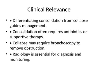
Clinical Relevance
• •Differentiating consolidation from collapse
guides management.
• • Consolidation often requires antibiotics or
supportive therapy.
• • Collapse may require bronchoscopy to
remove obstruction.
• • Radiology is essential for diagnosis and
monitoring.
10.
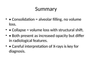
Summary
• • Consolidation= alveolar filling, no volume
loss.
• • Collapse = volume loss with structural shift.
• • Both present as increased opacity but differ
in radiological features.
• • Careful interpretation of X-rays is key for
diagnosis.
11.
Consolidation - InsertImage (Slide:
Radiological Features)
• Please insert a chest X-ray image showing consolidation here.
Recommended open-access images (download & insert):
1) X-ray lung consolidation — Wikimedia Commons (CC BY 2.0):
https://commons.wikimedia.org/wiki/File:X-ray_lung_consolidation.jpg
2) Pneumonia x-ray — Wikimedia Commons (public/CC):
https://commons.wikimedia.org/wiki/File:Pneumonia_x-ray.jpg
Suggested caption: 'Right middle lobe consolidation with air bronchograms. Source: Wikimedia Commons
(CC).'
After inserting the image, add a short interpretation: e.g., 'Air bronchograms present, no volume loss —
favors consolidation (pneumonia)'.
Right middle lobe consolidation with air bronchograms. Source: Wikimedia Commons (CC).
12.
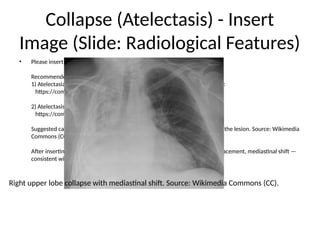
Collapse (Atelectasis) -Insert
Image (Slide: Radiological Features)
• Please insert a chest X-ray image showing lobar collapse here.
Recommended open-access images (download & insert):
1) Atelectasia1.jpg — Wikimedia Commons (Valencian Institute of Oncology):
https://commons.wikimedia.org/wiki/File:Atelectasia1.jpg
2) Atelectasis.png — Wikimedia Commons (CC BY-SA 4.0):
https://commons.wikimedia.org/wiki/File:Atelectasis.png
Suggested caption: 'Right upper lobe collapse with mediastinal shift towards the lesion. Source: Wikimedia
Commons (CC).'
After inserting the image, add interpretation: e.g., 'Volume loss, fissure displacement, mediastinal shift —
consistent with lobar collapse.'
Right upper lobe collapse with mediastinal shift. Source: Wikimedia Commons (CC).
13.
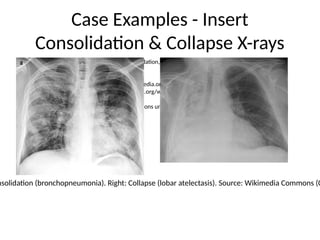
Case Examples -Insert
Consolidation & Collapse X-rays
• Insert two images side-by-side: (left) consolidation, (right) collapse.
Recommended images:
Left (consolidation): https://commons.wikimedia.org/wiki/File:X-ray_of_bronchopneumonia.png
Right (collapse): https://commons.wikimedia.org/wiki/File:Atelectasia1.jpg
Add boxed labels and brief bullet interpretations underneath each image.
nsolidation (bronchopneumonia). Right: Collapse (lobar atelectasis). Source: Wikimedia Commons (C


![CTEV [ clubfoot] DR ARUN LAL ,DR MOHAMED ASHRAF travancore medical college k...](https://cdn.slidesharecdn.com/ss_thumbnails/ctevclubfootdrarunlaldrmohamedashraftravancoremedicalcollegekollamkeralaindia-260208063247-18fc466c-thumbnail.jpg?width=640&height=640&fit=bounds)


