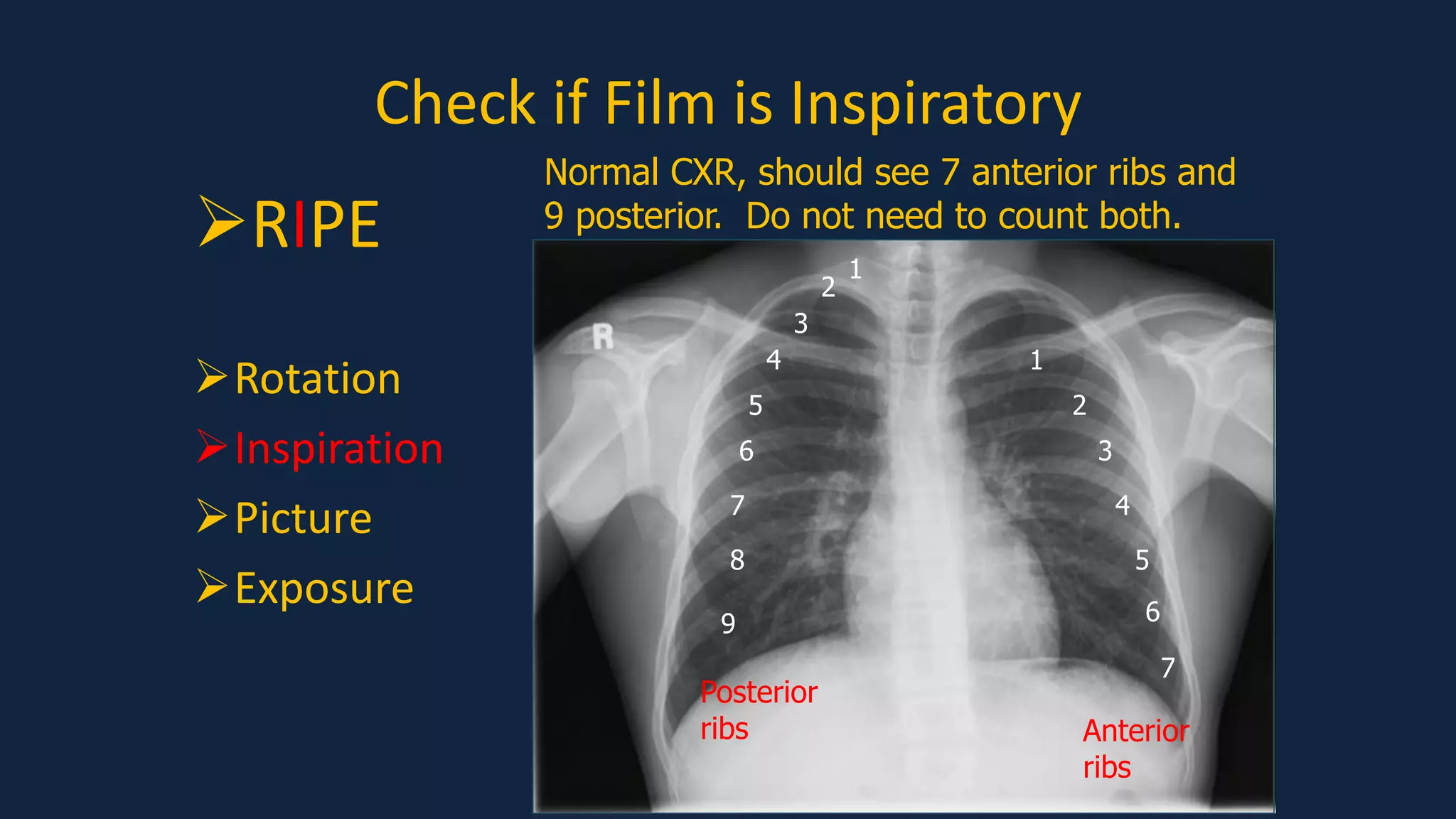
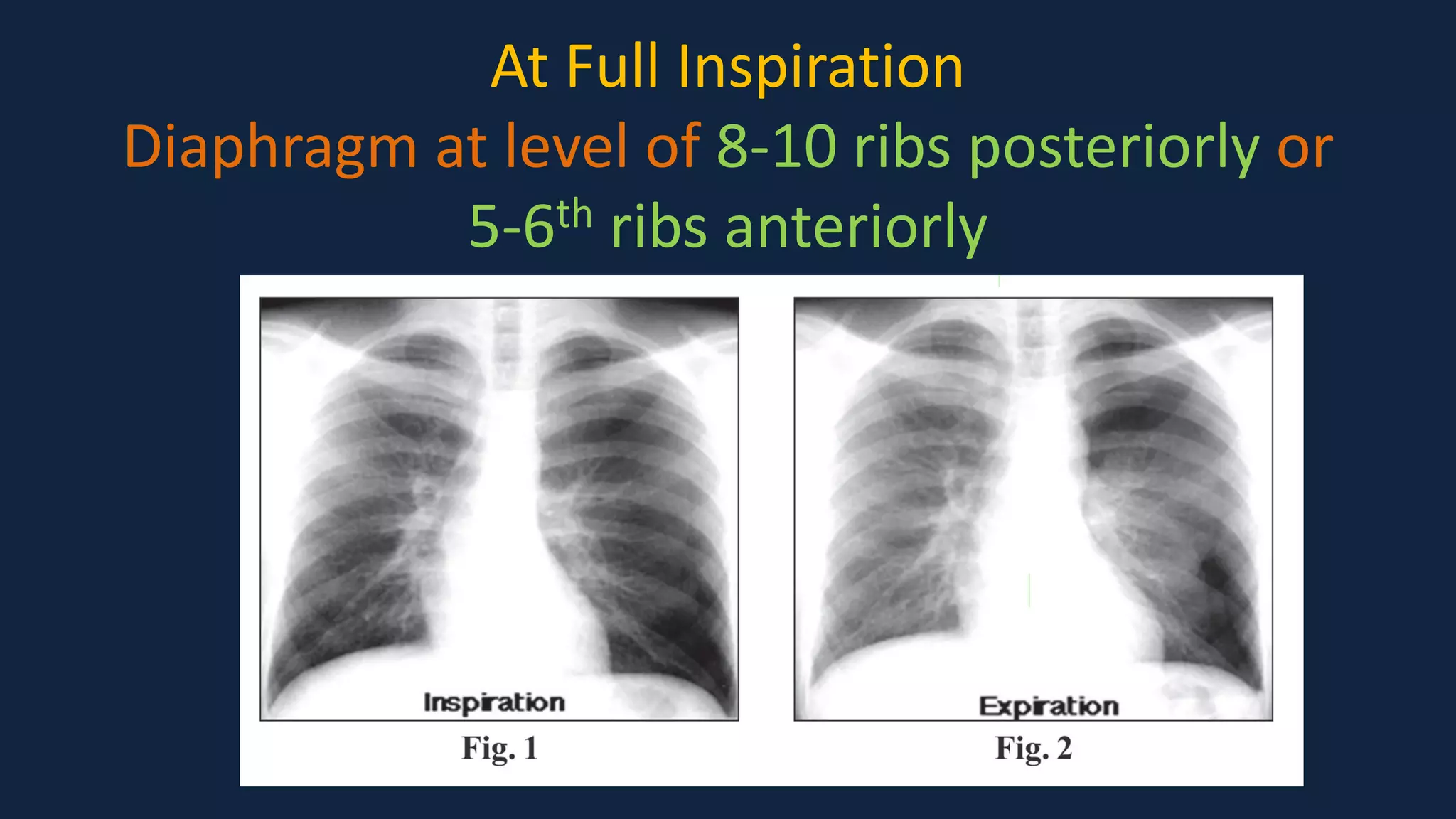
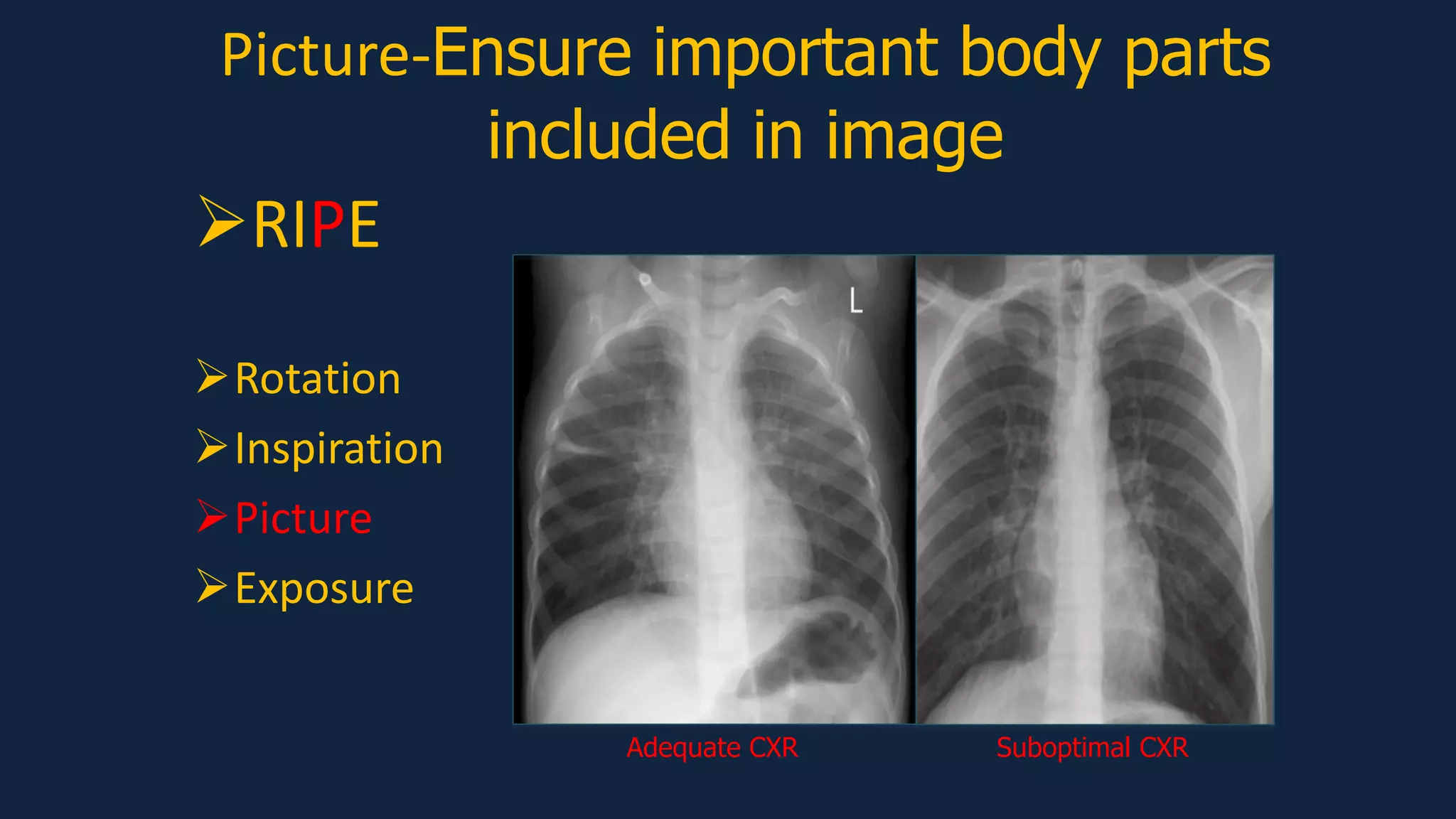
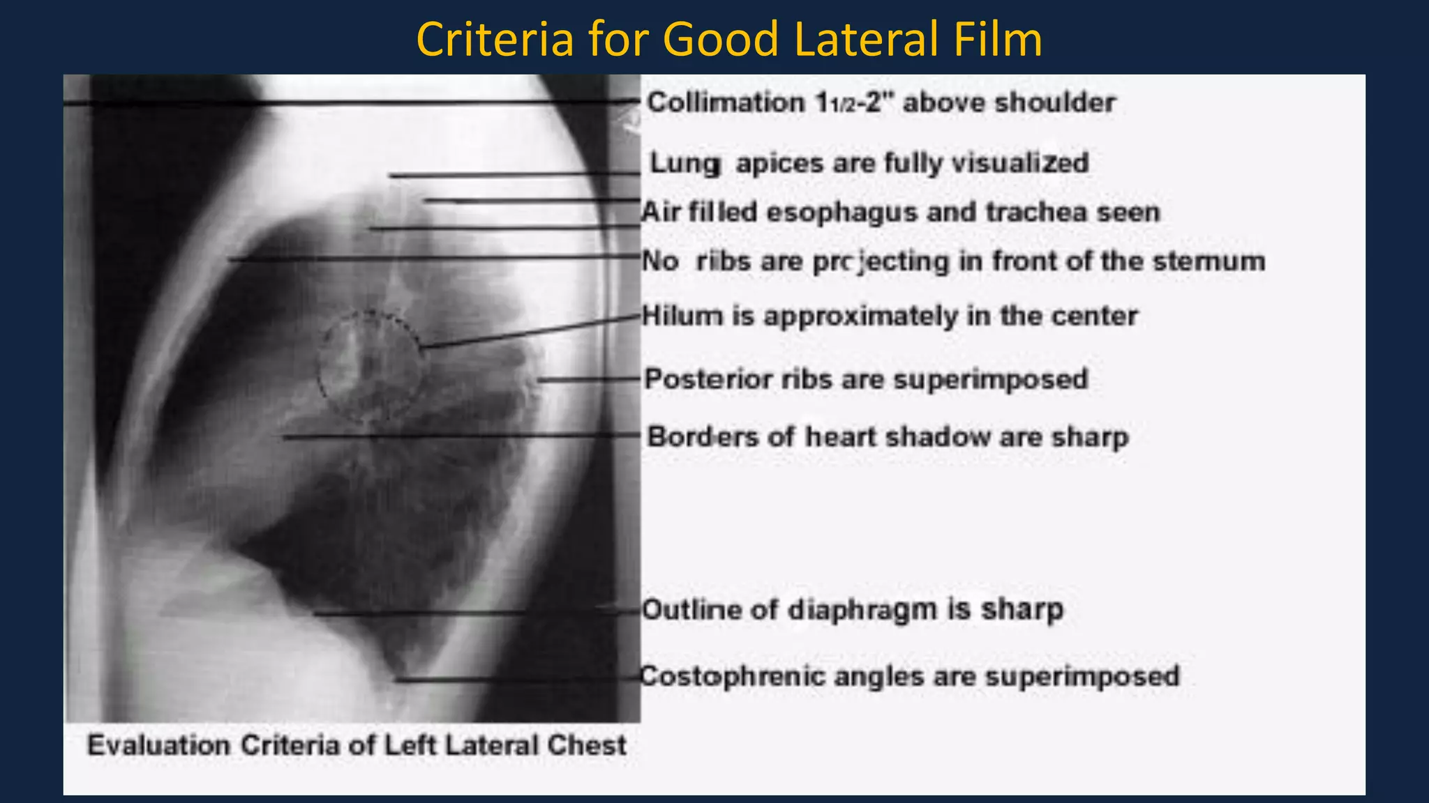
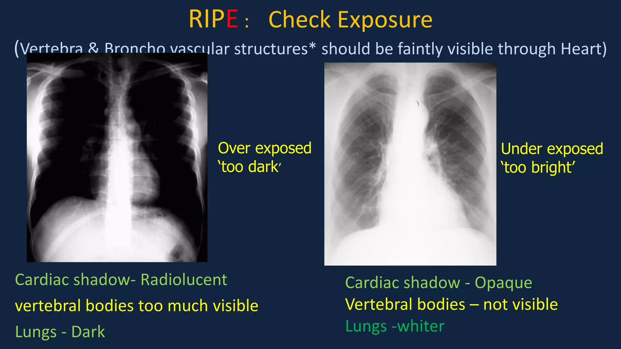
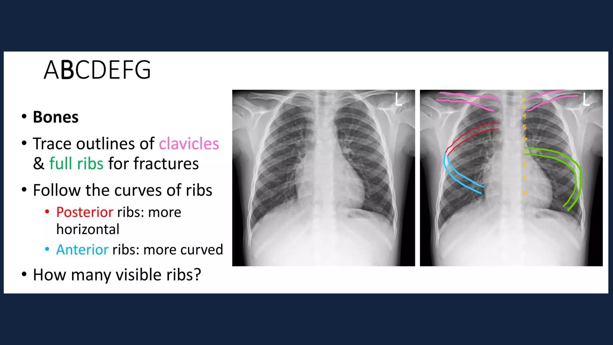
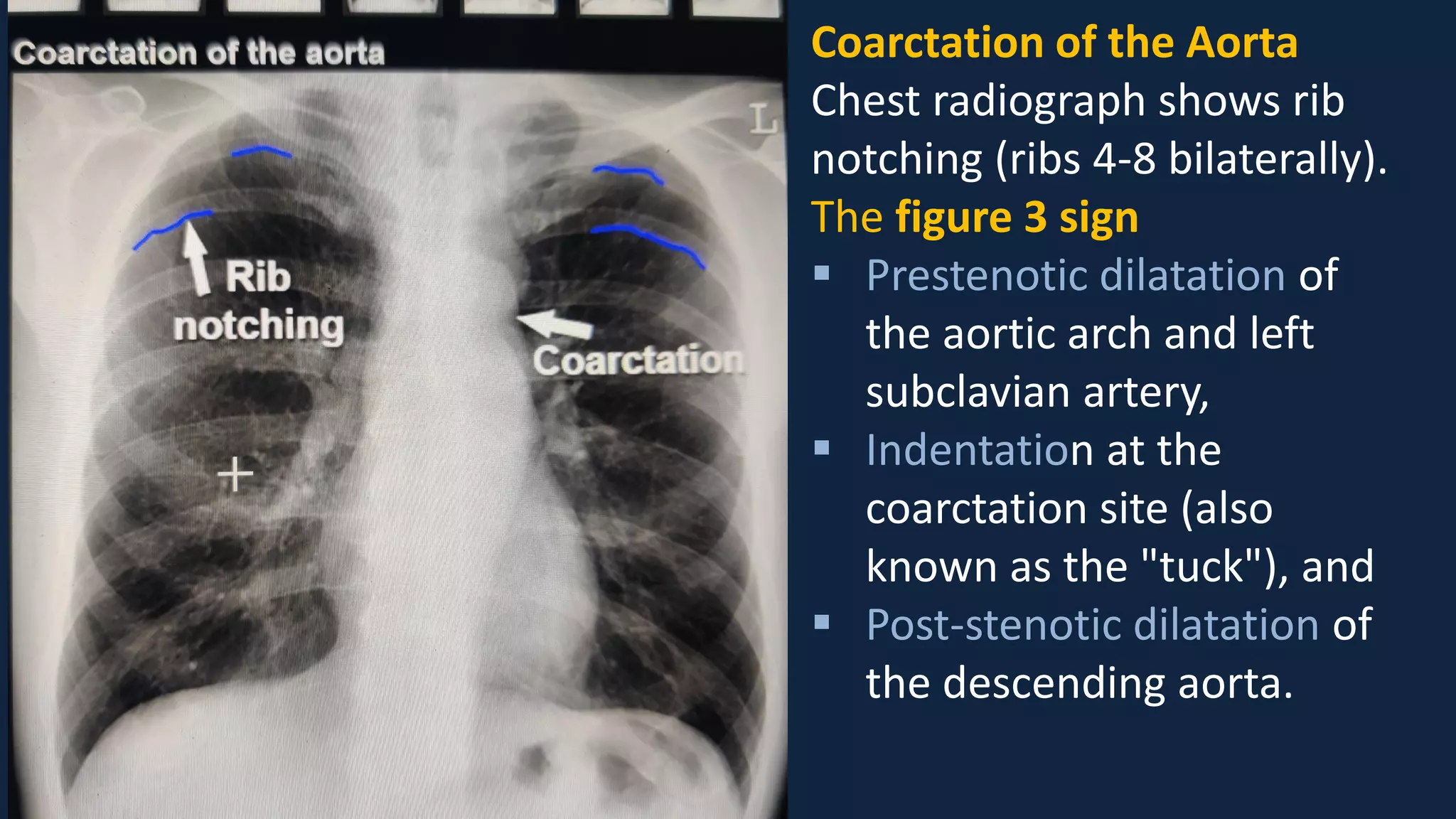
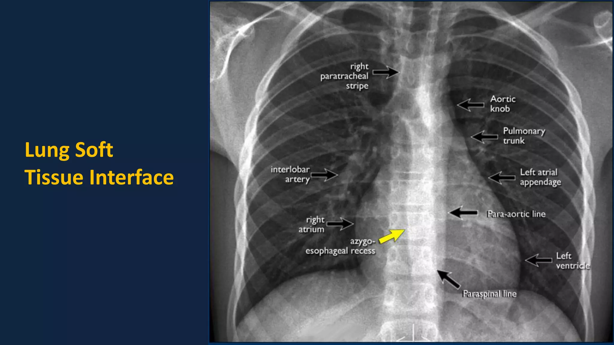
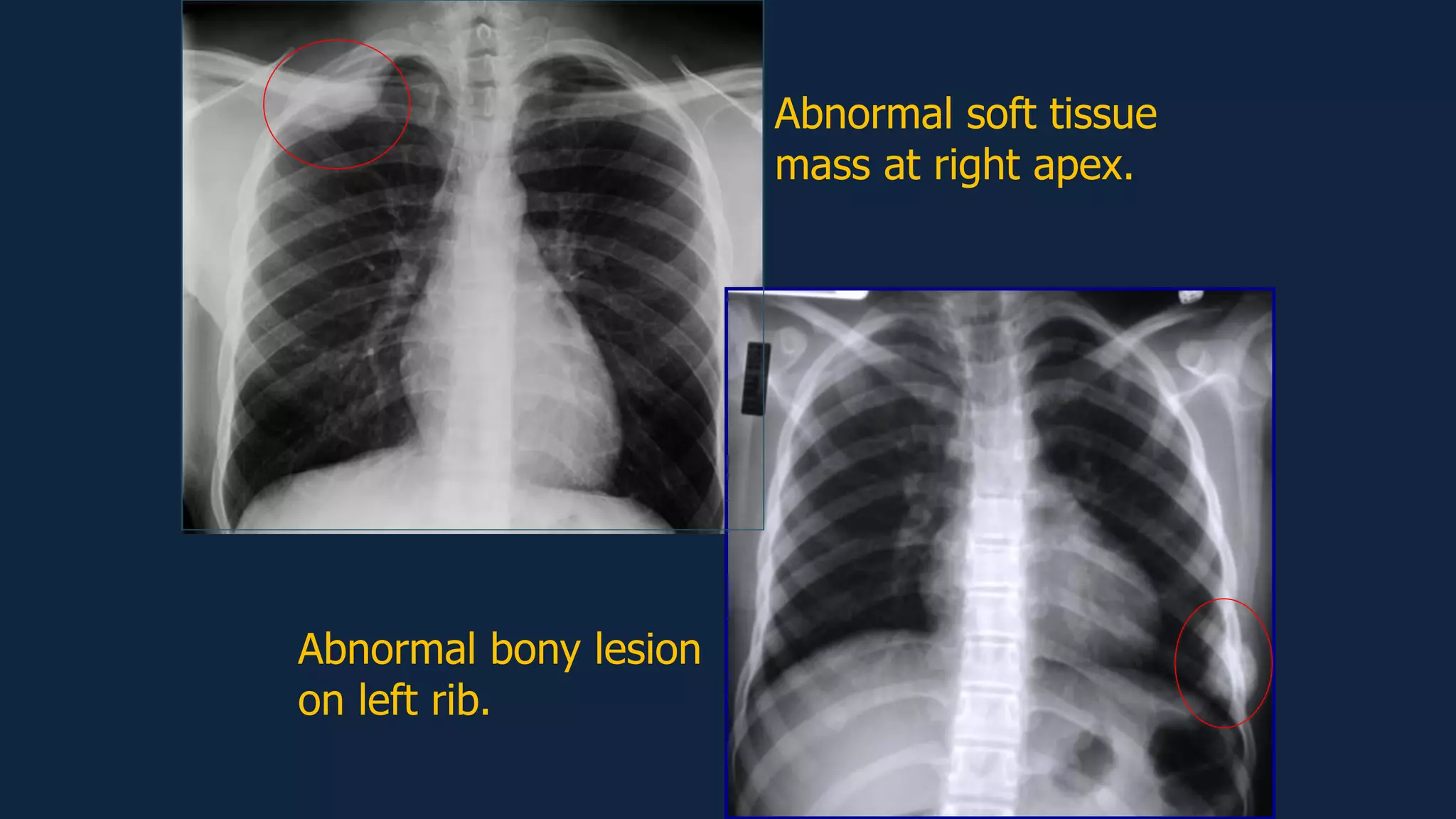
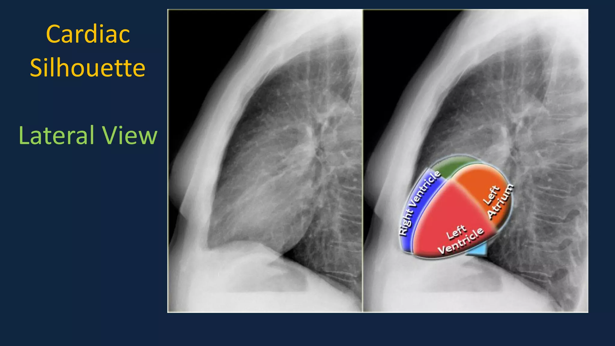
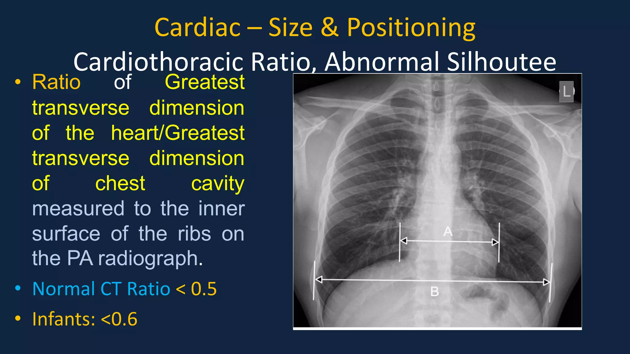
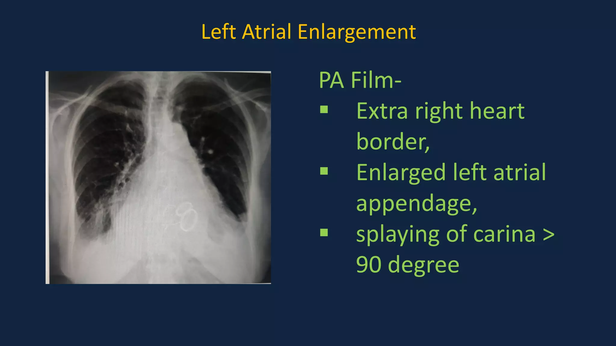
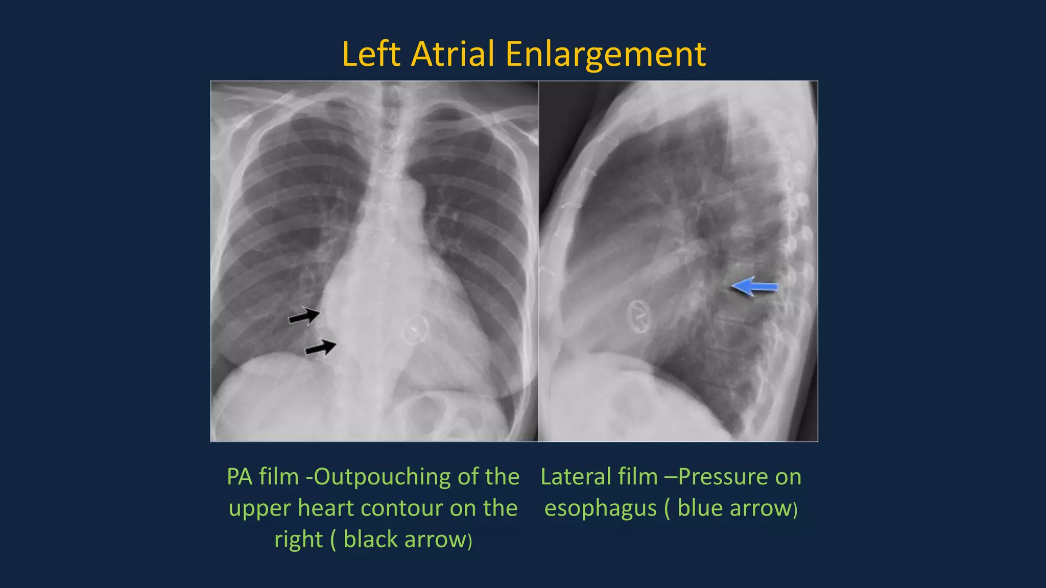
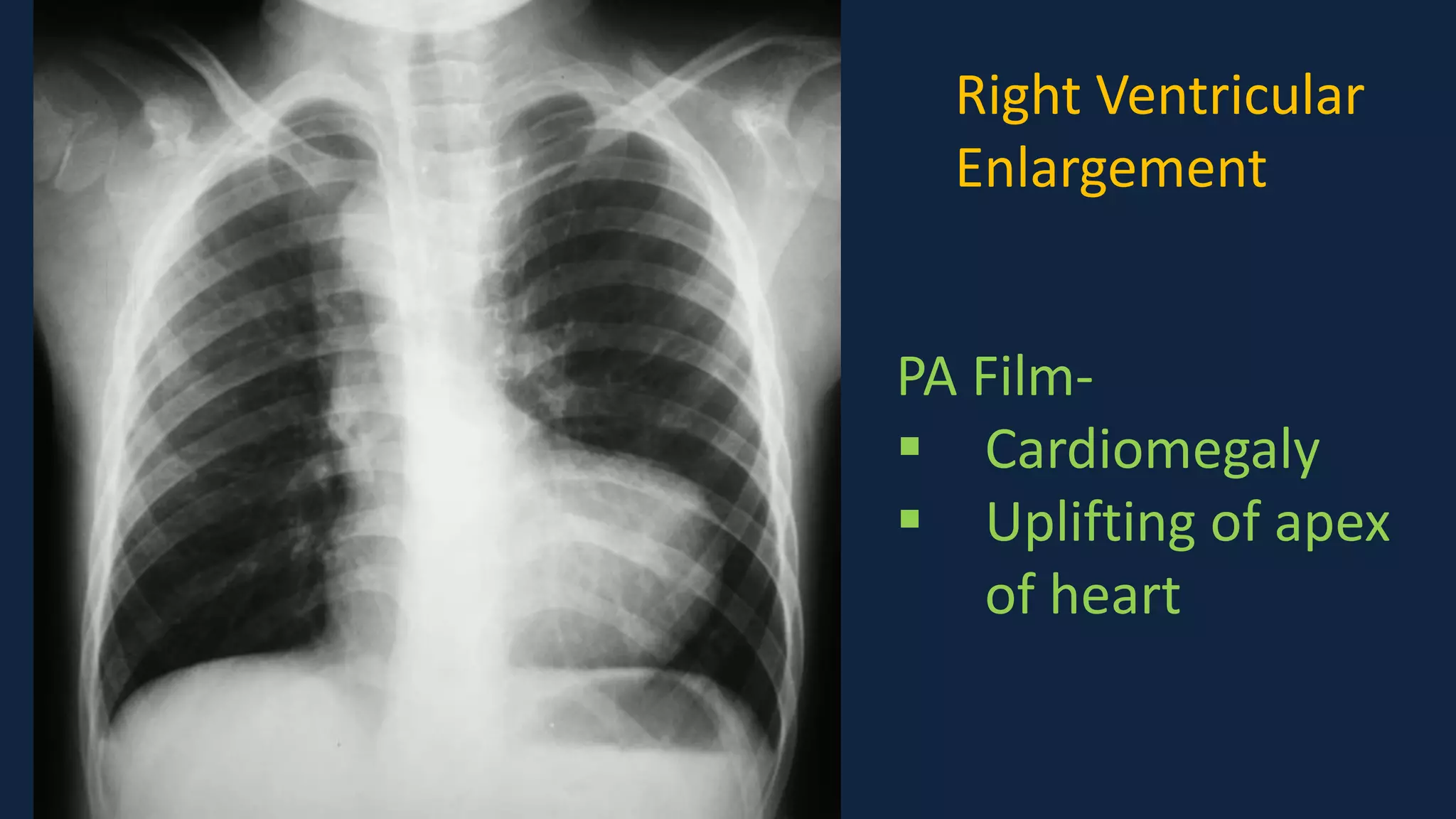
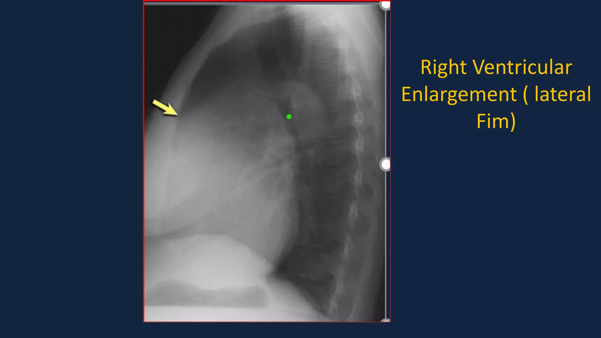
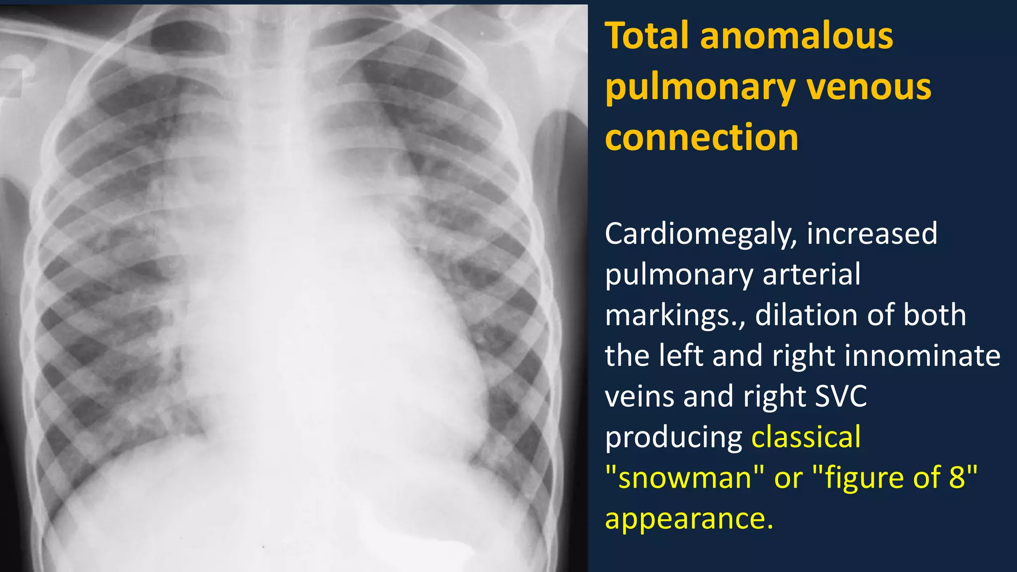
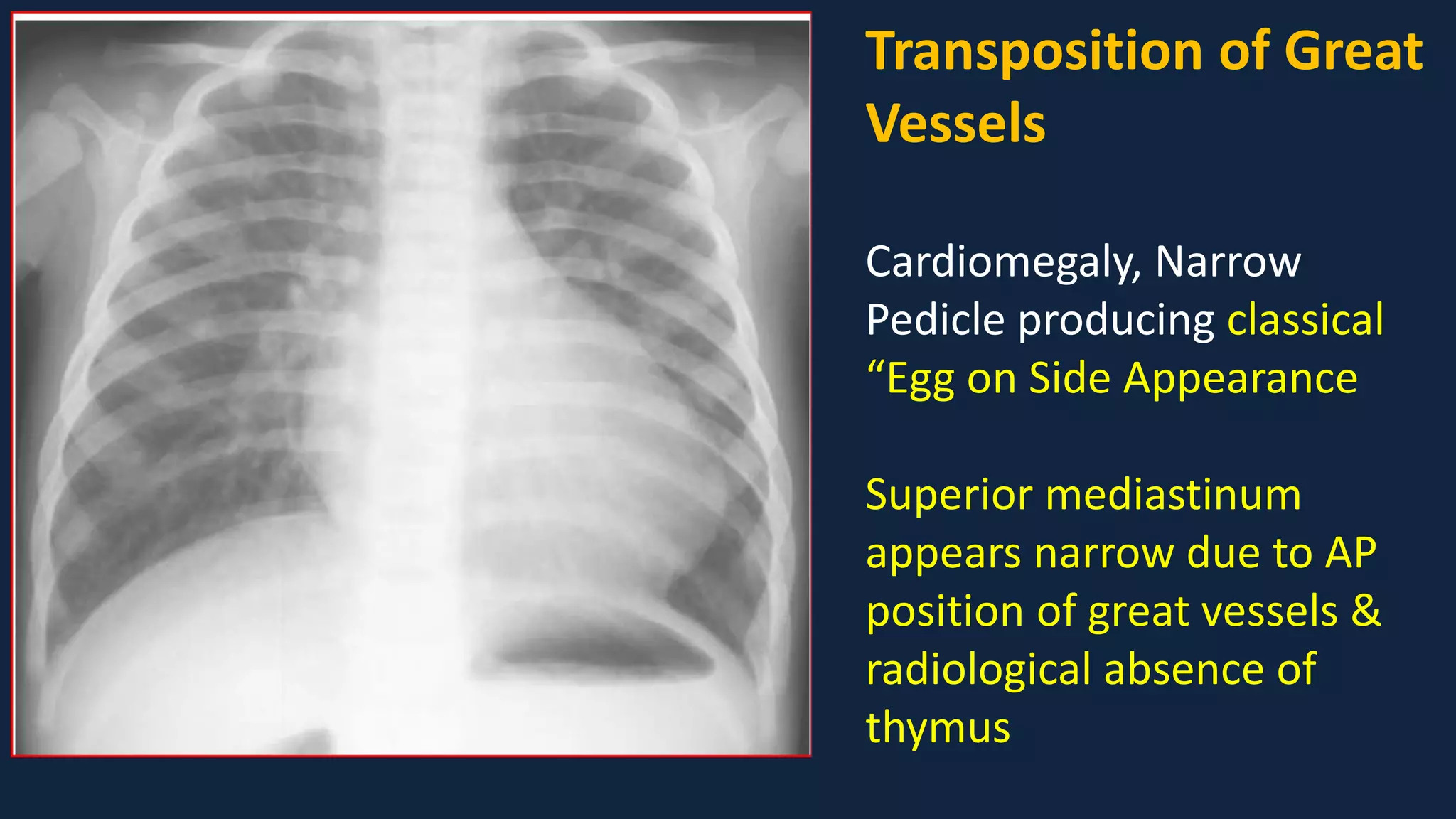
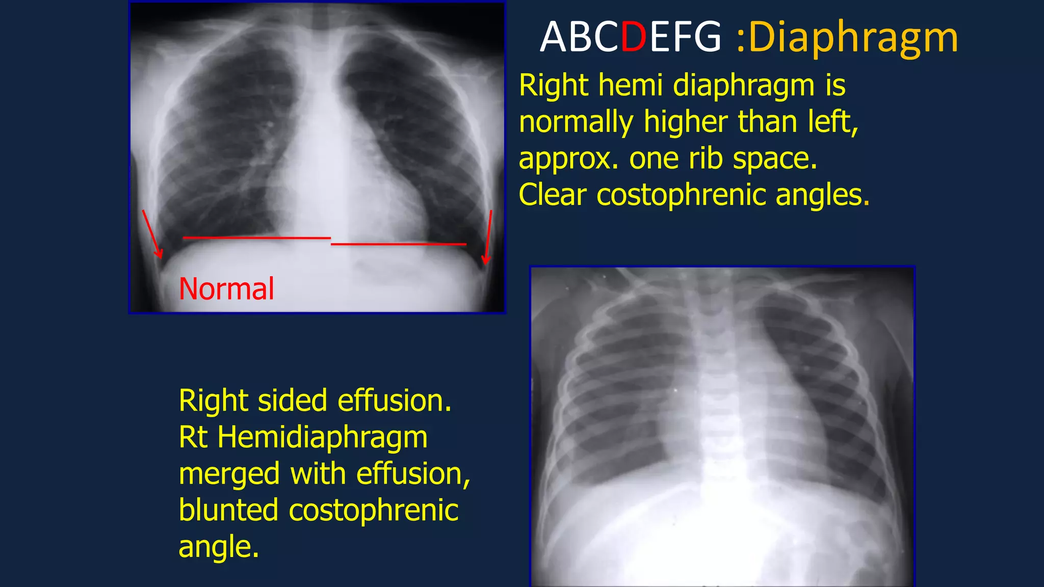
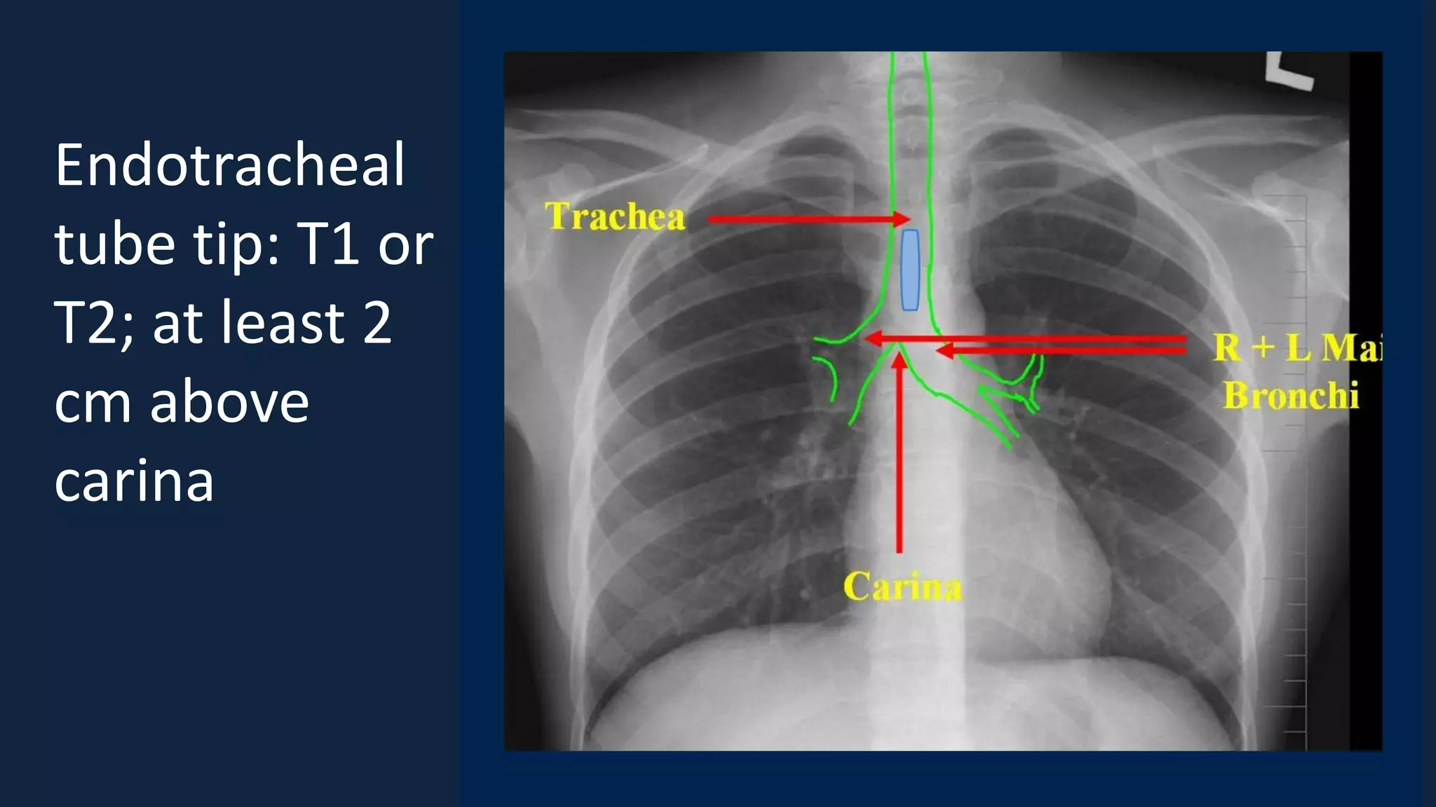
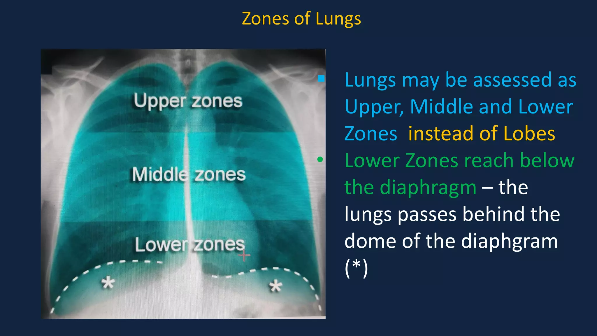
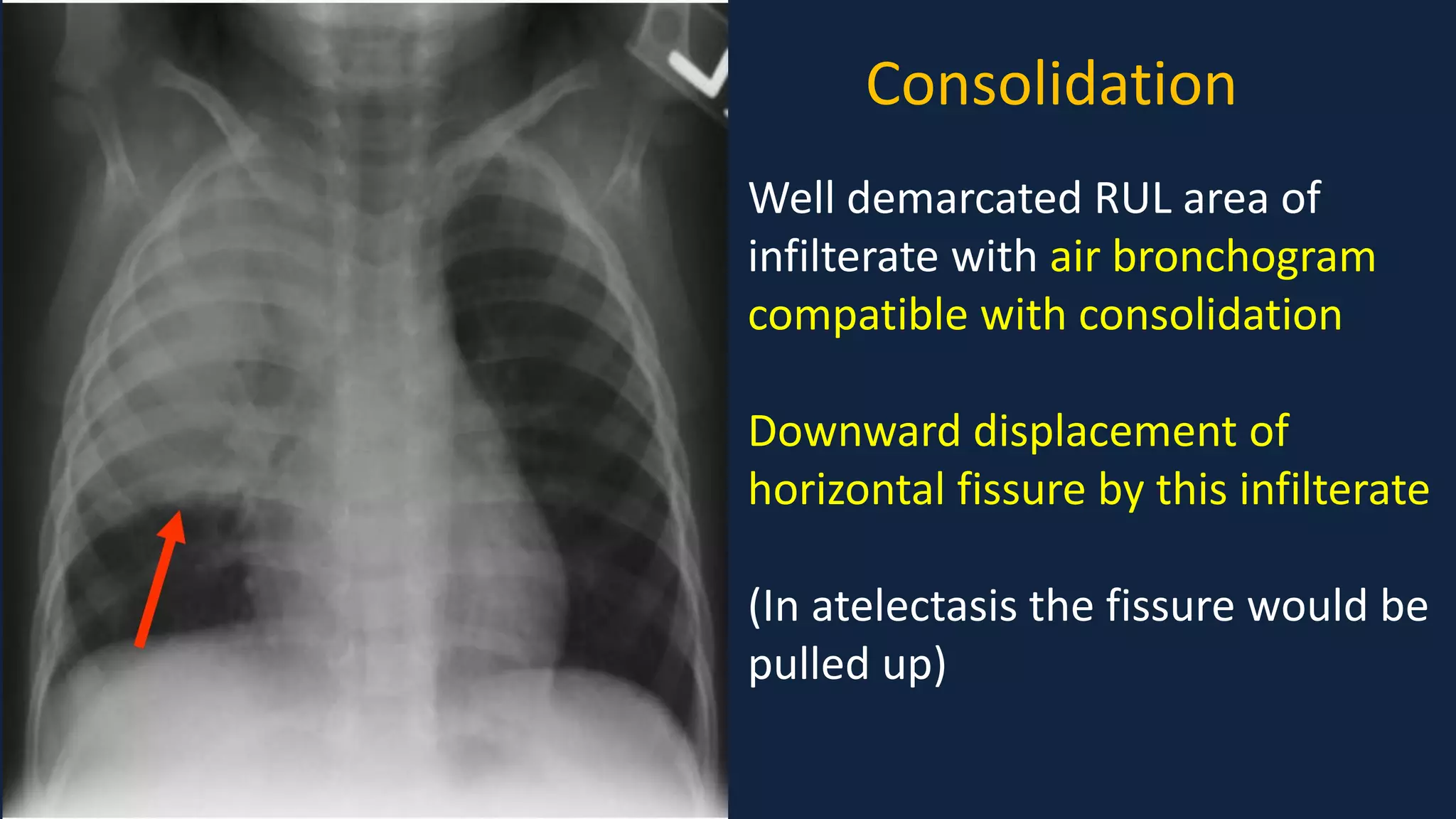
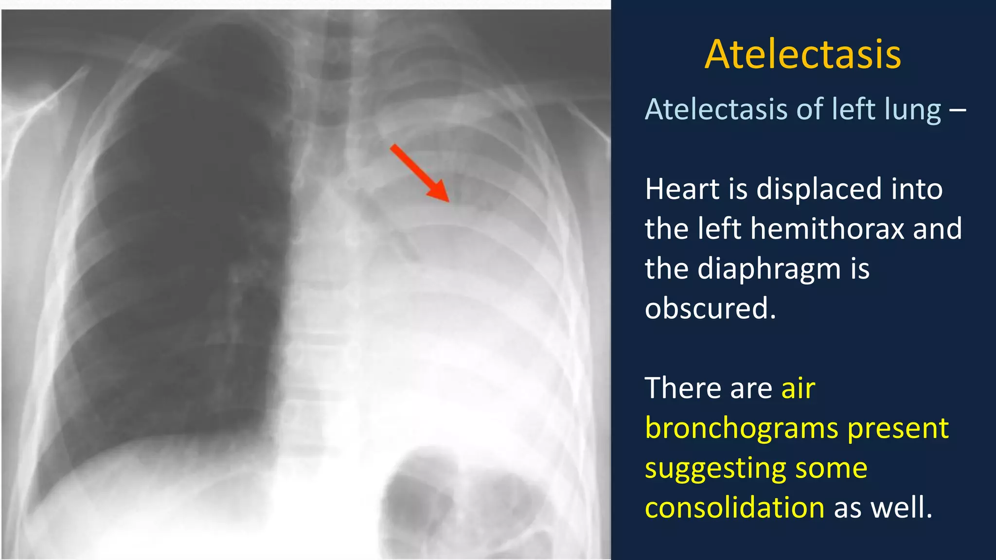
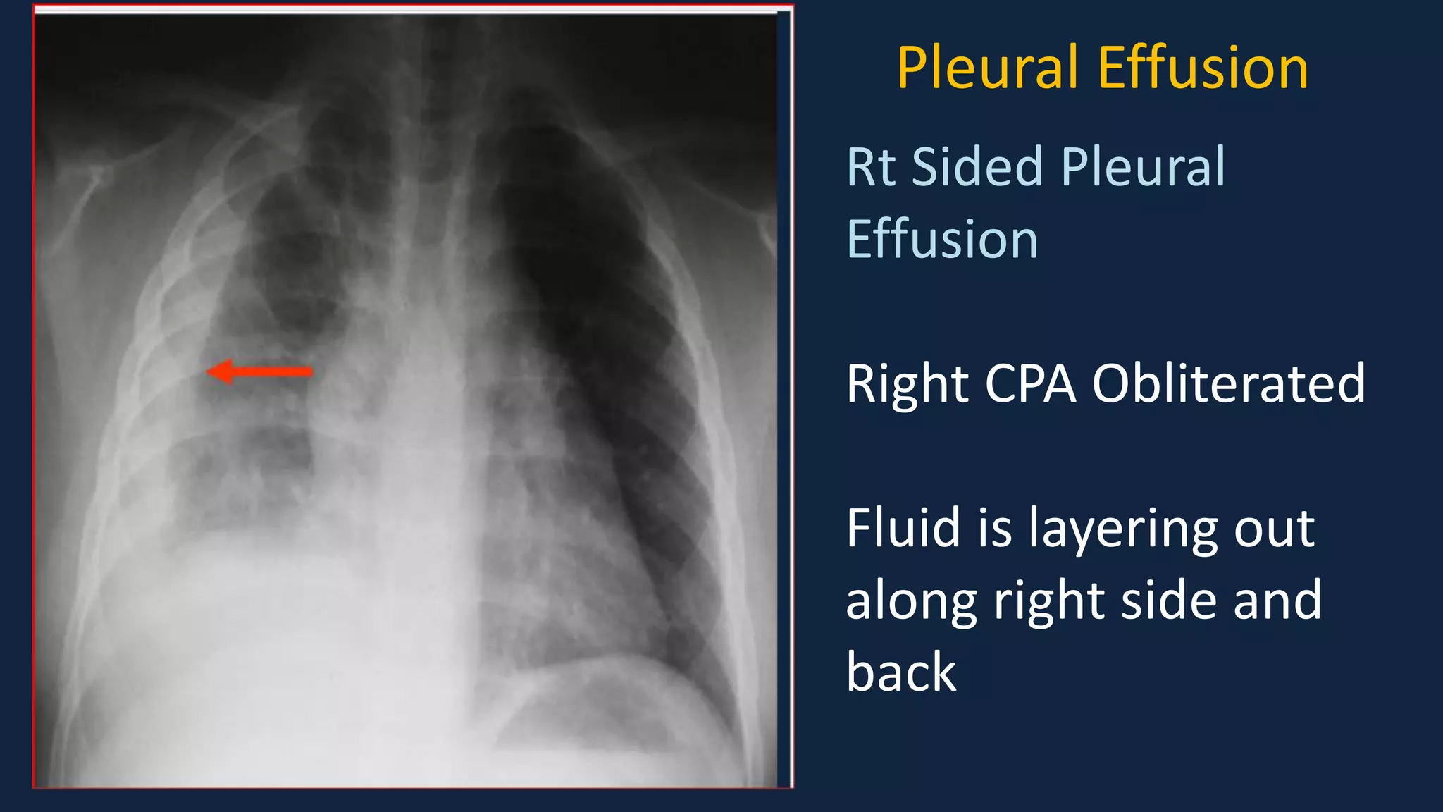
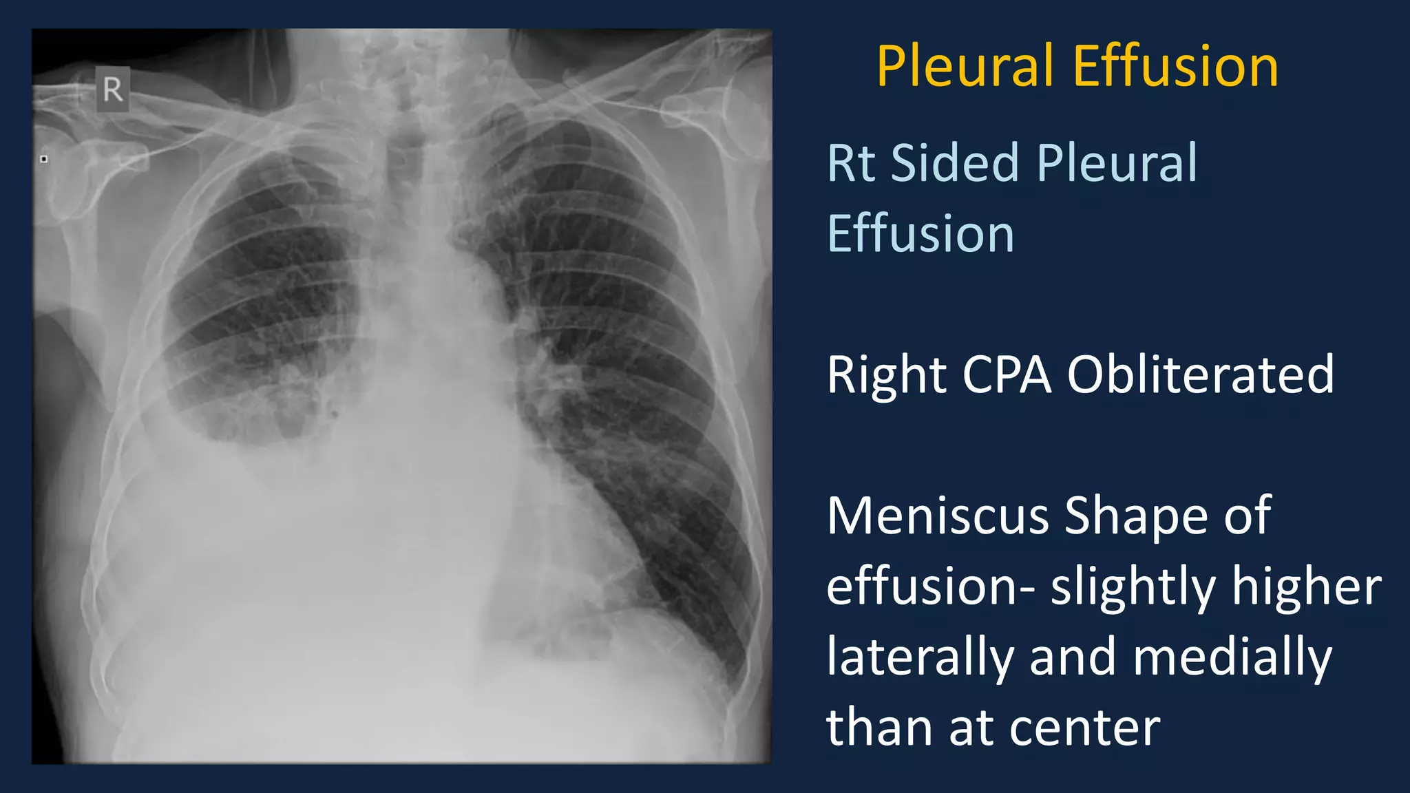
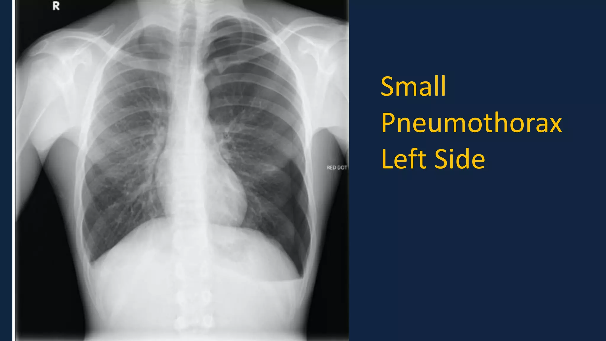
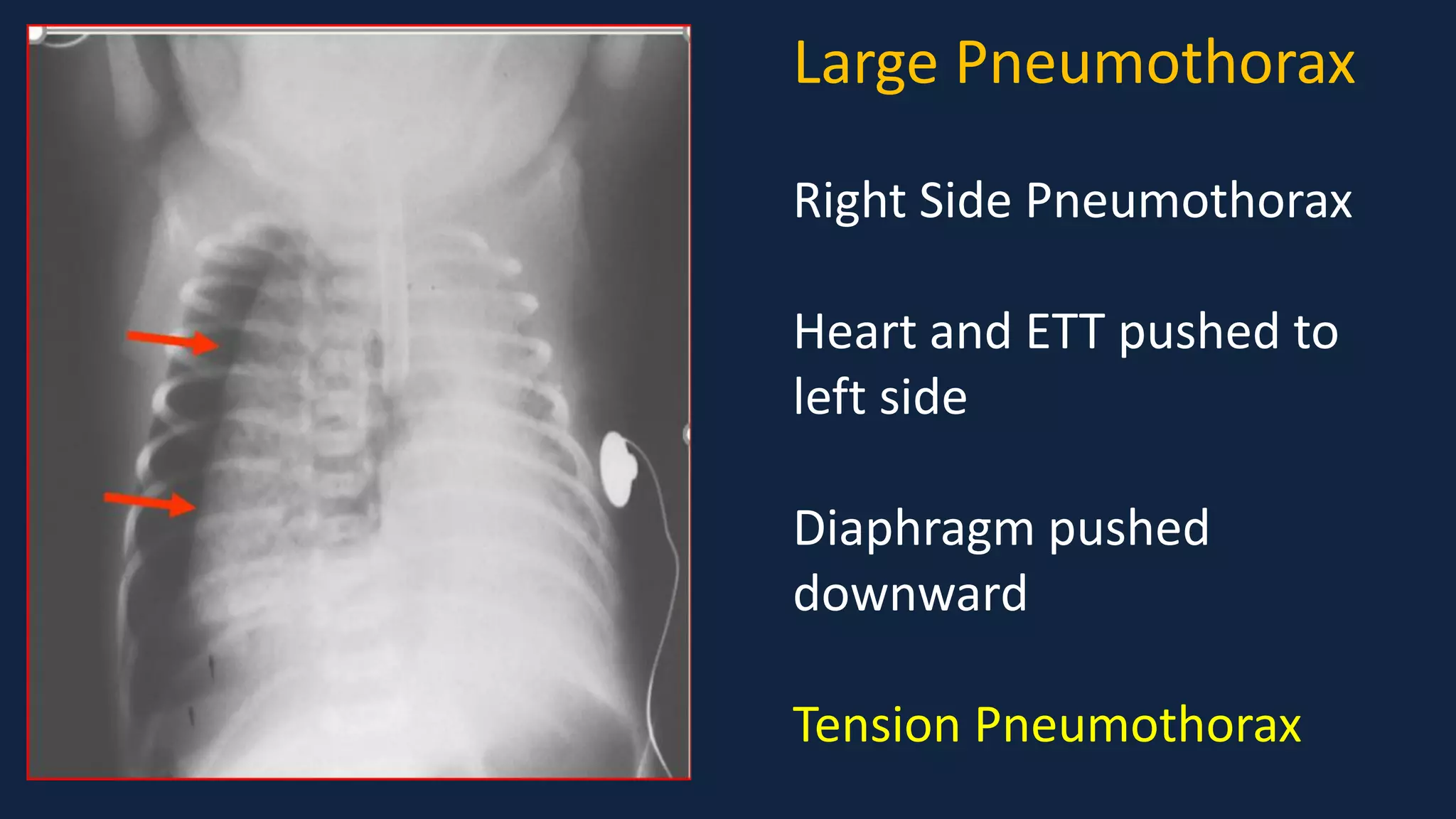
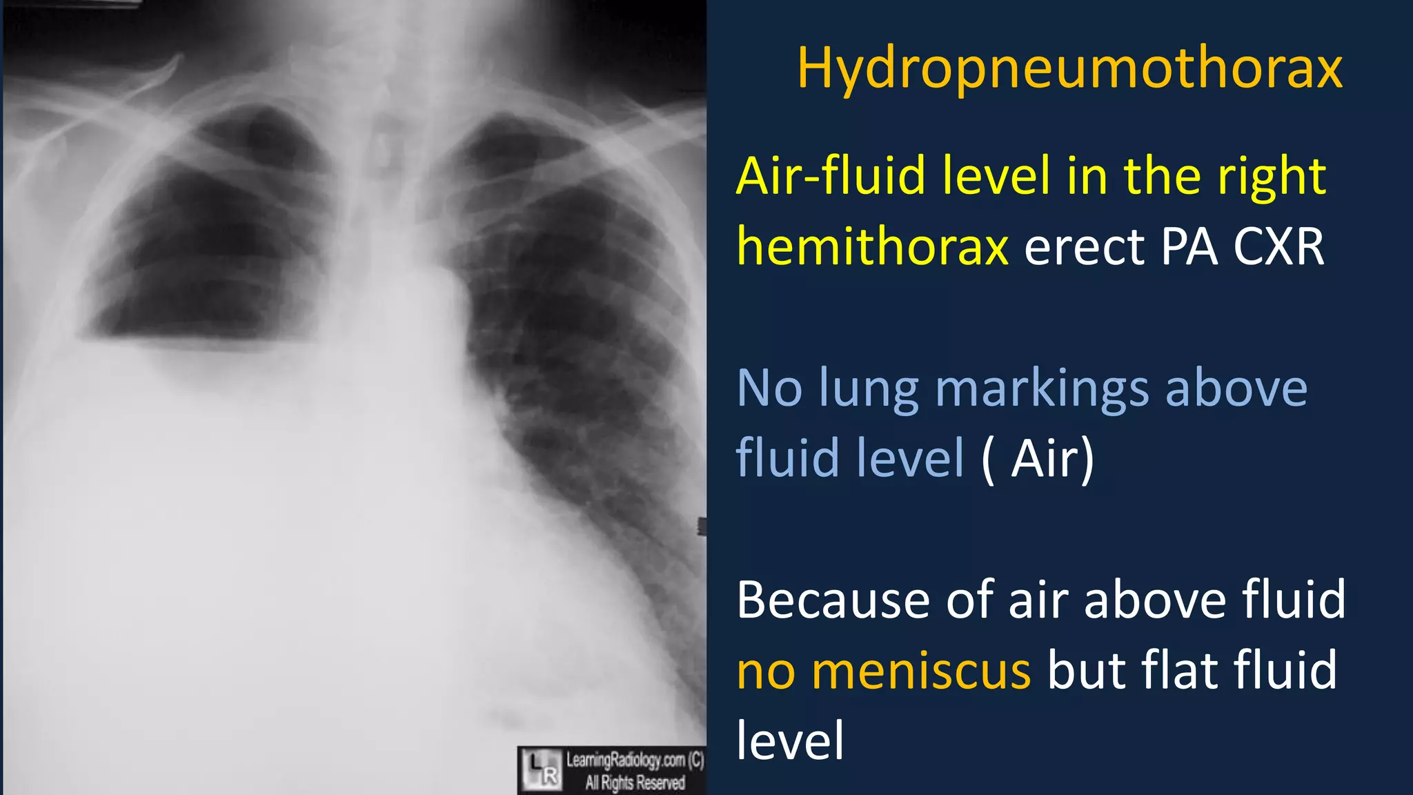
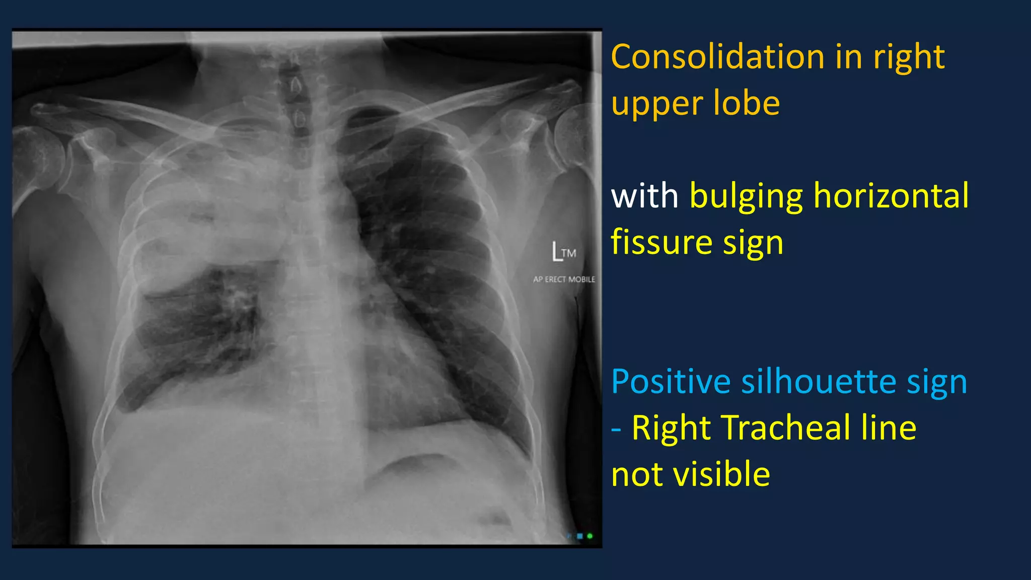
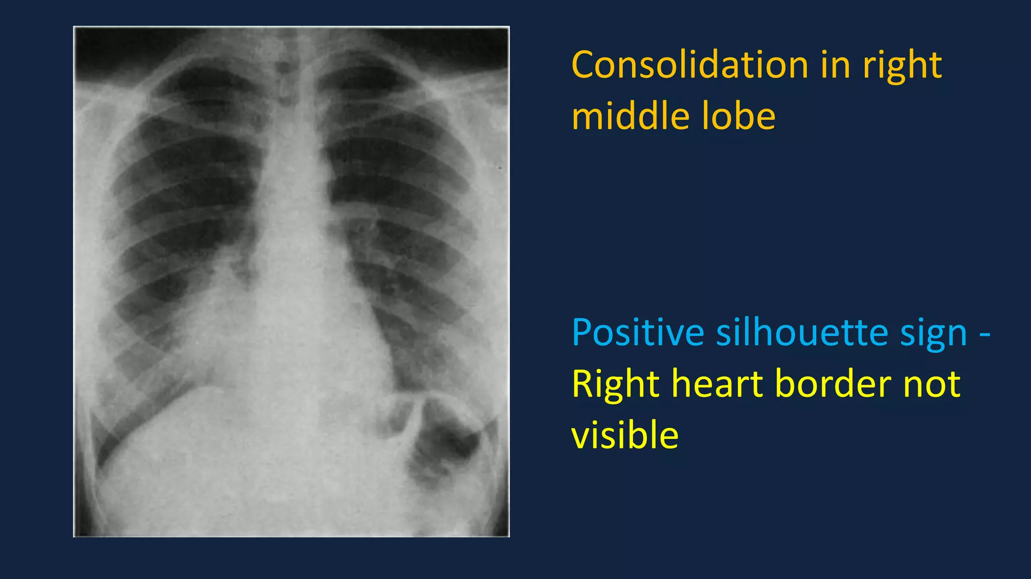
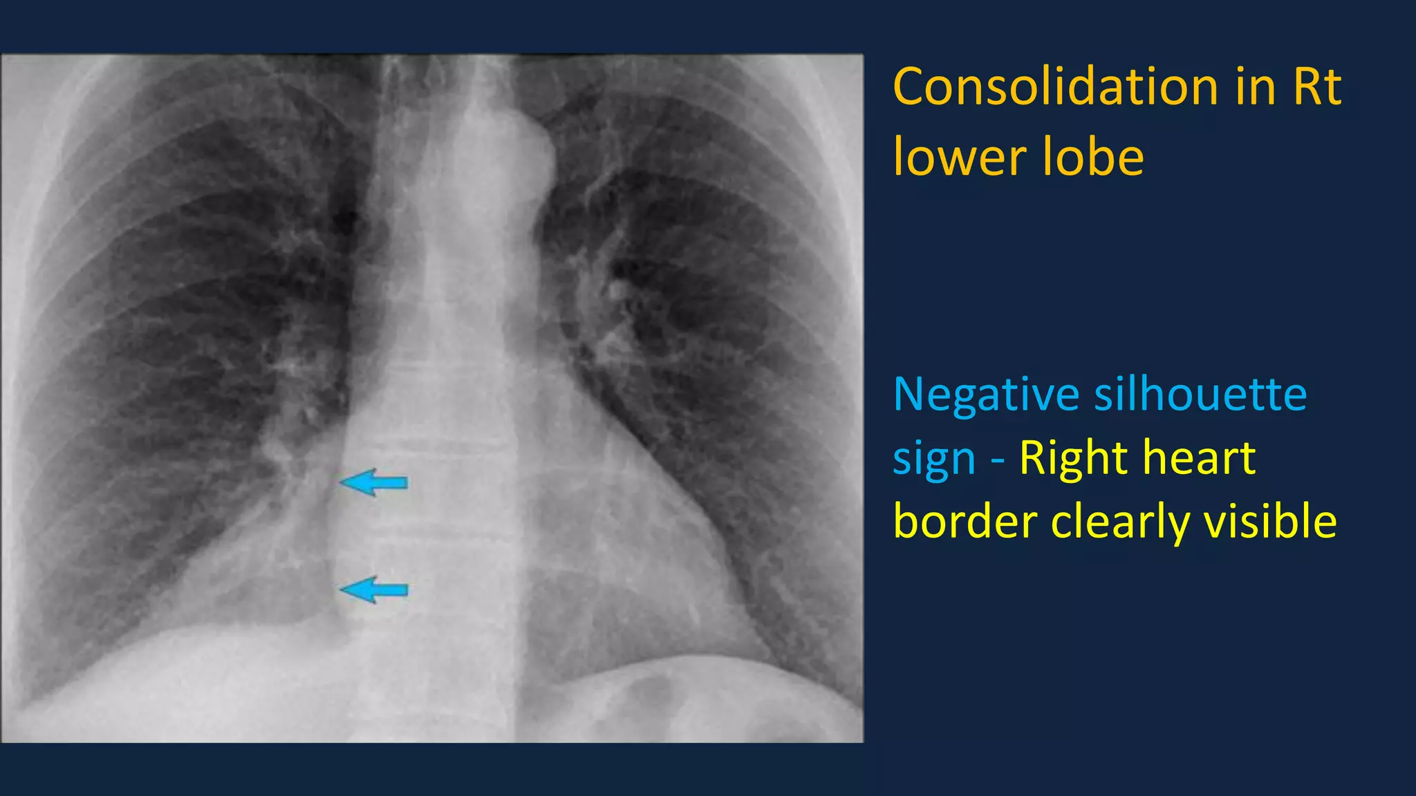
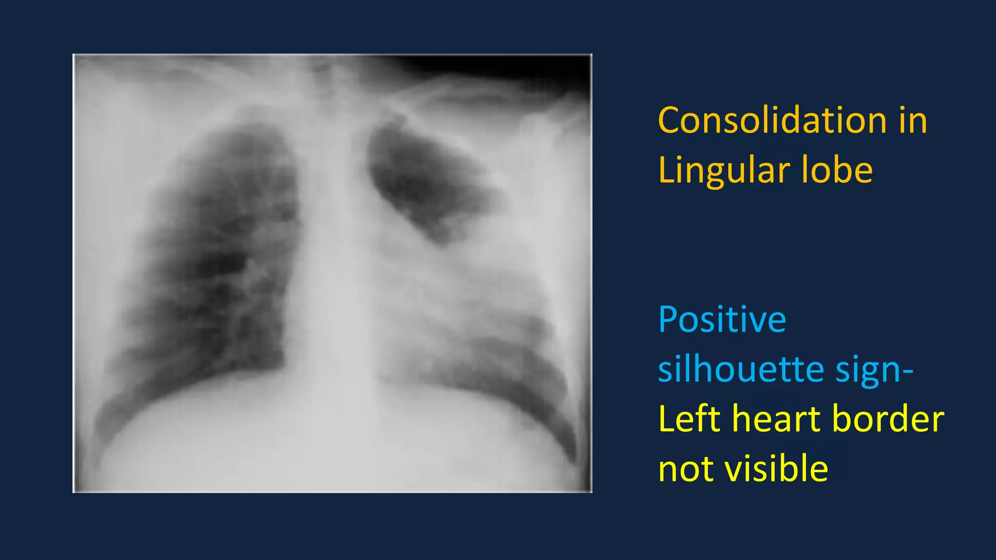
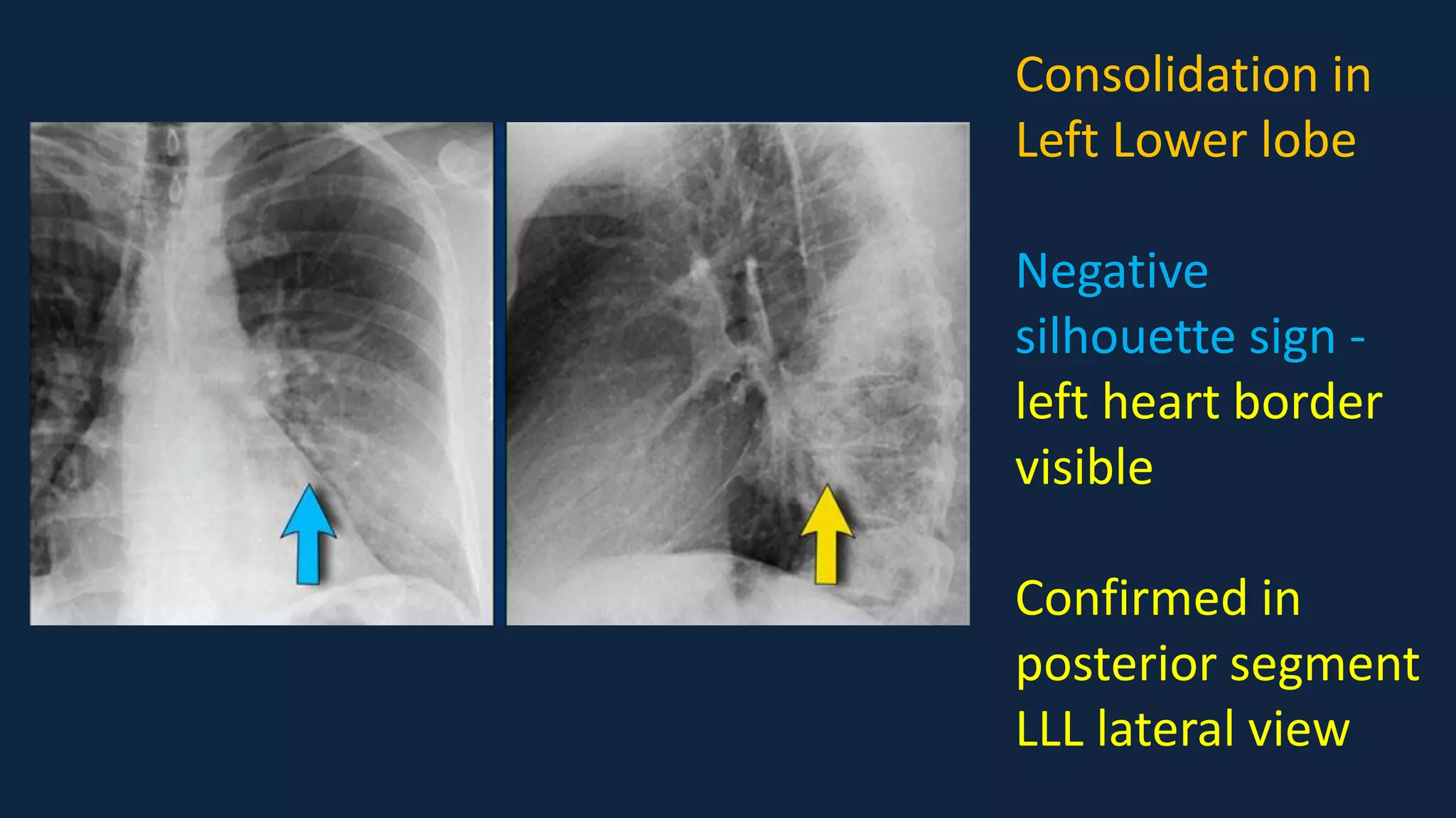
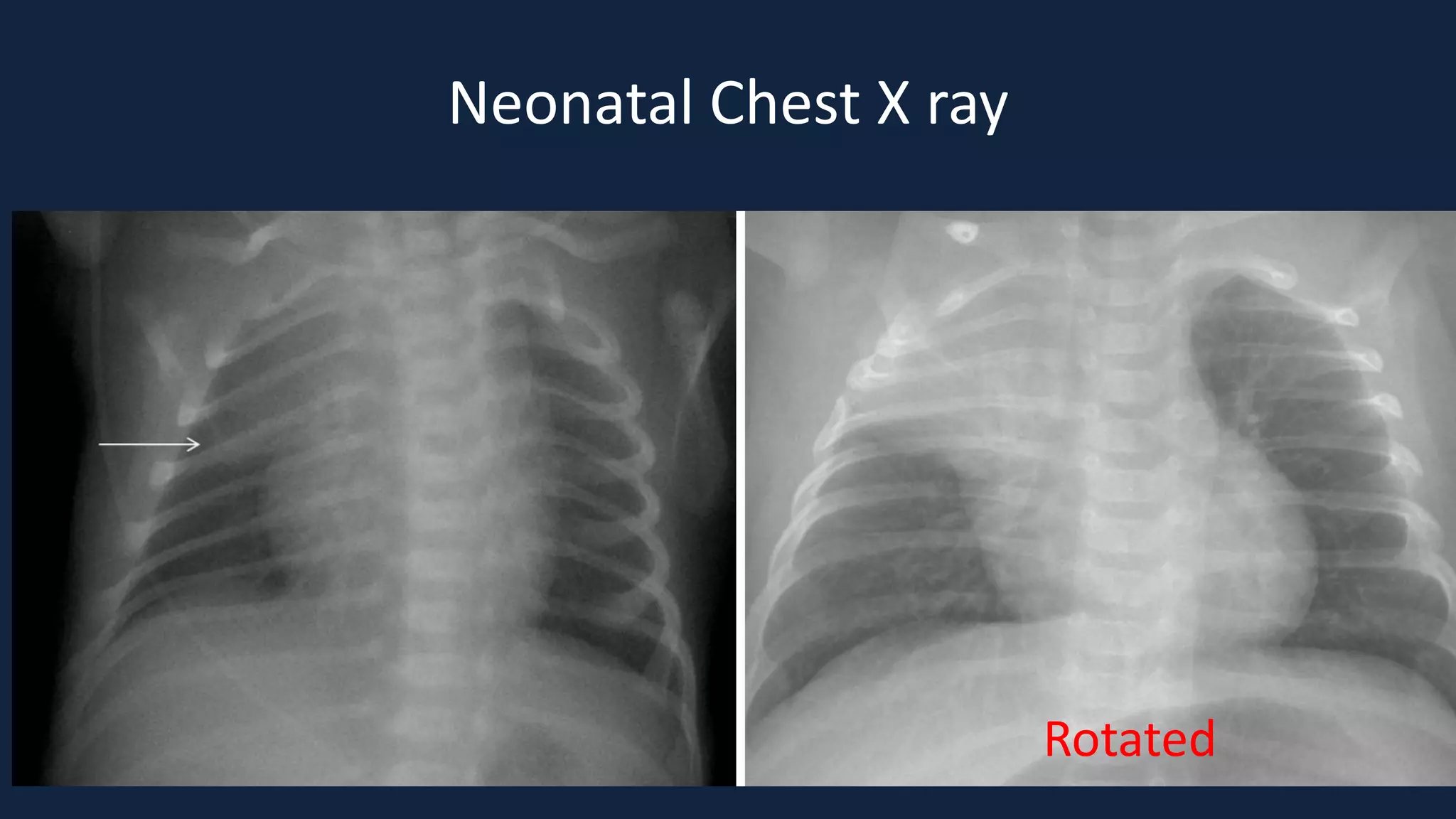
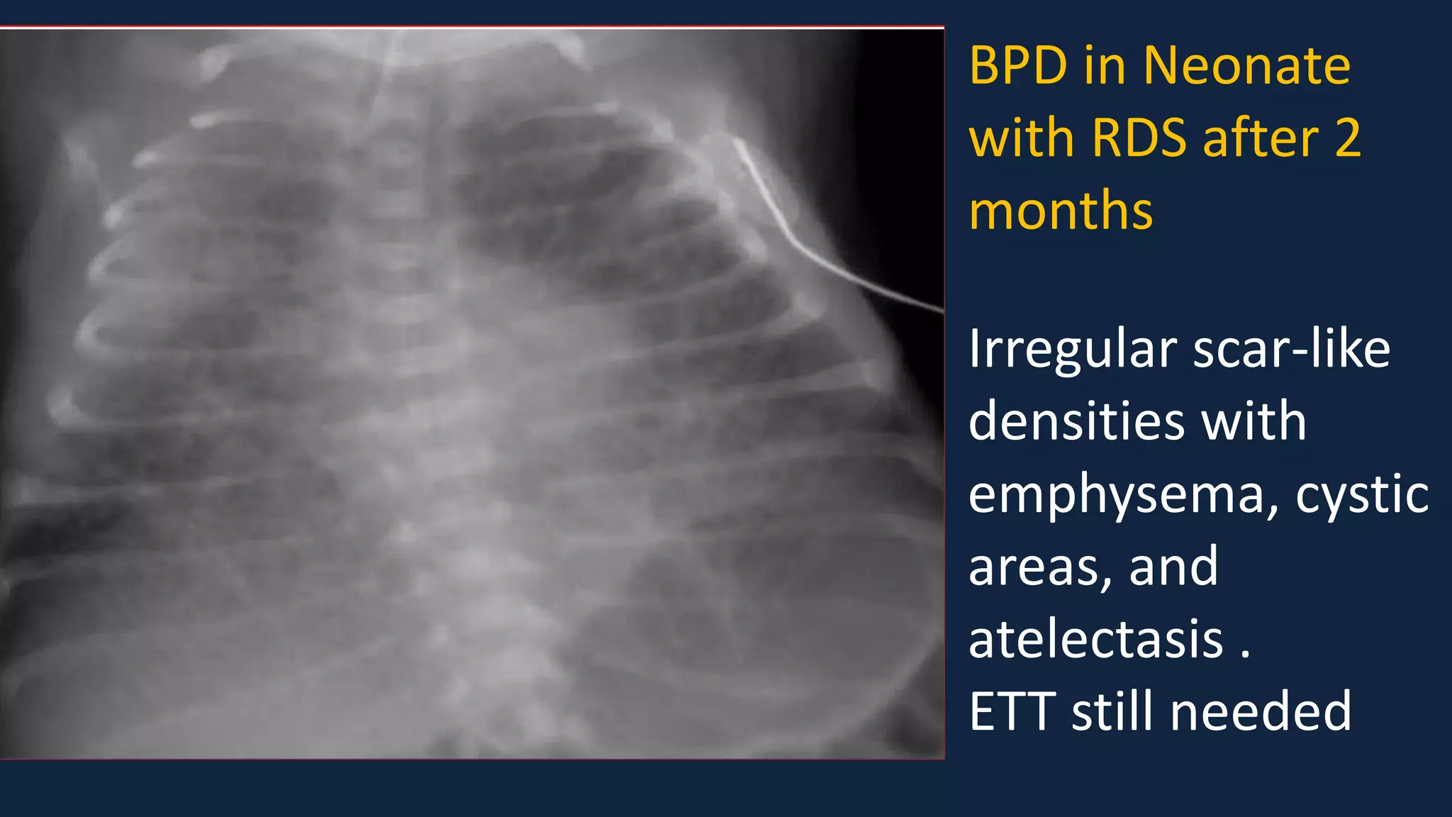
SlideShare a Scribd company logo
1 of 86
Approach to Interpret
Paediatric Chest X ray
DR ABHINAV GUPTA MD (RADIODIAGNOSIS)
Institute of Medical Sciences & SUM Hosp
Bhubaneswar, Odisha
PA Vs AP CXR ?
Perfect PA View Vs Rotated Film?
Inspiratory Vs Expiratory CXR Film?
Which is Wrong in these Films?
Any Difference between following X ray Films.
Which one is Technically Correct?
Any Difference between following Lateral X ray Films
Identify
Abnormality?
Orientation ?
Systematic Approach to Interpret
A Chest X ray Film
Termed: RIPE
Check Rotation
➢RIPE
➢Rotation
➢Inspiration
➢Picture
➢Exposure Identify the medial ends
of the clavicles – the
vertebral spinous
processes should be
equidistant between
them.
▪ Size and shape of the
mediastinal structures will be
affected.
▪ If the patient is rotated with the
left shoulder towards the x-ray
beam: -
- Heart appear larger (more left
ventricle will be seen)
- A normal thymus may look like
an upper lobe infiltrate.
Effect of Rotation
Normal Thymus -Thymus
Sail Sign ( Neonates &
Infants)
Rotated infant- pseudo
atelectasis RUL Thymic
shadow
Check if Film is Inspiratory
➢RIPE
➢Rotation
➢Inspiration
➢Picture
➢Exposure
1
2
3
4
5
6
7
8
9
1
2
3
4
5
6
7
Posterior
ribs Anterior
ribs
Normal CXR, should see 7 anterior ribs and
9 posterior. Do not need to count both.
At Full Inspiration
Diaphragm at level of 8-10 ribs posteriorly or
5-6th ribs anteriorly
Picture-Ensure important body parts
included in image
➢RIPE
➢Rotation
➢Inspiration
➢Picture
➢Exposure
Adequate CXR Suboptimal CXR
Criteria for Good Lateral Film
RIPE : Check Exposure
(Vertebra & Broncho vascular structures* should be faintly visible through Heart)
Cardiac shadow- Radiolucent
vertebral bodies too much visible
Lungs - Dark
Under exposed
‘too bright’
Over exposed
‘too dark’
Cardiac shadow - Opaque
Vertebral bodies – not visible
Lungs -whiter
& Soft Tissues
H
Hidden Areas
Coarctation of the Aorta
Chest radiograph shows rib
notching (ribs 4-8 bilaterally).
The figure 3 sign
▪ Prestenotic dilatation of
the aortic arch and left
subclavian artery,
▪ Indentation at the
coarctation site (also
known as the "tuck"), and
▪ Post-stenotic dilatation of
the descending aorta.
Examine all the Areas where the Lung Borders
the Diaphragm, the Heart and other Mediastinal structures.
Lung Soft Tissue Interface Results in:
•Line or stripe – e.g Right Para Tracheal stripe.
•Silhouette – e.g. Normal Silhouette of the Aortic knob or Left ventricle
These lines and silhouettes can be displaced or obscured with loss of the normal
silhouette. This is called the silhouette sign,
Displacement of Paraspinal line - Paravertebral abscess, hemorrhage due to a # or
metastases
Widening of the paratracheal line (> 2-3mm) -Lymphadenopathy, pleural
thickening, hemorrhage or fluid overload and heart failure.
Displacement of the para-aortic line - Elongation of the aorta, aneurysm, dissection
and rupture.
Lung Soft
Tissue Interface
Abnormal soft tissue
mass at right apex.
Abnormal bony lesion
on left rib.
Cardiac
Silhouette
PA View
Right Heart Border
▪ SVC
▪ Right atrium
▪ IVC
Left Heart Border
▪ Aortic knob
▪ Pulmonary trunk
▪ Left Atrial
Appendage
▪ Left Ventricle
Cardiac
Silhouette
Lateral View
Cardiac – Size & Positioning
Cardiothoracic Ratio, Abnormal Silhoutee
• Ratio of Greatest
transverse dimension
of the heart/Greatest
transverse dimension
of chest cavity
measured to the inner
surface of the ribs on
the PA radiograph.
• Normal CT Ratio < 0.5
• Infants: <0.6
Left Atrial Enlargement
PA Film-
▪ Extra right heart
border,
▪ Enlarged left atrial
appendage,
▪ splaying of carina >
90 degree
Left Atrial Enlargement
PA film -Outpouching of the
upper heart contour on the
right ( black arrow)
Lateral film –Pressure on
esophagus ( blue arrow)
PA Film-
▪ Cardiomegaly
▪ Uplifting of apex
of heart
Right Ventricular
Enlargement
Right Ventricular
Enlargement ( lateral
Fim)
Left Ventricular Enlargement
•Left heart border is displaced
leftward, inferiorly, or
posteriorly
•Rounding of the cardiac apex
Pulmonary Plethora Versus Pulmonary
Edema
Pulmonary plethora in a patient with a VSD.
Note the increased number and size of
discrete vessels without haziness.
Pulmonary oedema in a supine patient with cor triatriatum- A
(membranous obstruction to LA outflow). Note cardiomegaly,
perihilar alveolar haziness/consolidation and peribronchial
cuffing
Right Atrial Enlargement
Difficult to diagnose in CXR
Non-Specific signs:
•Enlarged globular heart
•Narrow vascular pedicle
•Increased convexity of lower
right heart border
•Rt Atrial margin > 5.5 cm from
midline
Pericardial Effusion
▪ Cardiomegaly
• Globular heart with
narrow pedicle.
• Widening of the
carinal angle in
posterior extension of
effusion
Tetralogy of Fallot
• Boot Shaped Heart with an
upturned cardiac apex due
to right ventricular
hypertrophy and
▪ Concave pulmonary arterial
segment
▪ Pulmonary oligemia
▪ A right-sided aortic arch is
seen in 25%.
Total anomalous
pulmonary venous
connection
Cardiomegaly, increased
pulmonary arterial
markings., dilation of both
the left and right innominate
veins and right SVC
producing classical
"snowman" or "figure of 8"
appearance.
Transposition of Great
Vessels
Cardiomegaly, Narrow
Pedicle producing classical
“Egg on Side Appearance
Superior mediastinum
appears narrow due to AP
position of great vessels &
radiological absence of
thymus
Normal
Right hemi diaphragm is
normally higher than left,
approx. one rib space.
Clear costophrenic angles.
Right sided effusion.
Rt Hemidiaphragm
merged with effusion,
blunted costophrenic
angle.
ABCDEFG :Diaphragm
Umbilical arterial
line • High:
Between T6 and
T9 thoracic
vertebrae • Low:
Between L3 and
L4 vertebrae
Umbilical venous
line: 0.5 cm to 1
cm above the
diaphragm
Endotracheal
tube tip: T1 or
T2; at least 2
cm above
carina
Pathology in following areas can easily
be overlooked.
These areas are also known as the
hidden areas:
•Apical zones
•Hilar zones
•Retrocardial zone
•Zone below the dome of diaphragm
Hidden areas
Zones of Lungs
▪ Lungs may be assessed as
Upper, Middle and Lower
Zones instead of Lobes
• Lower Zones reach below
the diaphragm – the
lungs passes behind the
dome of the diaphgram
(*)
Large lesion in the right lower lobe in
hidden area behind right dome of
diaphragm
Well demarcated RUL area of
infilterate with air bronchogram
compatible with consolidation
Downward displacement of
horizontal fissure by this infilterate
(In atelectasis the fissure would be
pulled up)
Consolidation
RUL, LUL atelectasis
The horizontal fissure is
displaced upwards.
The trachea is pulled to
the right side
Atelectasis
Atelectasis
Atelectasis of left lung –
Heart is displaced into
the left hemithorax and
the diaphragm is
obscured.
There are air
bronchograms present
suggesting some
consolidation as well.
Pleural Effusion
Rt Sided Pleural
Effusion
Right CPA Obliterated
Fluid is layering out
along right side and
back
Pleural Effusion
Rt Sided Pleural
Effusion
Right CPA Obliterated
Meniscus Shape of
effusion- slightly higher
laterally and medially
than at center
Small
Pneumothorax
Left Side
Large Pneumothorax
Right Side Pneumothorax
Heart and ETT pushed to
left side
Diaphragm pushed
downward
Tension Pneumothorax
Hydropneumothorax
Air-fluid level in the right
hemithorax erect PA CXR
No lung markings above
fluid level ( Air)
Because of air above fluid
no meniscus but flat fluid
level
Acute Pulmonary Edema
• Cardiomegaly
• Upper lobe pulmonary
venous diversion (Stag
Antler’s sign)
• Pulmomary Interstitial
edema (peribronchial
fluffing, kerley B lines)
• Bat wing Alveolar
Edema
Silhouette sign
Consolidation in right
upper lobe
with bulging horizontal
fissure sign
Positive silhouette sign
- Right Tracheal line
not visible
Consolidation in right
middle lobe
Positive silhouette sign -
Right heart border not
visible
Right Middle Lobe Consolidation
Consolidation in Rt
lower lobe
Negative silhouette
sign - Right heart
border clearly visible
Consolidation in
Lingular lobe
Positive
silhouette sign-
Left heart border
not visible
PA View
Positive silhouette
sign of the left heart
border. Consolidation
in Anterior Segment
Confirmed in lateral
view
Silhouette sign
Consolidation in
Left Lower lobe
Negative
silhouette sign -
left heart border
visible
Confirmed in
posterior segment
LLL lateral view
Pulmonary vessels
The left main pulmonary
artery (in purple) passes over
the left main bronchus and is
higher than the right
pulmonary artery (in blue)
which passes in front of the
right main bronchus.
Left to Right Shunt
Posteroanterior (PA)
chest radiograph
demonstrates
▪ Increase in
pulmonary arterial
markings with a
normal sized heart.
• Small Aortic knuckle
• Prominent
Pulmonary Artery
Prominent Convex
Dilated Pulmonary
Artery
Decreased Pulmonary
arteries markings
Eisenmenger
Syndrome/PAH
Coarctation of the Aorta
Chest radiograph shows rib
notching (ribs 4-8 bilaterally).
The figure 3 sign
▪ Prestenotic dilatation of
the aortic arch and left
subclavian artery,
▪ Indentation at the
coarctation site (also
known as the "tuck"), and
▪ Post-stenotic dilatation of
the descending aorta.
Findings in this PA Film ?
• A
• B
• C
• D
• E
• F
• G
Neonatal Chest X ray
Rotated
Skin fold artifact.
Curvilinear density is
seen in the left
costophrenic angle area
(arrowheads), which
should not be interpreted
as presence of
pneumothorax.
Neonatal RDS.
Granular lungs
with air
bronchograms in
the central
regions.
The volume of
the lung is
relatively small.
TTNB
Overaerated lungs
Radiating streaky
densities from the
hilum to the peripheral
lungs bilaterally.
Right minor fissure is
accentuated ( minor
fluid)
MAS
B/L Coarse irregular
opacities
Lungs overinflated left
costophrenic sulcus
shows an
emphysematous area
Right minor fissure is
accentuated ( minor
fluid)
Air leak ( Pneumo)
Right Lung is
partially collapsed
(arrows), which is
delineated by
pneumothorax.
BPD in Neonate
with RDS after 2
months
Irregular scar-like
densities with
emphysema, cystic
areas, and
atelectasis .
ETT still needed
Congenital
Diaphgrammatic Hernia
Air-filled bowel loops
occupy the left hemithorax,
compressing the heart to
the opposite side. The
trachea is also displaced to
the right side and the small
partially aerated left lung is
seen in left upper
hemithorax
Bibliography
• Liszewski et al Neonatal Lung Disorders, AJR, 2018; 210:964–975
• Chan M. Approach to interpreting Pediatric Chest X rays. Peds cases.
• Ruth Abelt. Chest X ray Interpretation: The ABC’s. Texas Children Hospital
• Ray SK. Paediatric Radiology Review. Indian J Pedaitrics, 2018: 7.1: 61-64
• Jain SN, Modi T, Varma RU. Decoding the neonatal chest radiograph: An insight into neonatal respiratory
distress. Indian J Radiol Imaging 2020;30:482-92
• Hye-Kyung Yoonn. Interpretation of Neonatal Chest Radiography. J Korean Soc Radiol 2016;74(5):279-290
• Kumar P. Neonatal Chest X-Ray interpretation. Neonatal unit, Department of Pediatrics, PGIMER, Chandigarh
• Menashe et al. Pediatric Chest Radiographs: Common and Less Common Errors. AJR 2016; 207:903–91
• Liam du Preez. Pediatric Chest X-rays. University of Vermont UVM ScholarWorks. 2020
• Rebeca Slagle. The NICU’s 50 Shades of Gray: X-ray interpretation. Texas Children Hospital
• William F Hook. X ray Film Reading Made easy. University of North Dakota School Of Medicine and Health
Sciences.
Author is thankful to authors of above publications/ web resources from which
presentation has been made for purpose of educating medical students and
postgraduates

More Related Content

What's hot

Congenital cystic adenomatoid malformation
Congenital cystic adenomatoid malformationCongenital cystic adenomatoid malformation
Congenital cystic adenomatoid malformationSneha Volvoikar
 
Collapse consolidation
Collapse consolidationCollapse consolidation
Collapse consolidationairwave12
 
Interpretation of the paediatric chest x ray 1
Interpretation of the paediatric chest x ray 1Interpretation of the paediatric chest x ray 1
Interpretation of the paediatric chest x ray 1Archita Goel
 
Case Study: Diaphragm Eventration with differtials
Case Study: Diaphragm Eventration with differtialsCase Study: Diaphragm Eventration with differtials
Case Study: Diaphragm Eventration with differtialsDr Saleem M Mansha
 
Chest x ray basic interpretation
Chest x ray basic interpretationChest x ray basic interpretation
Chest x ray basic interpretationVikram Patil
 
IMAGING IN ABDOMINAL TUBERCULOSIS
IMAGING IN ABDOMINAL TUBERCULOSISIMAGING IN ABDOMINAL TUBERCULOSIS
IMAGING IN ABDOMINAL TUBERCULOSISNavni Garg
 
Pneumomediastinum
PneumomediastinumPneumomediastinum
PneumomediastinumGamal Agmy
 
Collapse- RADIOLOGY
Collapse- RADIOLOGYCollapse- RADIOLOGY
Collapse- RADIOLOGYNavdeep Shah
 
Radiographic Presentation of Congenital Heart Disease
Radiographic Presentation of Congenital Heart DiseaseRadiographic Presentation of Congenital Heart Disease
Radiographic Presentation of Congenital Heart DiseaseTarique Ajij
 
chest xray of cardiovascular disease PPT
chest xray of cardiovascular disease PPTchest xray of cardiovascular disease PPT
chest xray of cardiovascular disease PPTNaba Kumar Barman
 
Clinical approach to congenital heart disease diagnosis
Clinical approach to congenital heart disease diagnosisClinical approach to congenital heart disease diagnosis
Clinical approach to congenital heart disease diagnosisikramdr01
 
basics of chest X- ray interpretation
basics of chest X- ray interpretationbasics of chest X- ray interpretation
basics of chest X- ray interpretationMaha Yousif
 
Congenital lung abnormalities
Congenital lung abnormalitiesCongenital lung abnormalities
Congenital lung abnormalitiesairwave12
 

What's hot (20)

Chest xrays
Chest xraysChest xrays
Chest xrays
 
Congenital cystic adenomatoid malformation
Congenital cystic adenomatoid malformationCongenital cystic adenomatoid malformation
Congenital cystic adenomatoid malformation
 
Collapse consolidation
Collapse consolidationCollapse consolidation
Collapse consolidation
 
Interpretation of the paediatric chest x ray 1
Interpretation of the paediatric chest x ray 1Interpretation of the paediatric chest x ray 1
Interpretation of the paediatric chest x ray 1
 
Holoprosencephaly
HoloprosencephalyHoloprosencephaly
Holoprosencephaly
 
Case Study: Diaphragm Eventration with differtials
Case Study: Diaphragm Eventration with differtialsCase Study: Diaphragm Eventration with differtials
Case Study: Diaphragm Eventration with differtials
 
Chest x ray basic interpretation
Chest x ray basic interpretationChest x ray basic interpretation
Chest x ray basic interpretation
 
IMAGING IN ABDOMINAL TUBERCULOSIS
IMAGING IN ABDOMINAL TUBERCULOSISIMAGING IN ABDOMINAL TUBERCULOSIS
IMAGING IN ABDOMINAL TUBERCULOSIS
 
Pneumomediastinum
PneumomediastinumPneumomediastinum
Pneumomediastinum
 
Collapse- RADIOLOGY
Collapse- RADIOLOGYCollapse- RADIOLOGY
Collapse- RADIOLOGY
 
Radiographic Presentation of Congenital Heart Disease
Radiographic Presentation of Congenital Heart DiseaseRadiographic Presentation of Congenital Heart Disease
Radiographic Presentation of Congenital Heart Disease
 
Chest xray
Chest xray  Chest xray
Chest xray
 
chest xray of cardiovascular disease PPT
chest xray of cardiovascular disease PPTchest xray of cardiovascular disease PPT
chest xray of cardiovascular disease PPT
 
Abnormal Chest xray
Abnormal Chest xray Abnormal Chest xray
Abnormal Chest xray
 
Clinical approach to congenital heart disease diagnosis
Clinical approach to congenital heart disease diagnosisClinical approach to congenital heart disease diagnosis
Clinical approach to congenital heart disease diagnosis
 
basics of chest X- ray interpretation
basics of chest X- ray interpretationbasics of chest X- ray interpretation
basics of chest X- ray interpretation
 
Congenital lung abnormalities
Congenital lung abnormalitiesCongenital lung abnormalities
Congenital lung abnormalities
 
Cxr congenital
Cxr  congenitalCxr  congenital
Cxr congenital
 
6 diseases of the airways
6 diseases of the airways6 diseases of the airways
6 diseases of the airways
 
Neurosonogram.. Dr.Padmesh
Neurosonogram.. Dr.PadmeshNeurosonogram.. Dr.Padmesh
Neurosonogram.. Dr.Padmesh
 

Similar to Pediatric Chest x ray Interpretation.pdf

chest radiology in cardiovascular disease
chest radiology in cardiovascular diseasechest radiology in cardiovascular disease
chest radiology in cardiovascular diseasejaxboss
 
Cardiac X-ray .pptx
Cardiac X-ray .pptxCardiac X-ray .pptx
Cardiac X-ray .pptxdesktoppc
 
Acquired Valvular Diseases Radiology
Acquired Valvular Diseases RadiologyAcquired Valvular Diseases Radiology
Acquired Valvular Diseases RadiologyDeepak M
 
Valvular heart diseases imaging
Valvular heart diseases imagingValvular heart diseases imaging
Valvular heart diseases imagingDev Lakhera
 
409855422-CHEST-X-RAY.pptx
409855422-CHEST-X-RAY.pptx409855422-CHEST-X-RAY.pptx
409855422-CHEST-X-RAY.pptxYurikoAndre
 
CARDIAC X RAY new.pptx
CARDIAC X RAY new.pptxCARDIAC X RAY new.pptx
CARDIAC X RAY new.pptxvikramthakur16
 
Radiographic approach to cardiac enlargement
Radiographic approach to cardiac enlargementRadiographic approach to cardiac enlargement
Radiographic approach to cardiac enlargementMilan Silwal
 
Chest XRay and other imaging investigations of chest, CT chest, HRCT Chest
Chest XRay and other imaging investigations of chest, CT chest, HRCT ChestChest XRay and other imaging investigations of chest, CT chest, HRCT Chest
Chest XRay and other imaging investigations of chest, CT chest, HRCT ChestBishnu Khatiwada
 
Chest x ray and other imaging investigations of chest by dr bishnu
Chest x ray and other imaging investigations of chest by dr bishnuChest x ray and other imaging investigations of chest by dr bishnu
Chest x ray and other imaging investigations of chest by dr bishnuMilan Silwal
 
Chest x ray in relation to cardiovascular evaluataion.pptx
Chest x ray in relation to  cardiovascular  evaluataion.pptxChest x ray in relation to  cardiovascular  evaluataion.pptx
Chest x ray in relation to cardiovascular evaluataion.pptxAnanya Goswami
 
Cxr revised 24 11-91
Cxr revised 24 11-91Cxr revised 24 11-91
Cxr revised 24 11-91aalmasi1970
 
Radiography of acquired Heart Disease.pptx
Radiography of acquired Heart Disease.pptxRadiography of acquired Heart Disease.pptx
Radiography of acquired Heart Disease.pptxssuser227d6b
 
Paediatric Chest X-ray Interpretation
Paediatric Chest X-ray Interpretation Paediatric Chest X-ray Interpretation
Paediatric Chest X-ray Interpretation Dr. Manidipa Barman
 
Xray in Congenital Heart Disease - .pptx
Xray in Congenital Heart Disease - .pptxXray in Congenital Heart Disease - .pptx
Xray in Congenital Heart Disease - .pptxruhailbhat
 
Chapter 6- Chest & Cardiovascular radiology.pdf
Chapter 6- Chest & Cardiovascular radiology.pdfChapter 6- Chest & Cardiovascular radiology.pdf
Chapter 6- Chest & Cardiovascular radiology.pdfBereketMathewosGeleb
 

Similar to Pediatric Chest x ray Interpretation.pdf (20)

chest radiology in cardiovascular disease
chest radiology in cardiovascular diseasechest radiology in cardiovascular disease
chest radiology in cardiovascular disease
 
Cardiac X-ray .pptx
Cardiac X-ray .pptxCardiac X-ray .pptx
Cardiac X-ray .pptx
 
Acquired Valvular Diseases Radiology
Acquired Valvular Diseases RadiologyAcquired Valvular Diseases Radiology
Acquired Valvular Diseases Radiology
 
Valvular heart diseases imaging
Valvular heart diseases imagingValvular heart diseases imaging
Valvular heart diseases imaging
 
409855422-CHEST-X-RAY.pptx
409855422-CHEST-X-RAY.pptx409855422-CHEST-X-RAY.pptx
409855422-CHEST-X-RAY.pptx
 
CARDIAC X RAY new.pptx
CARDIAC X RAY new.pptxCARDIAC X RAY new.pptx
CARDIAC X RAY new.pptx
 
Radiographic approach to cardiac enlargement
Radiographic approach to cardiac enlargementRadiographic approach to cardiac enlargement
Radiographic approach to cardiac enlargement
 
Chest XRay and other imaging investigations of chest, CT chest, HRCT Chest
Chest XRay and other imaging investigations of chest, CT chest, HRCT ChestChest XRay and other imaging investigations of chest, CT chest, HRCT Chest
Chest XRay and other imaging investigations of chest, CT chest, HRCT Chest
 
Chest x ray and other imaging investigations of chest by dr bishnu
Chest x ray and other imaging investigations of chest by dr bishnuChest x ray and other imaging investigations of chest by dr bishnu
Chest x ray and other imaging investigations of chest by dr bishnu
 
Chest x ray in relation to cardiovascular evaluataion.pptx
Chest x ray in relation to  cardiovascular  evaluataion.pptxChest x ray in relation to  cardiovascular  evaluataion.pptx
Chest x ray in relation to cardiovascular evaluataion.pptx
 
Cxr revised 24 11-91
Cxr revised 24 11-91Cxr revised 24 11-91
Cxr revised 24 11-91
 
Radiography of acquired Heart Disease.pptx
Radiography of acquired Heart Disease.pptxRadiography of acquired Heart Disease.pptx
Radiography of acquired Heart Disease.pptx
 
Paediatric Chest X-ray Interpretation
Paediatric Chest X-ray Interpretation Paediatric Chest X-ray Interpretation
Paediatric Chest X-ray Interpretation
 
imaging.pptx
imaging.pptximaging.pptx
imaging.pptx
 
Normal chest x ray
Normal chest x rayNormal chest x ray
Normal chest x ray
 
Xray in Congenital Heart Disease - .pptx
Xray in Congenital Heart Disease - .pptxXray in Congenital Heart Disease - .pptx
Xray in Congenital Heart Disease - .pptx
 
Chest X-rays Basic Interpretation
Chest X-rays Basic InterpretationChest X-rays Basic Interpretation
Chest X-rays Basic Interpretation
 
CXR.pptx
CXR.pptxCXR.pptx
CXR.pptx
 
Chapter 6- Chest & Cardiovascular radiology.pdf
Chapter 6- Chest & Cardiovascular radiology.pdfChapter 6- Chest & Cardiovascular radiology.pdf
Chapter 6- Chest & Cardiovascular radiology.pdf
 
Medicine- Xrays
Medicine- XraysMedicine- Xrays
Medicine- Xrays
 

More from Dr Anil Kumar Gupta, M.D. (Pediatrics) (7)

Oxygen Therapy in COVID-19.pptx
Oxygen Therapy in COVID-19.pptxOxygen Therapy in COVID-19.pptx
Oxygen Therapy in COVID-19.pptx
 
Recent Publications
Recent PublicationsRecent Publications
Recent Publications
 
Innovative achievements
Innovative achievementsInnovative achievements
Innovative achievements
 
Innovative achievements
Innovative achievementsInnovative achievements
Innovative achievements
 
HIV Sensitive Social Protection Schemes of Delhi Govt
HIV Sensitive Social Protection Schemes of Delhi GovtHIV Sensitive Social Protection Schemes of Delhi Govt
HIV Sensitive Social Protection Schemes of Delhi Govt
 
Early Infant Diagnosis of HIV-1
Early Infant Diagnosis of HIV-1Early Infant Diagnosis of HIV-1
Early Infant Diagnosis of HIV-1
 
Certificate Global School of Public Health UW
Certificate Global School of Public Health UWCertificate Global School of Public Health UW
Certificate Global School of Public Health UW
 

Recently uploaded

Alper Gobel In Media Res Media Component
Alper Gobel In Media Res Media ComponentAlper Gobel In Media Res Media Component
Alper Gobel In Media Res Media ComponentInMediaRes1
 
Presiding Officer Training module 2024 lok sabha elections
Presiding Officer Training module 2024 lok sabha electionsPresiding Officer Training module 2024 lok sabha elections
Presiding Officer Training module 2024 lok sabha electionsanshu789521
 
“Oh GOSH! Reflecting on Hackteria's Collaborative Practices in a Global Do-It...
“Oh GOSH! Reflecting on Hackteria's Collaborative Practices in a Global Do-It...“Oh GOSH! Reflecting on Hackteria's Collaborative Practices in a Global Do-It...
“Oh GOSH! Reflecting on Hackteria's Collaborative Practices in a Global Do-It...Marc Dusseiller Dusjagr
 
Interactive Powerpoint_How to Master effective communication
Interactive Powerpoint_How to Master effective communicationInteractive Powerpoint_How to Master effective communication
Interactive Powerpoint_How to Master effective communicationnomboosow
 
Mastering the Unannounced Regulatory Inspection
Mastering the Unannounced Regulatory InspectionMastering the Unannounced Regulatory Inspection
Mastering the Unannounced Regulatory InspectionSafetyChain Software
 
Contemporary philippine arts from the regions_PPT_Module_12 [Autosaved] (1).pptx
Contemporary philippine arts from the regions_PPT_Module_12 [Autosaved] (1).pptxContemporary philippine arts from the regions_PPT_Module_12 [Autosaved] (1).pptx
Contemporary philippine arts from the regions_PPT_Module_12 [Autosaved] (1).pptxRoyAbrique
 
Introduction to AI in Higher Education_draft.pptx
Introduction to AI in Higher Education_draft.pptxIntroduction to AI in Higher Education_draft.pptx
Introduction to AI in Higher Education_draft.pptxpboyjonauth
 
Paris 2024 Olympic Geographies - an activity
Paris 2024 Olympic Geographies - an activityParis 2024 Olympic Geographies - an activity
Paris 2024 Olympic Geographies - an activityGeoBlogs
 
Crayon Activity Handout For the Crayon A
Crayon Activity Handout For the Crayon ACrayon Activity Handout For the Crayon A
Crayon Activity Handout For the Crayon AUnboundStockton
 
mini mental status format.docx
mini    mental       status     format.docxmini    mental       status     format.docx
mini mental status format.docxPoojaSen20
 
CARE OF CHILD IN INCUBATOR..........pptx
CARE OF CHILD IN INCUBATOR..........pptxCARE OF CHILD IN INCUBATOR..........pptx
CARE OF CHILD IN INCUBATOR..........pptxGaneshChakor2
 
Science 7 - LAND and SEA BREEZE and its Characteristics
Science 7 - LAND and SEA BREEZE and its CharacteristicsScience 7 - LAND and SEA BREEZE and its Characteristics
Science 7 - LAND and SEA BREEZE and its CharacteristicsKarinaGenton
 
ECONOMIC CONTEXT - LONG FORM TV DRAMA - PPT
ECONOMIC CONTEXT - LONG FORM TV DRAMA - PPTECONOMIC CONTEXT - LONG FORM TV DRAMA - PPT
ECONOMIC CONTEXT - LONG FORM TV DRAMA - PPTiammrhaywood
 
A Critique of the Proposed National Education Policy Reform
A Critique of the Proposed National Education Policy ReformA Critique of the Proposed National Education Policy Reform
A Critique of the Proposed National Education Policy ReformChameera Dedduwage
 
Accessible design: Minimum effort, maximum impact
Accessible design: Minimum effort, maximum impactAccessible design: Minimum effort, maximum impact
Accessible design: Minimum effort, maximum impactdawncurless
 

Recently uploaded (20)

Código Creativo y Arte de Software | Unidad 1
Código Creativo y Arte de Software | Unidad 1Código Creativo y Arte de Software | Unidad 1
Código Creativo y Arte de Software | Unidad 1
 
Model Call Girl in Tilak Nagar Delhi reach out to us at 🔝9953056974🔝
Model Call Girl in Tilak Nagar Delhi reach out to us at 🔝9953056974🔝Model Call Girl in Tilak Nagar Delhi reach out to us at 🔝9953056974🔝
Model Call Girl in Tilak Nagar Delhi reach out to us at 🔝9953056974🔝
 
Alper Gobel In Media Res Media Component
Alper Gobel In Media Res Media ComponentAlper Gobel In Media Res Media Component
Alper Gobel In Media Res Media Component
 
Presiding Officer Training module 2024 lok sabha elections
Presiding Officer Training module 2024 lok sabha electionsPresiding Officer Training module 2024 lok sabha elections
Presiding Officer Training module 2024 lok sabha elections
 
TataKelola dan KamSiber Kecerdasan Buatan v022.pdf
TataKelola dan KamSiber Kecerdasan Buatan v022.pdfTataKelola dan KamSiber Kecerdasan Buatan v022.pdf
TataKelola dan KamSiber Kecerdasan Buatan v022.pdf
 
“Oh GOSH! Reflecting on Hackteria's Collaborative Practices in a Global Do-It...
“Oh GOSH! Reflecting on Hackteria's Collaborative Practices in a Global Do-It...“Oh GOSH! Reflecting on Hackteria's Collaborative Practices in a Global Do-It...
“Oh GOSH! Reflecting on Hackteria's Collaborative Practices in a Global Do-It...
 
Interactive Powerpoint_How to Master effective communication
Interactive Powerpoint_How to Master effective communicationInteractive Powerpoint_How to Master effective communication
Interactive Powerpoint_How to Master effective communication
 
9953330565 Low Rate Call Girls In Rohini Delhi NCR
9953330565 Low Rate Call Girls In Rohini  Delhi NCR9953330565 Low Rate Call Girls In Rohini  Delhi NCR
9953330565 Low Rate Call Girls In Rohini Delhi NCR
 
Mastering the Unannounced Regulatory Inspection
Mastering the Unannounced Regulatory InspectionMastering the Unannounced Regulatory Inspection
Mastering the Unannounced Regulatory Inspection
 
Contemporary philippine arts from the regions_PPT_Module_12 [Autosaved] (1).pptx
Contemporary philippine arts from the regions_PPT_Module_12 [Autosaved] (1).pptxContemporary philippine arts from the regions_PPT_Module_12 [Autosaved] (1).pptx
Contemporary philippine arts from the regions_PPT_Module_12 [Autosaved] (1).pptx
 
Introduction to AI in Higher Education_draft.pptx
Introduction to AI in Higher Education_draft.pptxIntroduction to AI in Higher Education_draft.pptx
Introduction to AI in Higher Education_draft.pptx
 
Paris 2024 Olympic Geographies - an activity
Paris 2024 Olympic Geographies - an activityParis 2024 Olympic Geographies - an activity
Paris 2024 Olympic Geographies - an activity
 
Crayon Activity Handout For the Crayon A
Crayon Activity Handout For the Crayon ACrayon Activity Handout For the Crayon A
Crayon Activity Handout For the Crayon A
 
mini mental status format.docx
mini    mental       status     format.docxmini    mental       status     format.docx
mini mental status format.docx
 
CARE OF CHILD IN INCUBATOR..........pptx
CARE OF CHILD IN INCUBATOR..........pptxCARE OF CHILD IN INCUBATOR..........pptx
CARE OF CHILD IN INCUBATOR..........pptx
 
Staff of Color (SOC) Retention Efforts DDSD
Staff of Color (SOC) Retention Efforts DDSDStaff of Color (SOC) Retention Efforts DDSD
Staff of Color (SOC) Retention Efforts DDSD
 
Science 7 - LAND and SEA BREEZE and its Characteristics
Science 7 - LAND and SEA BREEZE and its CharacteristicsScience 7 - LAND and SEA BREEZE and its Characteristics
Science 7 - LAND and SEA BREEZE and its Characteristics
 
ECONOMIC CONTEXT - LONG FORM TV DRAMA - PPT
ECONOMIC CONTEXT - LONG FORM TV DRAMA - PPTECONOMIC CONTEXT - LONG FORM TV DRAMA - PPT
ECONOMIC CONTEXT - LONG FORM TV DRAMA - PPT
 
A Critique of the Proposed National Education Policy Reform
A Critique of the Proposed National Education Policy ReformA Critique of the Proposed National Education Policy Reform
A Critique of the Proposed National Education Policy Reform
 
Accessible design: Minimum effort, maximum impact
Accessible design: Minimum effort, maximum impactAccessible design: Minimum effort, maximum impact
Accessible design: Minimum effort, maximum impact
 

Pediatric Chest x ray Interpretation.pdf

  • 1. Approach to Interpret Paediatric Chest X ray DR ABHINAV GUPTA MD (RADIODIAGNOSIS) Institute of Medical Sciences & SUM Hosp Bhubaneswar, Odisha
  • 2. PA Vs AP CXR ?
  • 3. Perfect PA View Vs Rotated Film?
  • 5. Which is Wrong in these Films?
  • 6. Any Difference between following X ray Films. Which one is Technically Correct?
  • 7. Any Difference between following Lateral X ray Films
  • 9. Systematic Approach to Interpret A Chest X ray Film Termed: RIPE
  • 10. Check Rotation ➢RIPE ➢Rotation ➢Inspiration ➢Picture ➢Exposure Identify the medial ends of the clavicles – the vertebral spinous processes should be equidistant between them.
  • 11. ▪ Size and shape of the mediastinal structures will be affected. ▪ If the patient is rotated with the left shoulder towards the x-ray beam: - - Heart appear larger (more left ventricle will be seen) - A normal thymus may look like an upper lobe infiltrate. Effect of Rotation
  • 12. Normal Thymus -Thymus Sail Sign ( Neonates & Infants) Rotated infant- pseudo atelectasis RUL Thymic shadow
  • 13. Check if Film is Inspiratory ➢RIPE ➢Rotation ➢Inspiration ➢Picture ➢Exposure 1 2 3 4 5 6 7 8 9 1 2 3 4 5 6 7 Posterior ribs Anterior ribs Normal CXR, should see 7 anterior ribs and 9 posterior. Do not need to count both.
  • 14. At Full Inspiration Diaphragm at level of 8-10 ribs posteriorly or 5-6th ribs anteriorly
  • 15. Picture-Ensure important body parts included in image ➢RIPE ➢Rotation ➢Inspiration ➢Picture ➢Exposure Adequate CXR Suboptimal CXR
  • 16. Criteria for Good Lateral Film
  • 17. RIPE : Check Exposure (Vertebra & Broncho vascular structures* should be faintly visible through Heart) Cardiac shadow- Radiolucent vertebral bodies too much visible Lungs - Dark Under exposed ‘too bright’ Over exposed ‘too dark’ Cardiac shadow - Opaque Vertebral bodies – not visible Lungs -whiter
  • 19.
  • 20.
  • 21. Coarctation of the Aorta Chest radiograph shows rib notching (ribs 4-8 bilaterally). The figure 3 sign ▪ Prestenotic dilatation of the aortic arch and left subclavian artery, ▪ Indentation at the coarctation site (also known as the "tuck"), and ▪ Post-stenotic dilatation of the descending aorta.
  • 22. Examine all the Areas where the Lung Borders the Diaphragm, the Heart and other Mediastinal structures. Lung Soft Tissue Interface Results in: •Line or stripe – e.g Right Para Tracheal stripe. •Silhouette – e.g. Normal Silhouette of the Aortic knob or Left ventricle These lines and silhouettes can be displaced or obscured with loss of the normal silhouette. This is called the silhouette sign, Displacement of Paraspinal line - Paravertebral abscess, hemorrhage due to a # or metastases Widening of the paratracheal line (> 2-3mm) -Lymphadenopathy, pleural thickening, hemorrhage or fluid overload and heart failure. Displacement of the para-aortic line - Elongation of the aorta, aneurysm, dissection and rupture.
  • 24. Abnormal soft tissue mass at right apex. Abnormal bony lesion on left rib.
  • 25. Cardiac Silhouette PA View Right Heart Border ▪ SVC ▪ Right atrium ▪ IVC Left Heart Border ▪ Aortic knob ▪ Pulmonary trunk ▪ Left Atrial Appendage ▪ Left Ventricle
  • 26.
  • 28. Cardiac – Size & Positioning Cardiothoracic Ratio, Abnormal Silhoutee • Ratio of Greatest transverse dimension of the heart/Greatest transverse dimension of chest cavity measured to the inner surface of the ribs on the PA radiograph. • Normal CT Ratio < 0.5 • Infants: <0.6
  • 29. Left Atrial Enlargement PA Film- ▪ Extra right heart border, ▪ Enlarged left atrial appendage, ▪ splaying of carina > 90 degree
  • 30. Left Atrial Enlargement PA film -Outpouching of the upper heart contour on the right ( black arrow) Lateral film –Pressure on esophagus ( blue arrow)
  • 31. PA Film- ▪ Cardiomegaly ▪ Uplifting of apex of heart Right Ventricular Enlargement
  • 33. Left Ventricular Enlargement •Left heart border is displaced leftward, inferiorly, or posteriorly •Rounding of the cardiac apex
  • 34. Pulmonary Plethora Versus Pulmonary Edema Pulmonary plethora in a patient with a VSD. Note the increased number and size of discrete vessels without haziness. Pulmonary oedema in a supine patient with cor triatriatum- A (membranous obstruction to LA outflow). Note cardiomegaly, perihilar alveolar haziness/consolidation and peribronchial cuffing
  • 35. Right Atrial Enlargement Difficult to diagnose in CXR Non-Specific signs: •Enlarged globular heart •Narrow vascular pedicle •Increased convexity of lower right heart border •Rt Atrial margin > 5.5 cm from midline
  • 36. Pericardial Effusion ▪ Cardiomegaly • Globular heart with narrow pedicle. • Widening of the carinal angle in posterior extension of effusion
  • 37. Tetralogy of Fallot • Boot Shaped Heart with an upturned cardiac apex due to right ventricular hypertrophy and ▪ Concave pulmonary arterial segment ▪ Pulmonary oligemia ▪ A right-sided aortic arch is seen in 25%.
  • 38. Total anomalous pulmonary venous connection Cardiomegaly, increased pulmonary arterial markings., dilation of both the left and right innominate veins and right SVC producing classical "snowman" or "figure of 8" appearance.
  • 39. Transposition of Great Vessels Cardiomegaly, Narrow Pedicle producing classical “Egg on Side Appearance Superior mediastinum appears narrow due to AP position of great vessels & radiological absence of thymus
  • 40. Normal Right hemi diaphragm is normally higher than left, approx. one rib space. Clear costophrenic angles. Right sided effusion. Rt Hemidiaphragm merged with effusion, blunted costophrenic angle. ABCDEFG :Diaphragm
  • 41.
  • 42. Umbilical arterial line • High: Between T6 and T9 thoracic vertebrae • Low: Between L3 and L4 vertebrae
  • 43. Umbilical venous line: 0.5 cm to 1 cm above the diaphragm
  • 44.
  • 45.
  • 46.
  • 47.
  • 48.
  • 49. Endotracheal tube tip: T1 or T2; at least 2 cm above carina
  • 50.
  • 51.
  • 52. Pathology in following areas can easily be overlooked. These areas are also known as the hidden areas: •Apical zones •Hilar zones •Retrocardial zone •Zone below the dome of diaphragm Hidden areas
  • 53. Zones of Lungs ▪ Lungs may be assessed as Upper, Middle and Lower Zones instead of Lobes • Lower Zones reach below the diaphragm – the lungs passes behind the dome of the diaphgram (*)
  • 54. Large lesion in the right lower lobe in hidden area behind right dome of diaphragm
  • 55. Well demarcated RUL area of infilterate with air bronchogram compatible with consolidation Downward displacement of horizontal fissure by this infilterate (In atelectasis the fissure would be pulled up) Consolidation
  • 56. RUL, LUL atelectasis The horizontal fissure is displaced upwards. The trachea is pulled to the right side Atelectasis
  • 57. Atelectasis Atelectasis of left lung – Heart is displaced into the left hemithorax and the diaphragm is obscured. There are air bronchograms present suggesting some consolidation as well.
  • 58. Pleural Effusion Rt Sided Pleural Effusion Right CPA Obliterated Fluid is layering out along right side and back
  • 59. Pleural Effusion Rt Sided Pleural Effusion Right CPA Obliterated Meniscus Shape of effusion- slightly higher laterally and medially than at center
  • 61. Large Pneumothorax Right Side Pneumothorax Heart and ETT pushed to left side Diaphragm pushed downward Tension Pneumothorax
  • 62. Hydropneumothorax Air-fluid level in the right hemithorax erect PA CXR No lung markings above fluid level ( Air) Because of air above fluid no meniscus but flat fluid level
  • 63. Acute Pulmonary Edema • Cardiomegaly • Upper lobe pulmonary venous diversion (Stag Antler’s sign) • Pulmomary Interstitial edema (peribronchial fluffing, kerley B lines) • Bat wing Alveolar Edema
  • 65. Consolidation in right upper lobe with bulging horizontal fissure sign Positive silhouette sign - Right Tracheal line not visible
  • 66. Consolidation in right middle lobe Positive silhouette sign - Right heart border not visible
  • 67. Right Middle Lobe Consolidation
  • 68. Consolidation in Rt lower lobe Negative silhouette sign - Right heart border clearly visible
  • 69. Consolidation in Lingular lobe Positive silhouette sign- Left heart border not visible
  • 70. PA View Positive silhouette sign of the left heart border. Consolidation in Anterior Segment Confirmed in lateral view Silhouette sign
  • 71. Consolidation in Left Lower lobe Negative silhouette sign - left heart border visible Confirmed in posterior segment LLL lateral view
  • 72.
  • 73. Pulmonary vessels The left main pulmonary artery (in purple) passes over the left main bronchus and is higher than the right pulmonary artery (in blue) which passes in front of the right main bronchus.
  • 74. Left to Right Shunt Posteroanterior (PA) chest radiograph demonstrates ▪ Increase in pulmonary arterial markings with a normal sized heart. • Small Aortic knuckle • Prominent Pulmonary Artery
  • 75. Prominent Convex Dilated Pulmonary Artery Decreased Pulmonary arteries markings Eisenmenger Syndrome/PAH
  • 76. Coarctation of the Aorta Chest radiograph shows rib notching (ribs 4-8 bilaterally). The figure 3 sign ▪ Prestenotic dilatation of the aortic arch and left subclavian artery, ▪ Indentation at the coarctation site (also known as the "tuck"), and ▪ Post-stenotic dilatation of the descending aorta.
  • 77. Findings in this PA Film ? • A • B • C • D • E • F • G
  • 78. Neonatal Chest X ray Rotated
  • 79. Skin fold artifact. Curvilinear density is seen in the left costophrenic angle area (arrowheads), which should not be interpreted as presence of pneumothorax.
  • 80. Neonatal RDS. Granular lungs with air bronchograms in the central regions. The volume of the lung is relatively small.
  • 81. TTNB Overaerated lungs Radiating streaky densities from the hilum to the peripheral lungs bilaterally. Right minor fissure is accentuated ( minor fluid)
  • 82. MAS B/L Coarse irregular opacities Lungs overinflated left costophrenic sulcus shows an emphysematous area Right minor fissure is accentuated ( minor fluid)
  • 83. Air leak ( Pneumo) Right Lung is partially collapsed (arrows), which is delineated by pneumothorax.
  • 84. BPD in Neonate with RDS after 2 months Irregular scar-like densities with emphysema, cystic areas, and atelectasis . ETT still needed
  • 85. Congenital Diaphgrammatic Hernia Air-filled bowel loops occupy the left hemithorax, compressing the heart to the opposite side. The trachea is also displaced to the right side and the small partially aerated left lung is seen in left upper hemithorax
  • 86. Bibliography • Liszewski et al Neonatal Lung Disorders, AJR, 2018; 210:964–975 • Chan M. Approach to interpreting Pediatric Chest X rays. Peds cases. • Ruth Abelt. Chest X ray Interpretation: The ABC’s. Texas Children Hospital • Ray SK. Paediatric Radiology Review. Indian J Pedaitrics, 2018: 7.1: 61-64 • Jain SN, Modi T, Varma RU. Decoding the neonatal chest radiograph: An insight into neonatal respiratory distress. Indian J Radiol Imaging 2020;30:482-92 • Hye-Kyung Yoonn. Interpretation of Neonatal Chest Radiography. J Korean Soc Radiol 2016;74(5):279-290 • Kumar P. Neonatal Chest X-Ray interpretation. Neonatal unit, Department of Pediatrics, PGIMER, Chandigarh • Menashe et al. Pediatric Chest Radiographs: Common and Less Common Errors. AJR 2016; 207:903–91 • Liam du Preez. Pediatric Chest X-rays. University of Vermont UVM ScholarWorks. 2020 • Rebeca Slagle. The NICU’s 50 Shades of Gray: X-ray interpretation. Texas Children Hospital • William F Hook. X ray Film Reading Made easy. University of North Dakota School Of Medicine and Health Sciences. Author is thankful to authors of above publications/ web resources from which presentation has been made for purpose of educating medical students and postgraduates