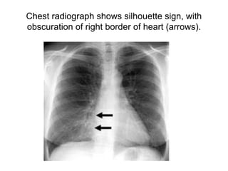
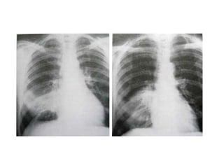
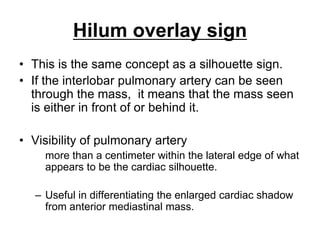
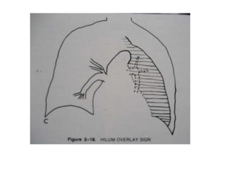
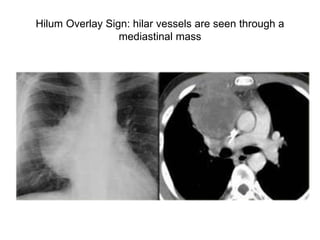
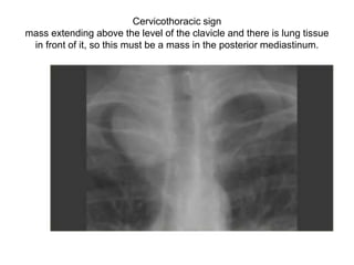
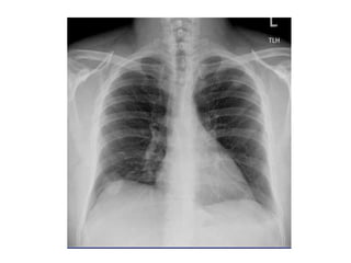
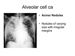
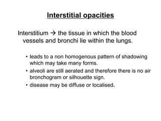
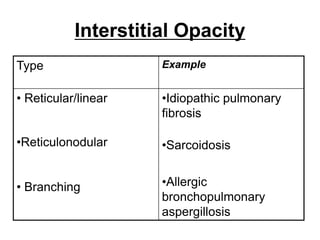
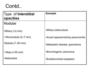
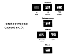
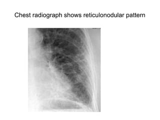
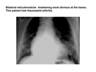
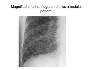
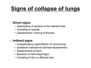
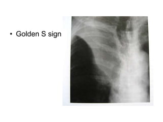
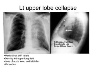
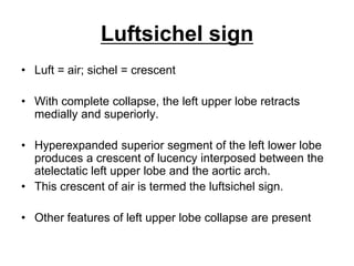
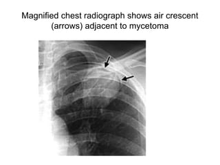
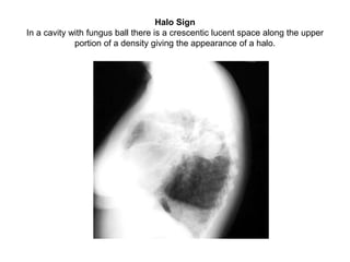
SlideShare a Scribd company logo
1 of 78
Abnormal Sign in Chest X-
ray
Silhouette sign:
Density difference ļƒ  delineation of the outline.
There are four basic densities in x-ray images:
gas
fat
water / soft tissue
bone / calcium
Loss of density difference of the adjacent
structures ļƒ  loss of silhouette.
Silhouette sign contdā€¦
ā€¢ The silhouette sign is the absence of depiction
of an anatomic soft-tissue border resulting
from the juxtaposition of structures of similar
radiographic attenuation.
ā€¢ The sign actually refers to the absence of a
silhouette.
ā€¢ If the borders are retained ļƒ 
ā€“ Not adjacent to each other or
ā€“ Of different radiographic densities.
Silhouette Sign
ā€¢ Cardiac margins are clearly seen because there
is contrast between the fluid density of the heart
and the adjacent air filled alveoli.
ā€¢ If the adjacent lung is devoid of air, the clarity of
the silhouette will be lost.
ā€¢ Pleura encircles the lung and diseases of the
pleura can also obliterate silhouettes. The same
is true for mediastinal masses.
Silhouette Adjacent
Lobe/Segment
Right diaphragm RLL/Basal segments
Right heart margin RML/Medial segment
Ascending aorta RUL/Anterior segment
Aortic knob LUL/Apicoposterior segment
Left heart margin Lingula/Inferior segment
Descending aorta LLL/Superior and medial
segments
Left diaphragm LLL/Basal segments
Chest radiograph shows silhouette sign, with
obscuration of right border of heart (arrows).
Lingular segment consolidation
Hilum overlay sign
ā€¢ This is the same concept as a silhouette sign.
ā€¢ If the interlobar pulmonary artery can be seen
through the mass, it means that the mass seen
is either in front of or behind it.
ā€¢ Visibility of pulmonary artery
more than a centimeter within the lateral edge of what
appears to be the cardiac silhouette.
ā€“ Useful in differentiating the enlarged cardiac shadow
from anterior mediastinal mass.
Hilum Overlay Sign: hilar vessels are seen through a
mediastinal mass
Hilum convergence sign
ā€¢ If pulmonary artery branches converge
towards the opacity ļƒ  enlarged artery
ā€¢ If converge towards heart (i.e., seen
through the opacity) ļƒ  hilar/mediastinal
mass
Cervico-thoracic sign
ā€“ If the thoracic lesion is in anatomic contact with soft tissues of
neck its contiguous border will be lost.
ā€¢ Lesion clearly visible above the clavicles ļƒ lies
posteriorly within the thorax.
ā€¢ If cephalic border of the lesion disappears as it
approaches the clavicle ļƒ  cervico-thoracic lesion
(partly in anterior mediastinum and partly in neck)
Cervicothoracic sign
mass extending above the level of the clavicle and there is lung tissue
in front of it, so this must be a mass in the posterior mediastinum.
Thoraco-abdominal sign
ā€¢ Posterior costophrenic sulcus extends
more caudally than anterior basilar lung.
ā€¢ Lesion extends below the dome of
diaphragm must be in posterior chest
whereas lesion terminates at dome must
be anterior.
Air bronchogram
ā€¢ Visualization of air-filled bronchi
surrounded by air-less lung.
Normal lung. Bronchi not seen Bronchi visible because the air
in the surrounding alveoli has
been replaced by fluid.
Air Bronchogram
ā€¢ In a normal chest x-ray, the tracheobronchial
tree is not visible beyond 4th order.
ā€¢ The lumen of bronchus contains air and the
surrounding alveoli contain air. Thus there is no
contrast to visualize bronchi.
ā€¢ The air column in bronchi beyond 4th order
becomes recognizable if the surrounding alveoli
is filled, providing a contrast = Air
bronchogram.
Chest x-ray of a patient with right upper lobe consolidation.
ā€¢ Indicates that the lesion is intrapulmonary.
ā€¢ Seen in
ā€“ Pneumonia
ā€“ Pulmonary edema
ā€“ Hyaline membrane disease
ā€“ Alveolar cell carcinoma
ā€“ Lymphoma
ā€“ Radiation pneumonitis
Bilateral upper lobe consolidation
ā€¢ Air alveologram
ā€“ Tiny areas of radiolucency within the
surrounding air-less lung
Patterns of Parenchymal Opacity
ā€¢ Shadowing in the lungs is due to:
ā€“ Alveolar disease
ā€“ Interstitial disease
ā€¢ This distinction is sometimes difficult and there is
often overlap between the two.
ā€¢ But it does help in interpretation and deciding
the possible cause of an abnormality. There are
often features of both.
ā€¢ Alveolar / Air-space / Acinar opacities
ā€“ Acinus
ā€¢ functional unit, distal to a terminal bronchiole
ā€¢ 6-8 mm in size
ā€¢ Communication via pores of Kohn and channels of
Lambert
Acinar Shadows
ā€¢ Features
ā€“ Acinar nodules ā€“ 6-8mm sized nodules
ā€“ Fluffy (ill-defined) margins
ā€“ Early coalescence ļƒ  consolidation
ā€“ Air bronchogram / alveologram
ā€“ Segmental / lobar distribution
ā€“ Bat's wing (butterfly) distribution
ā€“ Rapidly changing over time
ā€¢ Alveolar nodules
Alveolar cell ca
ā€¢ Acinar Nodules
ā€¢ Nodules of varying
size with irregular
margins
Interstitial opacities
Interstitium ļƒ  the tissue in which the blood
vessels and bronchi lie within the lungs.
ā€¢ leads to a non homogenous pattern of shadowing
which may take many forms.
ā€¢ alveoli are still aerated and therefore there is no air
bronchogram or silhouette sign.
ā€¢ disease may be diffuse or localised.
Interstitial Opacity
Type Example
ā€¢ Reticular/linear
ā€¢Reticulonodular
ā€¢ Branching
ā€¢Idiopathic pulmonary
fibrosis
ā€¢Sarcoidosis
ā€¢Allergic
bronchopulmonary
aspergillosis
Contd..
Type of Interstitial
opacities
Example
Nodular
ā€¢Miliary (<2 mm)
ā€¢ Micronodule (2ā€“7 mm)
ā€¢Nodule (7ā€“30 mm)
ā€¢ Mass (>30 mm)
ā€¢Atelectasis
ā€¢Miliary tuberculosis
ā€¢Acute hypersensitivity pneumonitis
ā€¢Metastatic disease, granuloma
ā€¢Bronchogenic carcinoma
ā€¢Endobronchial neoplasm
Patterns of Interstitial
Opacities in CXR
reticular pattern
ā€¢ a reticular pattern is a collection of
innumerable small linear opacities that, by
summation, produce an appearance
resembling a net.
Chest radiograph shows reticular pattern
reticulonodular pattern
ā€¢ A combined reticular and nodular pattern, the
reticulonodular pattern is usually the result of the
summation of points of intersection of
innumerable lines, creating the effect on chest
radiographs of superimposed micronodules.
ā€¢ The dimension of the nodules depends on the
size and number of linear or curvilinear
elements.
Chest radiograph shows reticulonodular pattern
Bilateral reticulonodular shadowing most obvious at the bases.
This patient had rheumatoid arthritis.
Magnified chest radiograph shows a nodular
pattern.
Kerley Lines
ā€¢ Indicative of interstitial disease.
ā€¢ A: Long wavy lines in upper and mid lung field
ā€¢ B: 2-3 cm long, 1-2mm thick, pleural based in
bases perpendicular to lateral chest; reliable,
thickening of interlobular septa.
ā€¢ C: Fine fibrillatory lines: You can imagine this on
most cases
Kerley C lines
Kerley A lines
Kerley B lines
Kerley lines
Causes of Kerleyā€™s lines (A,B & C)
ā€¢ Transient
ā€“ Common: Pulmonary edema
ā€“ Rare: Pneumonia, pulmonary hemorrhage, Transient
respiratory distress of newborn
ā€¢ Persistent
ā€“ Common:
ā€¢ Lymphangitic metastases
ā€¢ Pneumoconiosis
ā€¢ Rheumatic mitral valve disease
ā€“ Rare:
ā€¢ Alveolar cell carcinoma
ā€¢ Congenital heart disease
ā€¢ Interstitial fibrosis of any cause
ā€¢ Lymphoma
ā€¢ Mineral oil aspiration etc.
Signs of collapse of lungs
ā€“ Direct signs
ā€¢ Opacity/loss of aeration of the affected lobe
ā€¢ Crowding of vessels
ā€¢ Dispalcement / bowing of fissures
ā€“ Indirect signs
ā€¢ Compensatory hyperinflation of normal lung
ā€¢ Ipsilateral mediastinal/ tracheal displacement
ā€¢ Displacement of hilum
ā€¢ Elevation of hemi-diaphragm
ā€¢ Crowding of ribs on affected side
ā€¢ Lobar collapse
The lobes collapse in characteristic fashion:
ā€“ 1. The upper lobes collapse upwards,
medially and anteriorly
ā€“ 2. The middle lobe goes downwards and
medially
ā€“ 3. The lower lobes collapse posteriorly,
medially and downwards.
The lesser
fissure moves
upwards but
remains pivoted
at the R hilum
medially
Lateral view.
The upper lobe
collapses
upwards,
anteriorly, and
towards the
mediastinum.
Collapse of the R upper lobe.
Opacity in the upper lobe,
silhouette sign upper
mediastinum & upward
displacement of the lesser
fissure.
S Curve of Golden
ā€¢ When there is a mass adjacent to a
fissure, the fissure takes the shape of an
"S". The proximal convexity is due to a
mass, and the distal concavity is due to
atelectasis.
Posteroanterior radiograph of the chest demonstrates the
Golden S sign. Note the convexity (arrowhead) from the
mass and the concavity (arrow) of the minor fissure
ā€¢ Golden S sign
Lt upper lobe collapse
ā€¢Mediastinal shift to left
ā€¢Density left upper lung field
ā€¢Loss of aortic knob and left hilar
silhouettes
Bowing Sign
ā€¢ In LUL atelectasis or following resection,
the oblique fissure bows forwards (lateral
view).
ā€¢ Bowing sign refers to this feature.
Bowing sign
Luftsichel sign
ā€¢ Luft = air; sichel = crescent
ā€¢ With complete collapse, the left upper lobe retracts
medially and superiorly.
ā€¢ Hyperexpanded superior segment of the left lower lobe
produces a crescent of lucency interposed between the
atelectatic left upper lobe and the aortic arch.
ā€¢ This crescent of air is termed the luftsichel sign.
ā€¢ Other features of left upper lobe collapse are present
Luftsichel sign
Rt. middle lobe collapse. There
is a density next to the heart,
below the R hilum, which is
roughly triangular in shape
Lateral view shows the middle
lobe collapse more clearly. The
triangular opacity anteriorly is
the collapsed lobe
Atelectasis Right Lower Lobe
ā€¢ Density in right
lower lung field
ā€¢ Indistinct right
diaphragm
ā€¢ Right heart
silhouette retained
ā€¢ Transverse fissure
moved down
ā€¢ Right hilum
moved down
Left Lower Lobe Atelectasis
ā€¢Inhomogeneous cardiac
density
ā€¢Left hilum pulled down
ā€¢Non-visualization of left
diaphragm
ā€¢Triangular retrocardiac
atelectatic LLL
Plate (linear) atelectasis
ā€¢ Is a focal area of subsegmental atelectasis with a linear
configuration, almost always extending to the pleura.
ā€¢ Also known as discoid or plate like atelectasis.
ā€¢ It is commonly horizontal but sometimes oblique or
vertical.
ā€¢ The thickness of the atelectasis may range from a few
millimeters to more than 1 cm.
ā€¢ Seen in impaired diaphragmatic motion: thoracic trauma,
subphrenic disease, pathologic elevation of diaphragm,
normal hypersthenic individuals with a high diaphragm.
Chest radiograph shows basal linear atelectasis
ā€¢ Plate atelectasis
ā€¢ linear subsegmental atelectatic shadows
at the lung bases
Cut Off Sign
ā€¢ An abrupt ending of visualized bronchus =
"cut off sign".
ā€¢ It indicates an intrabronchial lesion.
ā€¢ This is useful to identify the etiology of
atelectasis .
Open Bronchus Sign / Alveolar
Atelectasis
ā€¢ Presence of air bronchogram in atelectatic
lung.
ā€¢ indicates that the airways are patent.
ā€¢ Commonly seen in adhesive alveolar
atelectasis.
Open bronchus sign
Close up view
Air crescent/ halo sign
ā€¢ An air crescent is a collection of air in a
crescentic shape that separates the wall of a
cavity from an inner mass.
ā€¢ The air crescent sign is often considered
characteristic of either Aspergillus colonization of
preexisting cavities or retraction of infarcted lung
in angioinvasive aspergillosis.
ā€¢ other causes:
ā€“ tuberculosis, Wegener granulomatosis, intracavitary
hemorrhage, lung cancer, lung gangrene
Magnified chest radiograph shows air crescent
(arrows) adjacent to mycetoma
Halo Sign
In a cavity with fungus ball there is a crescentic lucent space along the upper
portion of a density giving the appearance of a halo.
Double Density
ā€¢ Heart should be of uniform density, except over vertebra
and descending aorta.
ā€¢ If increased density in one portion compared to rest of
the heart, consider an abnormal density either in front or
behind the heart.
ā€¢ Left atrial enlargement can be recognized by the circular
double density.
ā€¢ LLL disease, hiatal hernia and posterior mediastinal
masses will give double density over left side of heart.
ā€¢ Esophageal disease, posterior mediastinal masses and
hiatal hernia can give double density over right side of
cardiac density.
Double density
Bulging Fissure Sign
Consolidation spreading rapidly, causing lobar expansion and
bulging of the adjacent fissure inferiorly .
The most common infective causative agents are
Klebsiella pneumoniae, Streptococcus pneumoniae,
Pseudomonas aeruginosa, Staphylococcus aureus
Continuous Diaphragm Sign
Continuous lucency outlining the
base of the heart, representing
pneumomediastinum .
Air in the mediastinum tracks
extrapleurally, between the heart
and diaphragm .
Pneumopericardium can have a
similar appearance but will show
air circumferentially outlining the
heart.
CT angiogram Sign
Identification of vessels within an
airless portion of lung on contrast-
enhanced CT .
The vessels are prominently seen
against a background of low-
attenuation material .
Associated with:
bronchoalveolar cell carcinoma
lymphoma
infectious pneumonias.
Deep Sulcus Sign
This sign refers to a deep
collection of intrapleural air
(pneumothorax) in the
costophrenic sulcus as seen
on the supine chest
radiograph .
Fallen Lung Sign
This sign refers to the appearance
of the collapsed lung occurring
with a fractured bronchus .
The bronchial fracture results in
the lung to fall away from the
hilum, either inferiorly and laterally
in an upright patient or posteriorly,
as seen on CT in a supine patient.
DD:
Pneumothorax causes a lung to
collapse inward toward the hilum.
Finger in Glove Sign
In allergic bronchopulmonary aspergillosis.
The impacted bronchi appear radiographically as opacities with distinctive
shapes.
Hampton Hump Sign
Pulmonary infarction secondary to pulmonary embolism produces
an abnormal area of opacification on the chest radiograph, which
is always in contact with the pleural surface.
Juxtaphrenic Peak Sign
This sign refers to a small triangular shadow that obscures the dome of the
diaphragm secondary to upper lobe atelectasis . The shadow is caused by
traction on the lower end of the major fissure, the inferior accessory fissure, or
the inferior pulmonary ligament.
THANK YOU
QUESTIONS
ā€¢ Silhouette Sign.
ā€¢ Hilum overlay sign.
ā€¢ Air bronchogram.
ā€¢ Patterns of parenchymal opacities.
ā€¢ Causes of kerelyā€™s line.
ā€¢ Signs of collapse of lung.
ā€¢ Luftsichel sign.
ā€¢ Air crescent sign.
ā€¢ Continous diaphragm sign.

More Related Content

What's hot

abnormal chest xray ppt
abnormal chest xray ppt abnormal chest xray ppt
abnormal chest xray ppt shyamsobti
Ā 
Role of Radiology in Pulmonary Tuberculosis
Role of Radiology in Pulmonary TuberculosisRole of Radiology in Pulmonary Tuberculosis
Role of Radiology in Pulmonary TuberculosisWaseem M.Nizamani
Ā 
Paediatric chest imaging
Paediatric chest imagingPaediatric chest imaging
Paediatric chest imagingSidra Afzal
Ā 
Solitary pulmonary nodule
Solitary pulmonary noduleSolitary pulmonary nodule
Solitary pulmonary noduleNavni Garg
Ā 
Apls Pediatric Emergency Radiology 2
Apls Pediatric Emergency Radiology 2Apls Pediatric Emergency Radiology 2
Apls Pediatric Emergency Radiology 2Dang Thanh Tuan
Ā 
Interstitial lung diseases- HRCT
Interstitial lung diseases- HRCTInterstitial lung diseases- HRCT
Interstitial lung diseases- HRCTNavdeep Shah
Ā 
lung hrct patterns
lung hrct patterns lung hrct patterns
lung hrct patterns Satish Naga
Ā 
Presentation1, radiological imaging of scimitar syndrome
Presentation1, radiological imaging of scimitar syndromePresentation1, radiological imaging of scimitar syndrome
Presentation1, radiological imaging of scimitar syndromeAbdellah Nazeer
Ā 
Diagnostic Imaging of Pulmonary infections
Diagnostic Imaging of Pulmonary infectionsDiagnostic Imaging of Pulmonary infections
Diagnostic Imaging of Pulmonary infectionsMohamed M.A. Zaitoun
Ā 
Presentation1.pptx, radiological signs in thoracic radiology.
Presentation1.pptx, radiological signs in thoracic radiology.Presentation1.pptx, radiological signs in thoracic radiology.
Presentation1.pptx, radiological signs in thoracic radiology.Abdellah Nazeer
Ā 
Ascites and Pleural Effusion
 Ascites and Pleural Effusion Ascites and Pleural Effusion
Ascites and Pleural EffusionMedia Genie
Ā 
Presentation1.pptx, radiological imaging of sarcoidosis.
Presentation1.pptx, radiological imaging of sarcoidosis.Presentation1.pptx, radiological imaging of sarcoidosis.
Presentation1.pptx, radiological imaging of sarcoidosis.Abdellah Nazeer
Ā 
Presentation1.pptx, radiological imaging of extra nodal lymphoma.
Presentation1.pptx, radiological imaging of extra nodal lymphoma.Presentation1.pptx, radiological imaging of extra nodal lymphoma.
Presentation1.pptx, radiological imaging of extra nodal lymphoma.Abdellah Nazeer
Ā 
Radiological signs in chest medicine part 2
Radiological signs in chest medicine part 2Radiological signs in chest medicine part 2
Radiological signs in chest medicine part 2Gamal Agmy
Ā 
chest xray of cardiovascular disease PPT
chest xray of cardiovascular disease PPTchest xray of cardiovascular disease PPT
chest xray of cardiovascular disease PPTNaba Kumar Barman
Ā 
Normal chest xray
Normal chest xrayNormal chest xray
Normal chest xraySai Kumar Sai
Ā 
Lung cancer radiology
Lung cancer radiologyLung cancer radiology
Lung cancer radiologydocaashishgupt
Ā 
Radiological features of Lung cancer Dr. Muhammad Bin Zulfiqar
Radiological features of Lung cancer Dr. Muhammad Bin ZulfiqarRadiological features of Lung cancer Dr. Muhammad Bin Zulfiqar
Radiological features of Lung cancer Dr. Muhammad Bin ZulfiqarDr. Muhammad Bin Zulfiqar
Ā 
Mediastinal lesions final
Mediastinal lesions   finalMediastinal lesions   final
Mediastinal lesions finalGobardhan Thapa
Ā 

What's hot (20)

abnormal chest xray ppt
abnormal chest xray ppt abnormal chest xray ppt
abnormal chest xray ppt
Ā 
Role of Radiology in Pulmonary Tuberculosis
Role of Radiology in Pulmonary TuberculosisRole of Radiology in Pulmonary Tuberculosis
Role of Radiology in Pulmonary Tuberculosis
Ā 
Paediatric chest imaging
Paediatric chest imagingPaediatric chest imaging
Paediatric chest imaging
Ā 
Solitary pulmonary nodule
Solitary pulmonary noduleSolitary pulmonary nodule
Solitary pulmonary nodule
Ā 
Apls Pediatric Emergency Radiology 2
Apls Pediatric Emergency Radiology 2Apls Pediatric Emergency Radiology 2
Apls Pediatric Emergency Radiology 2
Ā 
Interstitial lung diseases- HRCT
Interstitial lung diseases- HRCTInterstitial lung diseases- HRCT
Interstitial lung diseases- HRCT
Ā 
lung hrct patterns
lung hrct patterns lung hrct patterns
lung hrct patterns
Ā 
Presentation1, radiological imaging of scimitar syndrome
Presentation1, radiological imaging of scimitar syndromePresentation1, radiological imaging of scimitar syndrome
Presentation1, radiological imaging of scimitar syndrome
Ā 
Diagnostic Imaging of Pulmonary infections
Diagnostic Imaging of Pulmonary infectionsDiagnostic Imaging of Pulmonary infections
Diagnostic Imaging of Pulmonary infections
Ā 
Presentation1.pptx, radiological signs in thoracic radiology.
Presentation1.pptx, radiological signs in thoracic radiology.Presentation1.pptx, radiological signs in thoracic radiology.
Presentation1.pptx, radiological signs in thoracic radiology.
Ā 
Ascites and Pleural Effusion
 Ascites and Pleural Effusion Ascites and Pleural Effusion
Ascites and Pleural Effusion
Ā 
Presentation1.pptx, radiological imaging of sarcoidosis.
Presentation1.pptx, radiological imaging of sarcoidosis.Presentation1.pptx, radiological imaging of sarcoidosis.
Presentation1.pptx, radiological imaging of sarcoidosis.
Ā 
Presentation1.pptx, radiological imaging of extra nodal lymphoma.
Presentation1.pptx, radiological imaging of extra nodal lymphoma.Presentation1.pptx, radiological imaging of extra nodal lymphoma.
Presentation1.pptx, radiological imaging of extra nodal lymphoma.
Ā 
Radiological signs in chest medicine part 2
Radiological signs in chest medicine part 2Radiological signs in chest medicine part 2
Radiological signs in chest medicine part 2
Ā 
chest xray of cardiovascular disease PPT
chest xray of cardiovascular disease PPTchest xray of cardiovascular disease PPT
chest xray of cardiovascular disease PPT
Ā 
Normal chest xray
Normal chest xrayNormal chest xray
Normal chest xray
Ā 
Basics of CT Chest
Basics of CT Chest Basics of CT Chest
Basics of CT Chest
Ā 
Lung cancer radiology
Lung cancer radiologyLung cancer radiology
Lung cancer radiology
Ā 
Radiological features of Lung cancer Dr. Muhammad Bin Zulfiqar
Radiological features of Lung cancer Dr. Muhammad Bin ZulfiqarRadiological features of Lung cancer Dr. Muhammad Bin Zulfiqar
Radiological features of Lung cancer Dr. Muhammad Bin Zulfiqar
Ā 
Mediastinal lesions final
Mediastinal lesions   finalMediastinal lesions   final
Mediastinal lesions final
Ā 

Similar to Chest X-ray Silhouette Sign and Loss of Density Difference

Abnormal sign in chest X- Ray
Abnormal sign in chest X- RayAbnormal sign in chest X- Ray
Abnormal sign in chest X- RayUpakar Paudel
Ā 
Chest radiology part 1
Chest radiology part 1Chest radiology part 1
Chest radiology part 1Gamal Agmy
Ā 
Radiological presentation of chest diseases gamal agmy
Radiological presentation of chest diseases  gamal agmyRadiological presentation of chest diseases  gamal agmy
Radiological presentation of chest diseases gamal agmyGamal Agmy
Ā 
Radiological Presentation of Chest Diseases
Radiological Presentation of Chest Diseases  Radiological Presentation of Chest Diseases
Radiological Presentation of Chest Diseases Gamal Agmy
Ā 
Helpful radiological signs in cxr25 11-91
Helpful radiological signs in cxr25 11-91Helpful radiological signs in cxr25 11-91
Helpful radiological signs in cxr25 11-91aalmasi1970
Ā 
Radiological signs in chest medicine
Radiological signs in chest medicineRadiological signs in chest medicine
Radiological signs in chest medicineGamal Agmy
Ā 
Radiological Signs in Chest Medicine
Radiological Signs in Chest MedicineRadiological Signs in Chest Medicine
Radiological Signs in Chest MedicineGamal Agmy
Ā 
Chest x rays
Chest x raysChest x rays
Chest x raysPratik Kumar
Ā 
Medicine- Xrays
Medicine- XraysMedicine- Xrays
Medicine- XraysPratik Kumar
Ā 
Chest imaging
Chest imagingChest imaging
Chest imagingJANARDANCNMC
Ā 
Collapse consolidation
Collapse consolidationCollapse consolidation
Collapse consolidationairwave12
Ā 
Normal Chest X-Rays & Its Systemic Approach- Anatomy
Normal Chest X-Rays & Its Systemic Approach- AnatomyNormal Chest X-Rays & Its Systemic Approach- Anatomy
Normal Chest X-Rays & Its Systemic Approach- AnatomyDr.Bijay Yadav
Ā 
Interactive radiology case presentation
Interactive radiology case presentationInteractive radiology case presentation
Interactive radiology case presentationGamal Agmy
Ā 
Chest x. ray interpretation and teaching
Chest x. ray interpretation and teachingChest x. ray interpretation and teaching
Chest x. ray interpretation and teachingsamirelansary
Ā 
Chest x. ray interpretation and teaching
Chest x. ray interpretation and teachingChest x. ray interpretation and teaching
Chest x. ray interpretation and teachingsamirelansary
Ā 
XRAY
XRAYXRAY
XRAYSufindc
Ā 

Similar to Chest X-ray Silhouette Sign and Loss of Density Difference (20)

Abnormal sign in chest X- Ray
Abnormal sign in chest X- RayAbnormal sign in chest X- Ray
Abnormal sign in chest X- Ray
Ā 
Chest radiology part 1
Chest radiology part 1Chest radiology part 1
Chest radiology part 1
Ā 
Radiological presentation of chest diseases gamal agmy
Radiological presentation of chest diseases  gamal agmyRadiological presentation of chest diseases  gamal agmy
Radiological presentation of chest diseases gamal agmy
Ā 
Radiological Presentation of Chest Diseases
Radiological Presentation of Chest Diseases  Radiological Presentation of Chest Diseases
Radiological Presentation of Chest Diseases
Ā 
Helpful radiological signs in cxr25 11-91
Helpful radiological signs in cxr25 11-91Helpful radiological signs in cxr25 11-91
Helpful radiological signs in cxr25 11-91
Ā 
Radiological signs in chest medicine
Radiological signs in chest medicineRadiological signs in chest medicine
Radiological signs in chest medicine
Ā 
Radiological Signs in Chest Medicine
Radiological Signs in Chest MedicineRadiological Signs in Chest Medicine
Radiological Signs in Chest Medicine
Ā 
Chest x rays
Chest x raysChest x rays
Chest x rays
Ā 
Reading chest-x-rays
Reading chest-x-rays Reading chest-x-rays
Reading chest-x-rays
Ā 
Medicine- Xrays
Medicine- XraysMedicine- Xrays
Medicine- Xrays
Ā 
Xrays
XraysXrays
Xrays
Ā 
Abnormal Chest X rays
Abnormal Chest X raysAbnormal Chest X rays
Abnormal Chest X rays
Ā 
Chest imaging
Chest imagingChest imaging
Chest imaging
Ā 
Collapse consolidation
Collapse consolidationCollapse consolidation
Collapse consolidation
Ā 
Normal Chest X-Rays & Its Systemic Approach- Anatomy
Normal Chest X-Rays & Its Systemic Approach- AnatomyNormal Chest X-Rays & Its Systemic Approach- Anatomy
Normal Chest X-Rays & Its Systemic Approach- Anatomy
Ā 
Interactive radiology case presentation
Interactive radiology case presentationInteractive radiology case presentation
Interactive radiology case presentation
Ā 
Chest x. ray interpretation and teaching
Chest x. ray interpretation and teachingChest x. ray interpretation and teaching
Chest x. ray interpretation and teaching
Ā 
Chest x. ray interpretation and teaching
Chest x. ray interpretation and teachingChest x. ray interpretation and teaching
Chest x. ray interpretation and teaching
Ā 
Chest x. ray interpretation and teaching
Chest x. ray interpretation and teachingChest x. ray interpretation and teaching
Chest x. ray interpretation and teaching
Ā 
XRAY
XRAYXRAY
XRAY
Ā 

More from Milan Silwal

Urinary tract infections
Urinary tract infectionsUrinary tract infections
Urinary tract infectionsMilan Silwal
Ā 
Retroperitoneal masses
Retroperitoneal masses Retroperitoneal masses
Retroperitoneal masses Milan Silwal
Ā 
Renal artery Doppler and renal transplant
Renal artery Doppler and renal transplantRenal artery Doppler and renal transplant
Renal artery Doppler and renal transplantMilan Silwal
Ā 
RGU (Retrograde urethrogram), MCU (Micturating cystourethrogram) and its inte...
RGU (Retrograde urethrogram), MCU (Micturating cystourethrogram) and its inte...RGU (Retrograde urethrogram), MCU (Micturating cystourethrogram) and its inte...
RGU (Retrograde urethrogram), MCU (Micturating cystourethrogram) and its inte...Milan Silwal
Ā 
Intravenous urography
Intravenous urographyIntravenous urography
Intravenous urographyMilan Silwal
Ā 
Imaging of urinary bladder carcinoma
Imaging of urinary bladder carcinomaImaging of urinary bladder carcinoma
Imaging of urinary bladder carcinomaMilan Silwal
Ā 
Imaging in prostatic patholgy
Imaging in prostatic patholgyImaging in prostatic patholgy
Imaging in prostatic patholgyMilan Silwal
Ā 
Genito-urinary trauma
Genito-urinary traumaGenito-urinary trauma
Genito-urinary traumaMilan Silwal
Ā 
Endometrial abnormalities
Endometrial abnormalitiesEndometrial abnormalities
Endometrial abnormalitiesMilan Silwal
Ā 
Embryology of genitourinary system
Embryology of genitourinary systemEmbryology of genitourinary system
Embryology of genitourinary systemMilan Silwal
Ā 
Diseases of ureters
Diseases of uretersDiseases of ureters
Diseases of uretersMilan Silwal
Ā 
An apporach to ovarian pathology
An apporach to ovarian pathologyAn apporach to ovarian pathology
An apporach to ovarian pathologyMilan Silwal
Ā 
X ray c-spine
X ray c-spineX ray c-spine
X ray c-spineMilan Silwal
Ā 
Non traumatic Subarachnoid hemorrhage (SAH)
Non traumatic Subarachnoid hemorrhage (SAH)Non traumatic Subarachnoid hemorrhage (SAH)
Non traumatic Subarachnoid hemorrhage (SAH)Milan Silwal
Ā 
Imaging in orbital pathology
Imaging in orbital pathologyImaging in orbital pathology
Imaging in orbital pathologyMilan Silwal
Ā 
Neoplastic disorders of spinal cord
Neoplastic disorders of spinal cordNeoplastic disorders of spinal cord
Neoplastic disorders of spinal cordMilan Silwal
Ā 
Neoplastic disorders of spinal cord
Neoplastic disorders of spinal cordNeoplastic disorders of spinal cord
Neoplastic disorders of spinal cordMilan Silwal
Ā 
Neonatal transcranial USG
Neonatal transcranial USGNeonatal transcranial USG
Neonatal transcranial USGMilan Silwal
Ā 
Mastoid diseases imaging
Mastoid diseases imagingMastoid diseases imaging
Mastoid diseases imagingMilan Silwal
Ā 

More from Milan Silwal (20)

Urinary tract infections
Urinary tract infectionsUrinary tract infections
Urinary tract infections
Ā 
Retroperitoneal masses
Retroperitoneal masses Retroperitoneal masses
Retroperitoneal masses
Ā 
Renal artery Doppler and renal transplant
Renal artery Doppler and renal transplantRenal artery Doppler and renal transplant
Renal artery Doppler and renal transplant
Ā 
RGU (Retrograde urethrogram), MCU (Micturating cystourethrogram) and its inte...
RGU (Retrograde urethrogram), MCU (Micturating cystourethrogram) and its inte...RGU (Retrograde urethrogram), MCU (Micturating cystourethrogram) and its inte...
RGU (Retrograde urethrogram), MCU (Micturating cystourethrogram) and its inte...
Ā 
Mammography
MammographyMammography
Mammography
Ā 
Intravenous urography
Intravenous urographyIntravenous urography
Intravenous urography
Ā 
Imaging of urinary bladder carcinoma
Imaging of urinary bladder carcinomaImaging of urinary bladder carcinoma
Imaging of urinary bladder carcinoma
Ā 
Imaging in prostatic patholgy
Imaging in prostatic patholgyImaging in prostatic patholgy
Imaging in prostatic patholgy
Ā 
Genito-urinary trauma
Genito-urinary traumaGenito-urinary trauma
Genito-urinary trauma
Ā 
Endometrial abnormalities
Endometrial abnormalitiesEndometrial abnormalities
Endometrial abnormalities
Ā 
Embryology of genitourinary system
Embryology of genitourinary systemEmbryology of genitourinary system
Embryology of genitourinary system
Ā 
Diseases of ureters
Diseases of uretersDiseases of ureters
Diseases of ureters
Ā 
An apporach to ovarian pathology
An apporach to ovarian pathologyAn apporach to ovarian pathology
An apporach to ovarian pathology
Ā 
X ray c-spine
X ray c-spineX ray c-spine
X ray c-spine
Ā 
Non traumatic Subarachnoid hemorrhage (SAH)
Non traumatic Subarachnoid hemorrhage (SAH)Non traumatic Subarachnoid hemorrhage (SAH)
Non traumatic Subarachnoid hemorrhage (SAH)
Ā 
Imaging in orbital pathology
Imaging in orbital pathologyImaging in orbital pathology
Imaging in orbital pathology
Ā 
Neoplastic disorders of spinal cord
Neoplastic disorders of spinal cordNeoplastic disorders of spinal cord
Neoplastic disorders of spinal cord
Ā 
Neoplastic disorders of spinal cord
Neoplastic disorders of spinal cordNeoplastic disorders of spinal cord
Neoplastic disorders of spinal cord
Ā 
Neonatal transcranial USG
Neonatal transcranial USGNeonatal transcranial USG
Neonatal transcranial USG
Ā 
Mastoid diseases imaging
Mastoid diseases imagingMastoid diseases imaging
Mastoid diseases imaging
Ā 

Recently uploaded

Vip Call Girls Anna Salai Chennai šŸ‘‰ 8250192130 ā£ļøšŸ’Æ Top Class Girls Available
Vip Call Girls Anna Salai Chennai šŸ‘‰ 8250192130 ā£ļøšŸ’Æ Top Class Girls AvailableVip Call Girls Anna Salai Chennai šŸ‘‰ 8250192130 ā£ļøšŸ’Æ Top Class Girls Available
Vip Call Girls Anna Salai Chennai šŸ‘‰ 8250192130 ā£ļøšŸ’Æ Top Class Girls AvailableNehru place Escorts
Ā 
Artifacts in Nuclear Medicine with Identifying and resolving artifacts.
Artifacts in Nuclear Medicine with Identifying and resolving artifacts.Artifacts in Nuclear Medicine with Identifying and resolving artifacts.
Artifacts in Nuclear Medicine with Identifying and resolving artifacts.MiadAlsulami
Ā 
(šŸ‘‘VVIP ISHAAN ) Russian Call Girls Service Navi MumbaišŸ–•9920874524šŸ–•Independent...
(šŸ‘‘VVIP ISHAAN ) Russian Call Girls Service Navi MumbaišŸ–•9920874524šŸ–•Independent...(šŸ‘‘VVIP ISHAAN ) Russian Call Girls Service Navi MumbaišŸ–•9920874524šŸ–•Independent...
(šŸ‘‘VVIP ISHAAN ) Russian Call Girls Service Navi MumbaišŸ–•9920874524šŸ–•Independent...Taniya Sharma
Ā 
Bangalore Call Girls Marathahalli šŸ“ž 9907093804 High Profile Service 100% Safe
Bangalore Call Girls Marathahalli šŸ“ž 9907093804 High Profile Service 100% SafeBangalore Call Girls Marathahalli šŸ“ž 9907093804 High Profile Service 100% Safe
Bangalore Call Girls Marathahalli šŸ“ž 9907093804 High Profile Service 100% Safenarwatsonia7
Ā 
VIP Russian Call Girls in Varanasi Samaira 8250192130 Independent Escort Serv...
VIP Russian Call Girls in Varanasi Samaira 8250192130 Independent Escort Serv...VIP Russian Call Girls in Varanasi Samaira 8250192130 Independent Escort Serv...
VIP Russian Call Girls in Varanasi Samaira 8250192130 Independent Escort Serv...Neha Kaur
Ā 
Bangalore Call Girls Majestic šŸ“ž 9907093804 High Profile Service 100% Safe
Bangalore Call Girls Majestic šŸ“ž 9907093804 High Profile Service 100% SafeBangalore Call Girls Majestic šŸ“ž 9907093804 High Profile Service 100% Safe
Bangalore Call Girls Majestic šŸ“ž 9907093804 High Profile Service 100% Safenarwatsonia7
Ā 
Low Rate Call Girls Patna Anika 8250192130 Independent Escort Service Patna
Low Rate Call Girls Patna Anika 8250192130 Independent Escort Service PatnaLow Rate Call Girls Patna Anika 8250192130 Independent Escort Service Patna
Low Rate Call Girls Patna Anika 8250192130 Independent Escort Service Patnamakika9823
Ā 
VIP Call Girls Pune Vani 9907093804 Short 1500 Night 6000 Best call girls Ser...
VIP Call Girls Pune Vani 9907093804 Short 1500 Night 6000 Best call girls Ser...VIP Call Girls Pune Vani 9907093804 Short 1500 Night 6000 Best call girls Ser...
VIP Call Girls Pune Vani 9907093804 Short 1500 Night 6000 Best call girls Ser...Miss joya
Ā 
VIP Service Call Girls Sindhi Colony šŸ“³ 7877925207 For 18+ VIP Call Girl At Th...
VIP Service Call Girls Sindhi Colony šŸ“³ 7877925207 For 18+ VIP Call Girl At Th...VIP Service Call Girls Sindhi Colony šŸ“³ 7877925207 For 18+ VIP Call Girl At Th...
VIP Service Call Girls Sindhi Colony šŸ“³ 7877925207 For 18+ VIP Call Girl At Th...jageshsingh5554
Ā 
High Profile Call Girls Coimbatore Saanviā˜Žļø 8250192130 Independent Escort Se...
High Profile Call Girls Coimbatore Saanviā˜Žļø  8250192130 Independent Escort Se...High Profile Call Girls Coimbatore Saanviā˜Žļø  8250192130 Independent Escort Se...
High Profile Call Girls Coimbatore Saanviā˜Žļø 8250192130 Independent Escort Se...narwatsonia7
Ā 
Call Girl Coimbatore Prishaā˜Žļø 8250192130 Independent Escort Service Coimbatore
Call Girl Coimbatore Prishaā˜Žļø  8250192130 Independent Escort Service CoimbatoreCall Girl Coimbatore Prishaā˜Žļø  8250192130 Independent Escort Service Coimbatore
Call Girl Coimbatore Prishaā˜Žļø 8250192130 Independent Escort Service Coimbatorenarwatsonia7
Ā 
Russian Escorts Girls Nehru Place ZINATHI šŸ”9711199012 ā˜Ŗ 24/7 Call Girls Delhi
Russian Escorts Girls  Nehru Place ZINATHI šŸ”9711199012 ā˜Ŗ 24/7 Call Girls DelhiRussian Escorts Girls  Nehru Place ZINATHI šŸ”9711199012 ā˜Ŗ 24/7 Call Girls Delhi
Russian Escorts Girls Nehru Place ZINATHI šŸ”9711199012 ā˜Ŗ 24/7 Call Girls DelhiAlinaDevecerski
Ā 
Russian Call Girls in Pune Tanvi 9907093804 Short 1500 Night 6000 Best call g...
Russian Call Girls in Pune Tanvi 9907093804 Short 1500 Night 6000 Best call g...Russian Call Girls in Pune Tanvi 9907093804 Short 1500 Night 6000 Best call g...
Russian Call Girls in Pune Tanvi 9907093804 Short 1500 Night 6000 Best call g...Miss joya
Ā 
Premium Call Girls Cottonpet Whatsapp 7001035870 Independent Escort Service
Premium Call Girls Cottonpet Whatsapp 7001035870 Independent Escort ServicePremium Call Girls Cottonpet Whatsapp 7001035870 Independent Escort Service
Premium Call Girls Cottonpet Whatsapp 7001035870 Independent Escort Servicevidya singh
Ā 
Russian Call Girls in Pune Riya 9907093804 Short 1500 Night 6000 Best call gi...
Russian Call Girls in Pune Riya 9907093804 Short 1500 Night 6000 Best call gi...Russian Call Girls in Pune Riya 9907093804 Short 1500 Night 6000 Best call gi...
Russian Call Girls in Pune Riya 9907093804 Short 1500 Night 6000 Best call gi...Miss joya
Ā 
CALL ON āž„9907093804 šŸ” Call Girls Hadapsar ( Pune) Girls Service
CALL ON āž„9907093804 šŸ” Call Girls Hadapsar ( Pune)  Girls ServiceCALL ON āž„9907093804 šŸ” Call Girls Hadapsar ( Pune)  Girls Service
CALL ON āž„9907093804 šŸ” Call Girls Hadapsar ( Pune) Girls ServiceMiss joya
Ā 
Call Girl Number in Panvel MumbaišŸ“² 9833363713 šŸ’ž Full Night Enjoy
Call Girl Number in Panvel MumbaišŸ“² 9833363713 šŸ’ž Full Night EnjoyCall Girl Number in Panvel MumbaišŸ“² 9833363713 šŸ’ž Full Night Enjoy
Call Girl Number in Panvel MumbaišŸ“² 9833363713 šŸ’ž Full Night Enjoybabeytanya
Ā 
Call Girls Service Surat Samaira ā¤ļøšŸ‘ 8250192130 šŸ‘„ Independent Escort Service ...
Call Girls Service Surat Samaira ā¤ļøšŸ‘ 8250192130 šŸ‘„ Independent Escort Service ...Call Girls Service Surat Samaira ā¤ļøšŸ‘ 8250192130 šŸ‘„ Independent Escort Service ...
Call Girls Service Surat Samaira ā¤ļøšŸ‘ 8250192130 šŸ‘„ Independent Escort Service ...CALL GIRLS
Ā 
Call Girls Service Pune Vaishnavi 9907093804 Short 1500 Night 6000 Best call ...
Call Girls Service Pune Vaishnavi 9907093804 Short 1500 Night 6000 Best call ...Call Girls Service Pune Vaishnavi 9907093804 Short 1500 Night 6000 Best call ...
Call Girls Service Pune Vaishnavi 9907093804 Short 1500 Night 6000 Best call ...Miss joya
Ā 

Recently uploaded (20)

Vip Call Girls Anna Salai Chennai šŸ‘‰ 8250192130 ā£ļøšŸ’Æ Top Class Girls Available
Vip Call Girls Anna Salai Chennai šŸ‘‰ 8250192130 ā£ļøšŸ’Æ Top Class Girls AvailableVip Call Girls Anna Salai Chennai šŸ‘‰ 8250192130 ā£ļøšŸ’Æ Top Class Girls Available
Vip Call Girls Anna Salai Chennai šŸ‘‰ 8250192130 ā£ļøšŸ’Æ Top Class Girls Available
Ā 
Artifacts in Nuclear Medicine with Identifying and resolving artifacts.
Artifacts in Nuclear Medicine with Identifying and resolving artifacts.Artifacts in Nuclear Medicine with Identifying and resolving artifacts.
Artifacts in Nuclear Medicine with Identifying and resolving artifacts.
Ā 
(šŸ‘‘VVIP ISHAAN ) Russian Call Girls Service Navi MumbaišŸ–•9920874524šŸ–•Independent...
(šŸ‘‘VVIP ISHAAN ) Russian Call Girls Service Navi MumbaišŸ–•9920874524šŸ–•Independent...(šŸ‘‘VVIP ISHAAN ) Russian Call Girls Service Navi MumbaišŸ–•9920874524šŸ–•Independent...
(šŸ‘‘VVIP ISHAAN ) Russian Call Girls Service Navi MumbaišŸ–•9920874524šŸ–•Independent...
Ā 
Escort Service Call Girls In Sarita Vihar,, 99530Ā°56974 Delhi NCR
Escort Service Call Girls In Sarita Vihar,, 99530Ā°56974 Delhi NCREscort Service Call Girls In Sarita Vihar,, 99530Ā°56974 Delhi NCR
Escort Service Call Girls In Sarita Vihar,, 99530Ā°56974 Delhi NCR
Ā 
Bangalore Call Girls Marathahalli šŸ“ž 9907093804 High Profile Service 100% Safe
Bangalore Call Girls Marathahalli šŸ“ž 9907093804 High Profile Service 100% SafeBangalore Call Girls Marathahalli šŸ“ž 9907093804 High Profile Service 100% Safe
Bangalore Call Girls Marathahalli šŸ“ž 9907093804 High Profile Service 100% Safe
Ā 
VIP Russian Call Girls in Varanasi Samaira 8250192130 Independent Escort Serv...
VIP Russian Call Girls in Varanasi Samaira 8250192130 Independent Escort Serv...VIP Russian Call Girls in Varanasi Samaira 8250192130 Independent Escort Serv...
VIP Russian Call Girls in Varanasi Samaira 8250192130 Independent Escort Serv...
Ā 
Bangalore Call Girls Majestic šŸ“ž 9907093804 High Profile Service 100% Safe
Bangalore Call Girls Majestic šŸ“ž 9907093804 High Profile Service 100% SafeBangalore Call Girls Majestic šŸ“ž 9907093804 High Profile Service 100% Safe
Bangalore Call Girls Majestic šŸ“ž 9907093804 High Profile Service 100% Safe
Ā 
Low Rate Call Girls Patna Anika 8250192130 Independent Escort Service Patna
Low Rate Call Girls Patna Anika 8250192130 Independent Escort Service PatnaLow Rate Call Girls Patna Anika 8250192130 Independent Escort Service Patna
Low Rate Call Girls Patna Anika 8250192130 Independent Escort Service Patna
Ā 
VIP Call Girls Pune Vani 9907093804 Short 1500 Night 6000 Best call girls Ser...
VIP Call Girls Pune Vani 9907093804 Short 1500 Night 6000 Best call girls Ser...VIP Call Girls Pune Vani 9907093804 Short 1500 Night 6000 Best call girls Ser...
VIP Call Girls Pune Vani 9907093804 Short 1500 Night 6000 Best call girls Ser...
Ā 
VIP Service Call Girls Sindhi Colony šŸ“³ 7877925207 For 18+ VIP Call Girl At Th...
VIP Service Call Girls Sindhi Colony šŸ“³ 7877925207 For 18+ VIP Call Girl At Th...VIP Service Call Girls Sindhi Colony šŸ“³ 7877925207 For 18+ VIP Call Girl At Th...
VIP Service Call Girls Sindhi Colony šŸ“³ 7877925207 For 18+ VIP Call Girl At Th...
Ā 
High Profile Call Girls Coimbatore Saanviā˜Žļø 8250192130 Independent Escort Se...
High Profile Call Girls Coimbatore Saanviā˜Žļø  8250192130 Independent Escort Se...High Profile Call Girls Coimbatore Saanviā˜Žļø  8250192130 Independent Escort Se...
High Profile Call Girls Coimbatore Saanviā˜Žļø 8250192130 Independent Escort Se...
Ā 
Call Girl Coimbatore Prishaā˜Žļø 8250192130 Independent Escort Service Coimbatore
Call Girl Coimbatore Prishaā˜Žļø  8250192130 Independent Escort Service CoimbatoreCall Girl Coimbatore Prishaā˜Žļø  8250192130 Independent Escort Service Coimbatore
Call Girl Coimbatore Prishaā˜Žļø 8250192130 Independent Escort Service Coimbatore
Ā 
Russian Escorts Girls Nehru Place ZINATHI šŸ”9711199012 ā˜Ŗ 24/7 Call Girls Delhi
Russian Escorts Girls  Nehru Place ZINATHI šŸ”9711199012 ā˜Ŗ 24/7 Call Girls DelhiRussian Escorts Girls  Nehru Place ZINATHI šŸ”9711199012 ā˜Ŗ 24/7 Call Girls Delhi
Russian Escorts Girls Nehru Place ZINATHI šŸ”9711199012 ā˜Ŗ 24/7 Call Girls Delhi
Ā 
Russian Call Girls in Pune Tanvi 9907093804 Short 1500 Night 6000 Best call g...
Russian Call Girls in Pune Tanvi 9907093804 Short 1500 Night 6000 Best call g...Russian Call Girls in Pune Tanvi 9907093804 Short 1500 Night 6000 Best call g...
Russian Call Girls in Pune Tanvi 9907093804 Short 1500 Night 6000 Best call g...
Ā 
Premium Call Girls Cottonpet Whatsapp 7001035870 Independent Escort Service
Premium Call Girls Cottonpet Whatsapp 7001035870 Independent Escort ServicePremium Call Girls Cottonpet Whatsapp 7001035870 Independent Escort Service
Premium Call Girls Cottonpet Whatsapp 7001035870 Independent Escort Service
Ā 
Russian Call Girls in Pune Riya 9907093804 Short 1500 Night 6000 Best call gi...
Russian Call Girls in Pune Riya 9907093804 Short 1500 Night 6000 Best call gi...Russian Call Girls in Pune Riya 9907093804 Short 1500 Night 6000 Best call gi...
Russian Call Girls in Pune Riya 9907093804 Short 1500 Night 6000 Best call gi...
Ā 
CALL ON āž„9907093804 šŸ” Call Girls Hadapsar ( Pune) Girls Service
CALL ON āž„9907093804 šŸ” Call Girls Hadapsar ( Pune)  Girls ServiceCALL ON āž„9907093804 šŸ” Call Girls Hadapsar ( Pune)  Girls Service
CALL ON āž„9907093804 šŸ” Call Girls Hadapsar ( Pune) Girls Service
Ā 
Call Girl Number in Panvel MumbaišŸ“² 9833363713 šŸ’ž Full Night Enjoy
Call Girl Number in Panvel MumbaišŸ“² 9833363713 šŸ’ž Full Night EnjoyCall Girl Number in Panvel MumbaišŸ“² 9833363713 šŸ’ž Full Night Enjoy
Call Girl Number in Panvel MumbaišŸ“² 9833363713 šŸ’ž Full Night Enjoy
Ā 
Call Girls Service Surat Samaira ā¤ļøšŸ‘ 8250192130 šŸ‘„ Independent Escort Service ...
Call Girls Service Surat Samaira ā¤ļøšŸ‘ 8250192130 šŸ‘„ Independent Escort Service ...Call Girls Service Surat Samaira ā¤ļøšŸ‘ 8250192130 šŸ‘„ Independent Escort Service ...
Call Girls Service Surat Samaira ā¤ļøšŸ‘ 8250192130 šŸ‘„ Independent Escort Service ...
Ā 
Call Girls Service Pune Vaishnavi 9907093804 Short 1500 Night 6000 Best call ...
Call Girls Service Pune Vaishnavi 9907093804 Short 1500 Night 6000 Best call ...Call Girls Service Pune Vaishnavi 9907093804 Short 1500 Night 6000 Best call ...
Call Girls Service Pune Vaishnavi 9907093804 Short 1500 Night 6000 Best call ...
Ā 

Chest X-ray Silhouette Sign and Loss of Density Difference

  • 1. Abnormal Sign in Chest X- ray
  • 2. Silhouette sign: Density difference ļƒ  delineation of the outline. There are four basic densities in x-ray images: gas fat water / soft tissue bone / calcium Loss of density difference of the adjacent structures ļƒ  loss of silhouette.
  • 3. Silhouette sign contdā€¦ ā€¢ The silhouette sign is the absence of depiction of an anatomic soft-tissue border resulting from the juxtaposition of structures of similar radiographic attenuation. ā€¢ The sign actually refers to the absence of a silhouette. ā€¢ If the borders are retained ļƒ  ā€“ Not adjacent to each other or ā€“ Of different radiographic densities.
  • 4. Silhouette Sign ā€¢ Cardiac margins are clearly seen because there is contrast between the fluid density of the heart and the adjacent air filled alveoli. ā€¢ If the adjacent lung is devoid of air, the clarity of the silhouette will be lost. ā€¢ Pleura encircles the lung and diseases of the pleura can also obliterate silhouettes. The same is true for mediastinal masses.
  • 5. Silhouette Adjacent Lobe/Segment Right diaphragm RLL/Basal segments Right heart margin RML/Medial segment Ascending aorta RUL/Anterior segment Aortic knob LUL/Apicoposterior segment Left heart margin Lingula/Inferior segment Descending aorta LLL/Superior and medial segments Left diaphragm LLL/Basal segments
  • 6. Chest radiograph shows silhouette sign, with obscuration of right border of heart (arrows).
  • 7.
  • 9. Hilum overlay sign ā€¢ This is the same concept as a silhouette sign. ā€¢ If the interlobar pulmonary artery can be seen through the mass, it means that the mass seen is either in front of or behind it. ā€¢ Visibility of pulmonary artery more than a centimeter within the lateral edge of what appears to be the cardiac silhouette. ā€“ Useful in differentiating the enlarged cardiac shadow from anterior mediastinal mass.
  • 10.
  • 11. Hilum Overlay Sign: hilar vessels are seen through a mediastinal mass
  • 12. Hilum convergence sign ā€¢ If pulmonary artery branches converge towards the opacity ļƒ  enlarged artery ā€¢ If converge towards heart (i.e., seen through the opacity) ļƒ  hilar/mediastinal mass
  • 13.
  • 14. Cervico-thoracic sign ā€“ If the thoracic lesion is in anatomic contact with soft tissues of neck its contiguous border will be lost. ā€¢ Lesion clearly visible above the clavicles ļƒ lies posteriorly within the thorax. ā€¢ If cephalic border of the lesion disappears as it approaches the clavicle ļƒ  cervico-thoracic lesion (partly in anterior mediastinum and partly in neck)
  • 15. Cervicothoracic sign mass extending above the level of the clavicle and there is lung tissue in front of it, so this must be a mass in the posterior mediastinum.
  • 16. Thoraco-abdominal sign ā€¢ Posterior costophrenic sulcus extends more caudally than anterior basilar lung. ā€¢ Lesion extends below the dome of diaphragm must be in posterior chest whereas lesion terminates at dome must be anterior.
  • 17.
  • 18. Air bronchogram ā€¢ Visualization of air-filled bronchi surrounded by air-less lung. Normal lung. Bronchi not seen Bronchi visible because the air in the surrounding alveoli has been replaced by fluid.
  • 19. Air Bronchogram ā€¢ In a normal chest x-ray, the tracheobronchial tree is not visible beyond 4th order. ā€¢ The lumen of bronchus contains air and the surrounding alveoli contain air. Thus there is no contrast to visualize bronchi. ā€¢ The air column in bronchi beyond 4th order becomes recognizable if the surrounding alveoli is filled, providing a contrast = Air bronchogram.
  • 20. Chest x-ray of a patient with right upper lobe consolidation.
  • 21. ā€¢ Indicates that the lesion is intrapulmonary. ā€¢ Seen in ā€“ Pneumonia ā€“ Pulmonary edema ā€“ Hyaline membrane disease ā€“ Alveolar cell carcinoma ā€“ Lymphoma ā€“ Radiation pneumonitis
  • 22. Bilateral upper lobe consolidation
  • 23. ā€¢ Air alveologram ā€“ Tiny areas of radiolucency within the surrounding air-less lung
  • 24. Patterns of Parenchymal Opacity ā€¢ Shadowing in the lungs is due to: ā€“ Alveolar disease ā€“ Interstitial disease ā€¢ This distinction is sometimes difficult and there is often overlap between the two. ā€¢ But it does help in interpretation and deciding the possible cause of an abnormality. There are often features of both.
  • 25. ā€¢ Alveolar / Air-space / Acinar opacities ā€“ Acinus ā€¢ functional unit, distal to a terminal bronchiole ā€¢ 6-8 mm in size ā€¢ Communication via pores of Kohn and channels of Lambert
  • 26. Acinar Shadows ā€¢ Features ā€“ Acinar nodules ā€“ 6-8mm sized nodules ā€“ Fluffy (ill-defined) margins ā€“ Early coalescence ļƒ  consolidation ā€“ Air bronchogram / alveologram ā€“ Segmental / lobar distribution ā€“ Bat's wing (butterfly) distribution ā€“ Rapidly changing over time
  • 28. Alveolar cell ca ā€¢ Acinar Nodules ā€¢ Nodules of varying size with irregular margins
  • 29. Interstitial opacities Interstitium ļƒ  the tissue in which the blood vessels and bronchi lie within the lungs. ā€¢ leads to a non homogenous pattern of shadowing which may take many forms. ā€¢ alveoli are still aerated and therefore there is no air bronchogram or silhouette sign. ā€¢ disease may be diffuse or localised.
  • 30. Interstitial Opacity Type Example ā€¢ Reticular/linear ā€¢Reticulonodular ā€¢ Branching ā€¢Idiopathic pulmonary fibrosis ā€¢Sarcoidosis ā€¢Allergic bronchopulmonary aspergillosis
  • 31. Contd.. Type of Interstitial opacities Example Nodular ā€¢Miliary (<2 mm) ā€¢ Micronodule (2ā€“7 mm) ā€¢Nodule (7ā€“30 mm) ā€¢ Mass (>30 mm) ā€¢Atelectasis ā€¢Miliary tuberculosis ā€¢Acute hypersensitivity pneumonitis ā€¢Metastatic disease, granuloma ā€¢Bronchogenic carcinoma ā€¢Endobronchial neoplasm
  • 33. reticular pattern ā€¢ a reticular pattern is a collection of innumerable small linear opacities that, by summation, produce an appearance resembling a net.
  • 34. Chest radiograph shows reticular pattern
  • 35. reticulonodular pattern ā€¢ A combined reticular and nodular pattern, the reticulonodular pattern is usually the result of the summation of points of intersection of innumerable lines, creating the effect on chest radiographs of superimposed micronodules. ā€¢ The dimension of the nodules depends on the size and number of linear or curvilinear elements.
  • 36. Chest radiograph shows reticulonodular pattern
  • 37. Bilateral reticulonodular shadowing most obvious at the bases. This patient had rheumatoid arthritis.
  • 38. Magnified chest radiograph shows a nodular pattern.
  • 39. Kerley Lines ā€¢ Indicative of interstitial disease. ā€¢ A: Long wavy lines in upper and mid lung field ā€¢ B: 2-3 cm long, 1-2mm thick, pleural based in bases perpendicular to lateral chest; reliable, thickening of interlobular septa. ā€¢ C: Fine fibrillatory lines: You can imagine this on most cases
  • 40. Kerley C lines Kerley A lines Kerley B lines
  • 42. Causes of Kerleyā€™s lines (A,B & C) ā€¢ Transient ā€“ Common: Pulmonary edema ā€“ Rare: Pneumonia, pulmonary hemorrhage, Transient respiratory distress of newborn ā€¢ Persistent ā€“ Common: ā€¢ Lymphangitic metastases ā€¢ Pneumoconiosis ā€¢ Rheumatic mitral valve disease ā€“ Rare: ā€¢ Alveolar cell carcinoma ā€¢ Congenital heart disease ā€¢ Interstitial fibrosis of any cause ā€¢ Lymphoma ā€¢ Mineral oil aspiration etc.
  • 43. Signs of collapse of lungs ā€“ Direct signs ā€¢ Opacity/loss of aeration of the affected lobe ā€¢ Crowding of vessels ā€¢ Dispalcement / bowing of fissures ā€“ Indirect signs ā€¢ Compensatory hyperinflation of normal lung ā€¢ Ipsilateral mediastinal/ tracheal displacement ā€¢ Displacement of hilum ā€¢ Elevation of hemi-diaphragm ā€¢ Crowding of ribs on affected side
  • 44. ā€¢ Lobar collapse The lobes collapse in characteristic fashion: ā€“ 1. The upper lobes collapse upwards, medially and anteriorly ā€“ 2. The middle lobe goes downwards and medially ā€“ 3. The lower lobes collapse posteriorly, medially and downwards.
  • 45. The lesser fissure moves upwards but remains pivoted at the R hilum medially Lateral view. The upper lobe collapses upwards, anteriorly, and towards the mediastinum. Collapse of the R upper lobe. Opacity in the upper lobe, silhouette sign upper mediastinum & upward displacement of the lesser fissure.
  • 46. S Curve of Golden ā€¢ When there is a mass adjacent to a fissure, the fissure takes the shape of an "S". The proximal convexity is due to a mass, and the distal concavity is due to atelectasis.
  • 47. Posteroanterior radiograph of the chest demonstrates the Golden S sign. Note the convexity (arrowhead) from the mass and the concavity (arrow) of the minor fissure
  • 49. Lt upper lobe collapse ā€¢Mediastinal shift to left ā€¢Density left upper lung field ā€¢Loss of aortic knob and left hilar silhouettes
  • 50. Bowing Sign ā€¢ In LUL atelectasis or following resection, the oblique fissure bows forwards (lateral view). ā€¢ Bowing sign refers to this feature.
  • 52. Luftsichel sign ā€¢ Luft = air; sichel = crescent ā€¢ With complete collapse, the left upper lobe retracts medially and superiorly. ā€¢ Hyperexpanded superior segment of the left lower lobe produces a crescent of lucency interposed between the atelectatic left upper lobe and the aortic arch. ā€¢ This crescent of air is termed the luftsichel sign. ā€¢ Other features of left upper lobe collapse are present
  • 54. Rt. middle lobe collapse. There is a density next to the heart, below the R hilum, which is roughly triangular in shape Lateral view shows the middle lobe collapse more clearly. The triangular opacity anteriorly is the collapsed lobe
  • 55. Atelectasis Right Lower Lobe ā€¢ Density in right lower lung field ā€¢ Indistinct right diaphragm ā€¢ Right heart silhouette retained ā€¢ Transverse fissure moved down ā€¢ Right hilum moved down
  • 56. Left Lower Lobe Atelectasis ā€¢Inhomogeneous cardiac density ā€¢Left hilum pulled down ā€¢Non-visualization of left diaphragm ā€¢Triangular retrocardiac atelectatic LLL
  • 57. Plate (linear) atelectasis ā€¢ Is a focal area of subsegmental atelectasis with a linear configuration, almost always extending to the pleura. ā€¢ Also known as discoid or plate like atelectasis. ā€¢ It is commonly horizontal but sometimes oblique or vertical. ā€¢ The thickness of the atelectasis may range from a few millimeters to more than 1 cm. ā€¢ Seen in impaired diaphragmatic motion: thoracic trauma, subphrenic disease, pathologic elevation of diaphragm, normal hypersthenic individuals with a high diaphragm.
  • 58. Chest radiograph shows basal linear atelectasis
  • 59. ā€¢ Plate atelectasis ā€¢ linear subsegmental atelectatic shadows at the lung bases
  • 60. Cut Off Sign ā€¢ An abrupt ending of visualized bronchus = "cut off sign". ā€¢ It indicates an intrabronchial lesion. ā€¢ This is useful to identify the etiology of atelectasis .
  • 61. Open Bronchus Sign / Alveolar Atelectasis ā€¢ Presence of air bronchogram in atelectatic lung. ā€¢ indicates that the airways are patent. ā€¢ Commonly seen in adhesive alveolar atelectasis.
  • 64. Air crescent/ halo sign ā€¢ An air crescent is a collection of air in a crescentic shape that separates the wall of a cavity from an inner mass. ā€¢ The air crescent sign is often considered characteristic of either Aspergillus colonization of preexisting cavities or retraction of infarcted lung in angioinvasive aspergillosis. ā€¢ other causes: ā€“ tuberculosis, Wegener granulomatosis, intracavitary hemorrhage, lung cancer, lung gangrene
  • 65. Magnified chest radiograph shows air crescent (arrows) adjacent to mycetoma
  • 66. Halo Sign In a cavity with fungus ball there is a crescentic lucent space along the upper portion of a density giving the appearance of a halo.
  • 67. Double Density ā€¢ Heart should be of uniform density, except over vertebra and descending aorta. ā€¢ If increased density in one portion compared to rest of the heart, consider an abnormal density either in front or behind the heart. ā€¢ Left atrial enlargement can be recognized by the circular double density. ā€¢ LLL disease, hiatal hernia and posterior mediastinal masses will give double density over left side of heart. ā€¢ Esophageal disease, posterior mediastinal masses and hiatal hernia can give double density over right side of cardiac density.
  • 69. Bulging Fissure Sign Consolidation spreading rapidly, causing lobar expansion and bulging of the adjacent fissure inferiorly . The most common infective causative agents are Klebsiella pneumoniae, Streptococcus pneumoniae, Pseudomonas aeruginosa, Staphylococcus aureus
  • 70. Continuous Diaphragm Sign Continuous lucency outlining the base of the heart, representing pneumomediastinum . Air in the mediastinum tracks extrapleurally, between the heart and diaphragm . Pneumopericardium can have a similar appearance but will show air circumferentially outlining the heart.
  • 71. CT angiogram Sign Identification of vessels within an airless portion of lung on contrast- enhanced CT . The vessels are prominently seen against a background of low- attenuation material . Associated with: bronchoalveolar cell carcinoma lymphoma infectious pneumonias.
  • 72. Deep Sulcus Sign This sign refers to a deep collection of intrapleural air (pneumothorax) in the costophrenic sulcus as seen on the supine chest radiograph .
  • 73. Fallen Lung Sign This sign refers to the appearance of the collapsed lung occurring with a fractured bronchus . The bronchial fracture results in the lung to fall away from the hilum, either inferiorly and laterally in an upright patient or posteriorly, as seen on CT in a supine patient. DD: Pneumothorax causes a lung to collapse inward toward the hilum.
  • 74. Finger in Glove Sign In allergic bronchopulmonary aspergillosis. The impacted bronchi appear radiographically as opacities with distinctive shapes.
  • 75. Hampton Hump Sign Pulmonary infarction secondary to pulmonary embolism produces an abnormal area of opacification on the chest radiograph, which is always in contact with the pleural surface.
  • 76. Juxtaphrenic Peak Sign This sign refers to a small triangular shadow that obscures the dome of the diaphragm secondary to upper lobe atelectasis . The shadow is caused by traction on the lower end of the major fissure, the inferior accessory fissure, or the inferior pulmonary ligament.
  • 78. QUESTIONS ā€¢ Silhouette Sign. ā€¢ Hilum overlay sign. ā€¢ Air bronchogram. ā€¢ Patterns of parenchymal opacities. ā€¢ Causes of kerelyā€™s line. ā€¢ Signs of collapse of lung. ā€¢ Luftsichel sign. ā€¢ Air crescent sign. ā€¢ Continous diaphragm sign.