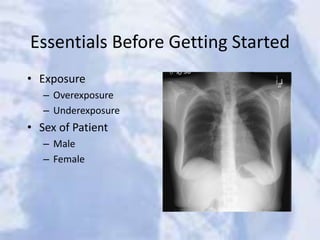
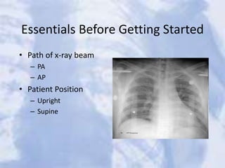
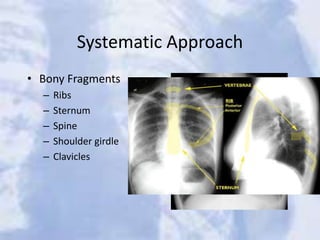
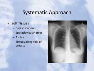
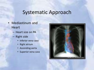
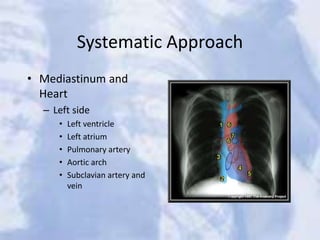
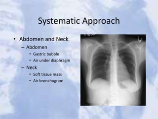
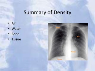
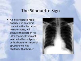
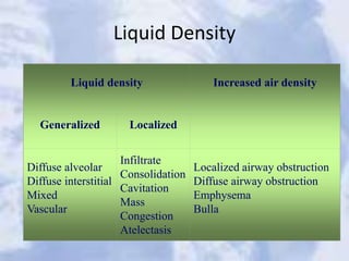
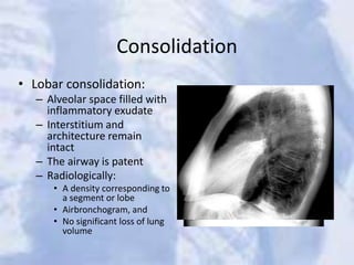
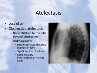
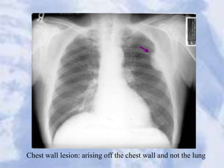
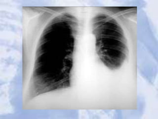
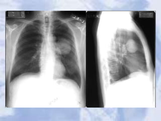
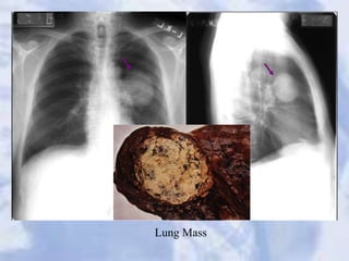
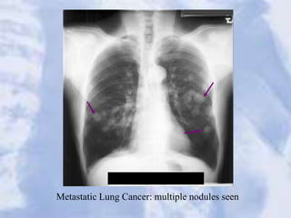
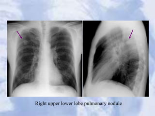
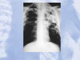
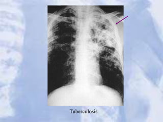
This document provides guidance on interpreting chest x-rays. It discusses essentials like exposure, patient positioning, and breathing technique. It describes a systematic approach to analyzing different parts of the chest x-ray like bones, soft tissues, lungs, heart, and abdomen. Key anatomical structures are identified. Common abnormalities like consolidation, atelectasis, cavitation, and masses are explained. A variety of case examples are presented and analyzed. The goal is to equip radiologists with the knowledge to systematically evaluate chest x-rays and identify and characterize any pathological findings.