1. Chest X-ray is a commonly used and inexpensive imaging test that provides important information to evaluate clinical questions.
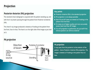
2. It is important to thoroughly examine all aspects of the X-ray such as quality, projections, inspiration and anatomy to identify any abnormalities.
3. Descriptions of abnormalities should include their location, size and other relevant characteristics compared to the surrounding normal structures.


![1. Radiology masterclass.[Online] Accessed [30
May 15]. Available
from:http://www.radiologymasterclass.co.uk
2. Corne J, Pointon K. Chest X-Ray Made Easy 3rd
Ed. Churchill Livingstone. 2010](https://image.slidesharecdn.com/chest-x-ray-231026041053-904d0127/85/chest-x-ray-pptx-61-320.jpg)


![Radiological_diagnosis_of_TB_ECHO_MOH[1].pptx](https://cdn.slidesharecdn.com/ss_thumbnails/radiologicaldiagnosisoftbechomoh1-240905083452-eb26e5f9-thumbnail.jpg?width=640&height=640&fit=bounds)


