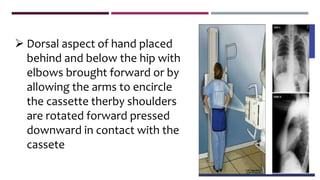
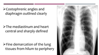
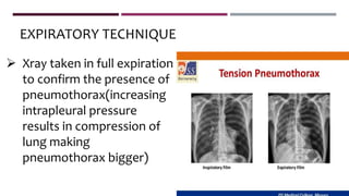
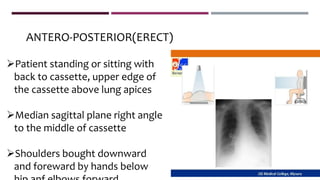
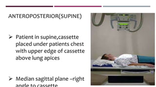
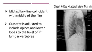
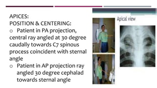
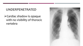
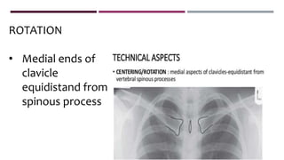
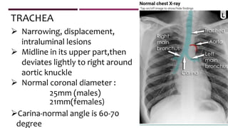
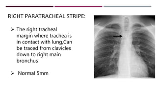
NORMAL CHEST XRAY
BY
DR.PRABHA JOSEPH
CONTENTS
 RECOMMENTED PROJECTIONS
• Position & centering
• Penetration
• Rotation
 MEDIASTINUM
• Junctional lines
Lungs
Hidden areas
Diaphragm
Soft tissues
bones
RECOMMENDED PROJECTIONS
 BASIC POSTERO-ANTERIOR(ERECT)
 ALTERNATIVE ANTERO-POSTERIOR(ERECT)
ANTERO-POSTERIOR(SUPINE)
ANTERO-POSTERIOR(SEMI ERECT)
 SUPPLEMENTAR LATERAL
POSTERO-ANTERIOR(EXPIRATION)
APICES
LATERAL-UPPER ANTERIOR REGION
DECUBITUS WITH HORIZONTAL BEAM
TOMOGRAPHY
POSITIONING:
 A 35 x 45 cm cassette is selected
 Patient positioned facing the cassette, with chin extended
and centered to the middle of the top of the cassette
 Feet paced slightly apart so patient remains steady
 Dorsal aspect of hand placed
behind and below the hip with
elbows brought forward or by
allowing the arms to encircle
the cassette therby shoulders
are rotated forward pressed
downward in contact with the
cassete
DIRECTION AND CENTERING:
 Horizontal central beam at right angles to the cassette
at the level of 8th thoracic vertebrae(spinous process
of T7)
 Surface marking of T7 is inferior angle of scapula
 Exposure is made in full normal arrested inspiration
ESSENTIAL IMAGE CHARECTERISTICS:
• Full lung field with scapula projecting laterally away
from lung field
• Clavicles symmetrical and equidistant from spinous
process and not obscuring the lung apices
• Well inflated(ie anterior six and posterior 10 ribs)
Costophrenic angles and
diaphragm outlined clearly
The mediastinum and heart
central and sharply defined
Fine demarcation of the lung
tissues from hilum to periphery
 Xray taken in full expiration
to confirm the presence of
pneumothorax(increasing
intrapleural pressure
results in compression of
lung making
pneumothorax bigger)
EXPIRATORY TECHNIQUE
ANTERO-POSTERIOR(ERECT)
Patient standing or sitting with
back to cassette, upper edge of
the cassette above lung apices
Median sagittal plane right angle
to the middle of cassette
Shoulders bought downward
and foreward by hands below
CENTERING
 Horizontal ray, right angle to
cassette towards sternal
notch
 Exposure taken in full
inspiration
ANTEROPOSTERIOR(SUPINE)
 Patient in supine,cassette
placed under patients chest
with upper edge of cassette
above lung apices
 Median sagittal plane –right
 Arms rotated laterally and supported by the side
 Head supported on pillow &chin raised
CENTERING:
• Right angled towards sternal notch
ANTERO POSTERIOR(SEMI ERECT)
 Patient in semi recumbent
position facing xray tube
 Cassette supported against
the back with upper edge
above apices
 Cassette should be parallel to
coronal plane
 Median sagittal plane right angle and to midline of the
cassette
 Arms are rotated medially with shoulders brought forward to
bring scapula clear to lung fields
 CENTERING:
 Towards the sternal notch
LATERAL VIEW
 Patient to be turned to bring
side of investigation in contact
with the cassette
 Median sagittal plane is parallel
to the cassette
 Arms are folded over the head
or raised above the head
 Mid axillary line coincident
with middle of the film
 Cassette is adjusted to
include apices and lower
lobes to the level of 1st
lumbar vertebrae
CENTERING
 Horizontal ray at right angle to the middle of
the cassette at the mid axillary line
APICES:
POSITION & CENTERING:
o Patient in PA projection,
central ray angled at 30 degree
caudally towards C7 spinous
process coincident with sternal
angle
o Patient in AP projection ray
angled 30 degree cephalad
towards sternal angle
o Patient reclining, and the coronal palne at 30
degree to cassette.nape of neck rests against
upper border of cassette,ray directed towars
sternal angle
UPPER ANTERIOR REGION-LATERAL
POSITION & CENTERING:
o Patient positioned with median sagittal palne
parallel to the cassette
o Centred at the level of shoulder of the side under
examination
o Shoulders drawn backward and arms extended
to move the shoulders clear of retrosternal space
LORDOTIC
Used to demonstrate right
middle lobe collapse or
interlobar pleural effusion
 Patient in PA
projection,clasping the
sides of vertical bucky
patient bends backwards
at the waist(30-40 degree)
PENETRATION:
 Vertebral bodies and disc spaces just visible down
to T8/9 level through cardiac shadow
 Overpenetration / underpenetration
 Lungs appear more
darker
 Intervertebral dics spaces
are clearly seen
OVERPENETRATION
UNDERPENETRATED
Cardiac shadow is opaque
with no visibility of thoracic
vertebra
ROTATION
• Medial ends of
clavicle
equidistand from
spinous process
 Narrowing, displacement,
intraluminal lesions
 Midline in its upper part,then
deviates lightly to right around
aortic knuckle
 Normal coronal diameter :
25mm (males)
21mm(females)
Carina-normal angle is 60-70
degree
TRACHEA
 The right tracheal
margin where trachea is
in contact with lung,Can
be traced from clavicles
down to right main
bronchus
 Normal 5mm
RIGHT PARATRACHEAL STRIPE:
Widening occurs in :
• Mediastinal
lymphadenopathy
• Tracheal malignancy
• Mediastinal tumours
• Mediastinitis
• Pleural effusion
Azygos vein :
 Lies in the angle between right
main bronchus & trachea
 Should be less than 10mm
Enlarged in :
o Enlarged subcarinal nodes
o Pregnancy
o Portal hypertension
o IVC, SVC obstruction
o Right heart failure
o Constrictive pericarditis
MEDIASTINUM & HEART
 2/3rd of cardiac shadow lies to left anf 1/3rd lies to right
 CARDIOTHORACIC RATIO:
Maximum tansverse diameter of the heart
and maximum width of thorax at CP angle,
measured from inner edge of ribs
CRT=(a+b)/c
A-Right heart border to
midline
B-left heart border to midline
C-max thoracic diameter
above CP angle from inner
border of rib
 normal less than 50%(PA)
60%(AP)
 All borders are clearly defined except where the
heart sits on the left hemidiaphragm.
 Right Sup. Mediastinal shadow – SVC &
innominate vessels, dilated aorta
 Left sup. Mediastinal shadow – subclavian artery
above aortic knuckle
ANTERIOR JUNCTION LINE
• Formed by lungs meeting
anterior to ascending aorta
• 1mm thick
• Overlying tracheal
translucency
• Runs down from below
suprasternal notch slightly
JUNCTION LINES
 Where the lungs meet posteriorly behind the
esophagus
 Straight/curved line convex to the left
 2mm wide
 Extends from lung apices to the aortic knuckle or
POSTERIOR JUNCTION LINE
o Inverted hockey stick shape
o From diaphragm on left of midline up & to the
right extending to tracheobronchial angle where
the azygos V drains into IVC.
AZYGO-OESOPHAGEAL INTERFACE
 Formed by lung & right wall of
esophagus
 From lung apex to azygos
 Only visualised if esophagus contain
air.
PLEURO-OESOPHAGEAL STRIPE
• Adjacent to vertebral bodies
• Left : <10mm wide(due to descending thoracic
aorta)
• Right : <3mm
• Enlargement occurs in tortuous aorta,
osteophytes, [paravertebral hematoma ,etc.
PARASPINAL LINE
 Seen in babies & young children
 Triangular Sail shaped structure
 Well defined borders projecting from one or both
sides of mediastinum
THYMUS
 Borders may be wavy in
outline – Wave sign of
mulvey – due to
indentation by costal
cartilages
THYMIC SIGNS
THYMIC SNAIL SIGN
 Triangular shaped inferior
margin of normal thymus
 More commonly seen on
right
THYMIC NOTCH SIGN
• The inferior border
of the thymus
blends with the
border of cardiac
silhouette
LUNGS
 DIVIDED INTO
UPPER
MID
LOWER
FISSURES
MAIN FISSURES
 Separates lobes of lung;Usually incomplete allowing
collateral air drift to occur between adjacent lobes
 Visualized when the xray is tangential
• From the hilum to the
6th rib in the axillary
line
• Straight / slight
downward curve
HORIZONTAL/MINOR
FISSURE:
• Both commence
posteriorly at T4/5 level
passing through hilum
• Left is steeper finishes
5cm behind anterior
costophrenic angle
• Right ends just behind
the angle
OBLIQUE FISSURE
 Comma shaped with triangular base peripherally
 Always Right sided
 Forms in apex of lung
 Consists of paired folds of parietal & visceral pleura,
azygos Vein which failed to migrate
ACCESSORY FISSURES
AZYGOES FISSURE:
 When left sided contains an accessory hemiazygos Vein
 Separates apical from basal segments of lower
lobes
 Common on right side
 Resembles horizontal fissure on PA film
 Differentiated on lateral film as it runs
posteriorly from hilum
SUPERIOR ACCESSORY FISSURE
• As an oblique line running
cranially from cardiophrenic
angle to hilum
• Separates medial basal from
other basal segments
• Common on right side
INFERIOR ACCESSORY
FISSURE
• Separates lingula from
other upper lobe segments
• Rare
LEFT SIDED HORIZONTAL FISSURE
INFERIOR PULMONARY LIGAMENT
 Double layer of pleura extending caudally from lower
margin of inferior pulmonary vein in hilum which may or
may not attached to diaphragm and attaches lower
lobe to mediastinum
 Frequently seen in CT
HIDDEN AREAS
 THE APICES
 MEDIASTINUM AND HILA
 DIAPHRAGM
 BONES
HILA
 Left hilum higher than
right
 Clearly defined concave
lateral borders
 Similar size
• Pulmonary artery contribute significantly to the hilar shadow
• Left Pulmonary Artery lies above the left main bronchus
• Right Pulmonary artery is anterior to the bronchus resulting
in right hilum being lower
• Normal lymph nodes not seen
DIAPHRAGM
 2-3mm thick
 Right higher than left
 Due to heart depressing left side &
not due to liver pushing up the
right side
 Left is higher if stomach or splenic
flexure is distended with gas
 Difference >3cm is significant
 On inspiration domes are at level of 6th rib anteriorly &
at or below 10th rib posteriorly
 Upper borders clearly visualized except on left side
where heart is in contact & in cardiophrenic angles
when there are prominent fat pads
 Loss of outline – indicates adjacent lung tissue does
not contain air. eg: consolidation or pleural disease
Are acute & well defined
Obliterated when diaphragms are
flat
Frequently contain low density ill
defined opacity caused by fat
pads.
COSTOPHRENIC ANGLE
SOFT TISSUES
• Include Chest wall, shoulders & lower neck
• Breast shadows
• Skin folds – seen in old age & in babies
BONES
o Sternum
o Clavicles
o Scapulae
o Ribs:
 Pathological rib notching
seen in aortic coartation
 Central homogenous coastal
cartilage calcification in
females,curvilinear marginal
calcification in males
o Spine
LATERAL VIEW
• The clear spaces-Retrosternal and Retrocardiac
• Obliteration of retrosternal space : in thymoma,
aneurysms of aorta, nodal masses
• Widening : emphysema
VERTEBRAL TRANSLUCENCY:
o Vertebral bodies more
translucent caudally
o Loss of this is seen in
Posterior basal
consolidation
THANK YOU

Normal chest xray

  • 1.
  • 2.
    CONTENTS  RECOMMENTED PROJECTIONS •Position & centering • Penetration • Rotation  MEDIASTINUM • Junctional lines
  • 3.
  • 4.
    RECOMMENDED PROJECTIONS  BASICPOSTERO-ANTERIOR(ERECT)  ALTERNATIVE ANTERO-POSTERIOR(ERECT) ANTERO-POSTERIOR(SUPINE) ANTERO-POSTERIOR(SEMI ERECT)  SUPPLEMENTAR LATERAL POSTERO-ANTERIOR(EXPIRATION) APICES LATERAL-UPPER ANTERIOR REGION DECUBITUS WITH HORIZONTAL BEAM TOMOGRAPHY
  • 5.
    POSITIONING:  A 35x 45 cm cassette is selected  Patient positioned facing the cassette, with chin extended and centered to the middle of the top of the cassette  Feet paced slightly apart so patient remains steady
  • 6.
     Dorsal aspectof hand placed behind and below the hip with elbows brought forward or by allowing the arms to encircle the cassette therby shoulders are rotated forward pressed downward in contact with the cassete
  • 7.
    DIRECTION AND CENTERING: Horizontal central beam at right angles to the cassette at the level of 8th thoracic vertebrae(spinous process of T7)  Surface marking of T7 is inferior angle of scapula  Exposure is made in full normal arrested inspiration
  • 8.
    ESSENTIAL IMAGE CHARECTERISTICS: •Full lung field with scapula projecting laterally away from lung field • Clavicles symmetrical and equidistant from spinous process and not obscuring the lung apices • Well inflated(ie anterior six and posterior 10 ribs)
  • 9.
    Costophrenic angles and diaphragmoutlined clearly The mediastinum and heart central and sharply defined Fine demarcation of the lung tissues from hilum to periphery
  • 10.
     Xray takenin full expiration to confirm the presence of pneumothorax(increasing intrapleural pressure results in compression of lung making pneumothorax bigger) EXPIRATORY TECHNIQUE
  • 11.
    ANTERO-POSTERIOR(ERECT) Patient standing orsitting with back to cassette, upper edge of the cassette above lung apices Median sagittal plane right angle to the middle of cassette Shoulders bought downward and foreward by hands below
  • 12.
    CENTERING  Horizontal ray,right angle to cassette towards sternal notch  Exposure taken in full inspiration
  • 13.
    ANTEROPOSTERIOR(SUPINE)  Patient insupine,cassette placed under patients chest with upper edge of cassette above lung apices  Median sagittal plane –right
  • 14.
     Arms rotatedlaterally and supported by the side  Head supported on pillow &chin raised CENTERING: • Right angled towards sternal notch
  • 15.
    ANTERO POSTERIOR(SEMI ERECT) Patient in semi recumbent position facing xray tube  Cassette supported against the back with upper edge above apices  Cassette should be parallel to coronal plane
  • 16.
     Median sagittalplane right angle and to midline of the cassette  Arms are rotated medially with shoulders brought forward to bring scapula clear to lung fields  CENTERING:  Towards the sternal notch
  • 17.
    LATERAL VIEW  Patientto be turned to bring side of investigation in contact with the cassette  Median sagittal plane is parallel to the cassette  Arms are folded over the head or raised above the head
  • 18.
     Mid axillaryline coincident with middle of the film  Cassette is adjusted to include apices and lower lobes to the level of 1st lumbar vertebrae
  • 19.
    CENTERING  Horizontal rayat right angle to the middle of the cassette at the mid axillary line
  • 20.
    APICES: POSITION & CENTERING: oPatient in PA projection, central ray angled at 30 degree caudally towards C7 spinous process coincident with sternal angle o Patient in AP projection ray angled 30 degree cephalad towards sternal angle
  • 21.
    o Patient reclining,and the coronal palne at 30 degree to cassette.nape of neck rests against upper border of cassette,ray directed towars sternal angle
  • 22.
    UPPER ANTERIOR REGION-LATERAL POSITION& CENTERING: o Patient positioned with median sagittal palne parallel to the cassette o Centred at the level of shoulder of the side under examination o Shoulders drawn backward and arms extended to move the shoulders clear of retrosternal space
  • 23.
    LORDOTIC Used to demonstrateright middle lobe collapse or interlobar pleural effusion  Patient in PA projection,clasping the sides of vertical bucky patient bends backwards at the waist(30-40 degree)
  • 24.
    PENETRATION:  Vertebral bodiesand disc spaces just visible down to T8/9 level through cardiac shadow  Overpenetration / underpenetration
  • 25.
     Lungs appearmore darker  Intervertebral dics spaces are clearly seen OVERPENETRATION
  • 26.
    UNDERPENETRATED Cardiac shadow isopaque with no visibility of thoracic vertebra
  • 27.
    ROTATION • Medial endsof clavicle equidistand from spinous process
  • 28.
     Narrowing, displacement, intraluminallesions  Midline in its upper part,then deviates lightly to right around aortic knuckle  Normal coronal diameter : 25mm (males) 21mm(females) Carina-normal angle is 60-70 degree TRACHEA
  • 29.
     The righttracheal margin where trachea is in contact with lung,Can be traced from clavicles down to right main bronchus  Normal 5mm RIGHT PARATRACHEAL STRIPE:
  • 30.
    Widening occurs in: • Mediastinal lymphadenopathy • Tracheal malignancy • Mediastinal tumours • Mediastinitis • Pleural effusion
  • 31.
    Azygos vein : Lies in the angle between right main bronchus & trachea  Should be less than 10mm Enlarged in : o Enlarged subcarinal nodes o Pregnancy o Portal hypertension o IVC, SVC obstruction o Right heart failure o Constrictive pericarditis
  • 32.
    MEDIASTINUM & HEART 2/3rd of cardiac shadow lies to left anf 1/3rd lies to right  CARDIOTHORACIC RATIO: Maximum tansverse diameter of the heart and maximum width of thorax at CP angle, measured from inner edge of ribs
  • 33.
    CRT=(a+b)/c A-Right heart borderto midline B-left heart border to midline C-max thoracic diameter above CP angle from inner border of rib  normal less than 50%(PA) 60%(AP)
  • 34.
     All bordersare clearly defined except where the heart sits on the left hemidiaphragm.  Right Sup. Mediastinal shadow – SVC & innominate vessels, dilated aorta  Left sup. Mediastinal shadow – subclavian artery above aortic knuckle
  • 35.
    ANTERIOR JUNCTION LINE •Formed by lungs meeting anterior to ascending aorta • 1mm thick • Overlying tracheal translucency • Runs down from below suprasternal notch slightly JUNCTION LINES
  • 36.
     Where thelungs meet posteriorly behind the esophagus  Straight/curved line convex to the left  2mm wide  Extends from lung apices to the aortic knuckle or POSTERIOR JUNCTION LINE
  • 38.
    o Inverted hockeystick shape o From diaphragm on left of midline up & to the right extending to tracheobronchial angle where the azygos V drains into IVC. AZYGO-OESOPHAGEAL INTERFACE
  • 40.
     Formed bylung & right wall of esophagus  From lung apex to azygos  Only visualised if esophagus contain air. PLEURO-OESOPHAGEAL STRIPE
  • 41.
    • Adjacent tovertebral bodies • Left : <10mm wide(due to descending thoracic aorta) • Right : <3mm • Enlargement occurs in tortuous aorta, osteophytes, [paravertebral hematoma ,etc. PARASPINAL LINE
  • 43.
     Seen inbabies & young children  Triangular Sail shaped structure  Well defined borders projecting from one or both sides of mediastinum THYMUS
  • 44.
     Borders maybe wavy in outline – Wave sign of mulvey – due to indentation by costal cartilages THYMIC SIGNS
  • 45.
    THYMIC SNAIL SIGN Triangular shaped inferior margin of normal thymus  More commonly seen on right
  • 46.
    THYMIC NOTCH SIGN •The inferior border of the thymus blends with the border of cardiac silhouette
  • 47.
  • 52.
    FISSURES MAIN FISSURES  Separateslobes of lung;Usually incomplete allowing collateral air drift to occur between adjacent lobes  Visualized when the xray is tangential
  • 53.
    • From thehilum to the 6th rib in the axillary line • Straight / slight downward curve HORIZONTAL/MINOR FISSURE:
  • 54.
    • Both commence posteriorlyat T4/5 level passing through hilum • Left is steeper finishes 5cm behind anterior costophrenic angle • Right ends just behind the angle OBLIQUE FISSURE
  • 55.
     Comma shapedwith triangular base peripherally  Always Right sided  Forms in apex of lung  Consists of paired folds of parietal & visceral pleura, azygos Vein which failed to migrate ACCESSORY FISSURES AZYGOES FISSURE:
  • 56.
     When leftsided contains an accessory hemiazygos Vein
  • 57.
     Separates apicalfrom basal segments of lower lobes  Common on right side  Resembles horizontal fissure on PA film  Differentiated on lateral film as it runs posteriorly from hilum SUPERIOR ACCESSORY FISSURE
  • 58.
    • As anoblique line running cranially from cardiophrenic angle to hilum • Separates medial basal from other basal segments • Common on right side INFERIOR ACCESSORY FISSURE
  • 59.
    • Separates lingulafrom other upper lobe segments • Rare LEFT SIDED HORIZONTAL FISSURE
  • 60.
    INFERIOR PULMONARY LIGAMENT Double layer of pleura extending caudally from lower margin of inferior pulmonary vein in hilum which may or may not attached to diaphragm and attaches lower lobe to mediastinum  Frequently seen in CT
  • 61.
    HIDDEN AREAS  THEAPICES  MEDIASTINUM AND HILA  DIAPHRAGM  BONES
  • 63.
    HILA  Left hilumhigher than right  Clearly defined concave lateral borders  Similar size
  • 64.
    • Pulmonary arterycontribute significantly to the hilar shadow • Left Pulmonary Artery lies above the left main bronchus • Right Pulmonary artery is anterior to the bronchus resulting in right hilum being lower • Normal lymph nodes not seen
  • 65.
    DIAPHRAGM  2-3mm thick Right higher than left  Due to heart depressing left side & not due to liver pushing up the right side  Left is higher if stomach or splenic flexure is distended with gas  Difference >3cm is significant
  • 66.
     On inspirationdomes are at level of 6th rib anteriorly & at or below 10th rib posteriorly  Upper borders clearly visualized except on left side where heart is in contact & in cardiophrenic angles when there are prominent fat pads  Loss of outline – indicates adjacent lung tissue does not contain air. eg: consolidation or pleural disease
  • 67.
    Are acute &well defined Obliterated when diaphragms are flat Frequently contain low density ill defined opacity caused by fat pads. COSTOPHRENIC ANGLE
  • 68.
    SOFT TISSUES • IncludeChest wall, shoulders & lower neck • Breast shadows • Skin folds – seen in old age & in babies
  • 70.
  • 71.
    o Ribs:  Pathologicalrib notching seen in aortic coartation  Central homogenous coastal cartilage calcification in females,curvilinear marginal calcification in males o Spine
  • 72.
    LATERAL VIEW • Theclear spaces-Retrosternal and Retrocardiac • Obliteration of retrosternal space : in thymoma, aneurysms of aorta, nodal masses • Widening : emphysema
  • 73.
    VERTEBRAL TRANSLUCENCY: o Vertebralbodies more translucent caudally o Loss of this is seen in Posterior basal consolidation
  • 74.