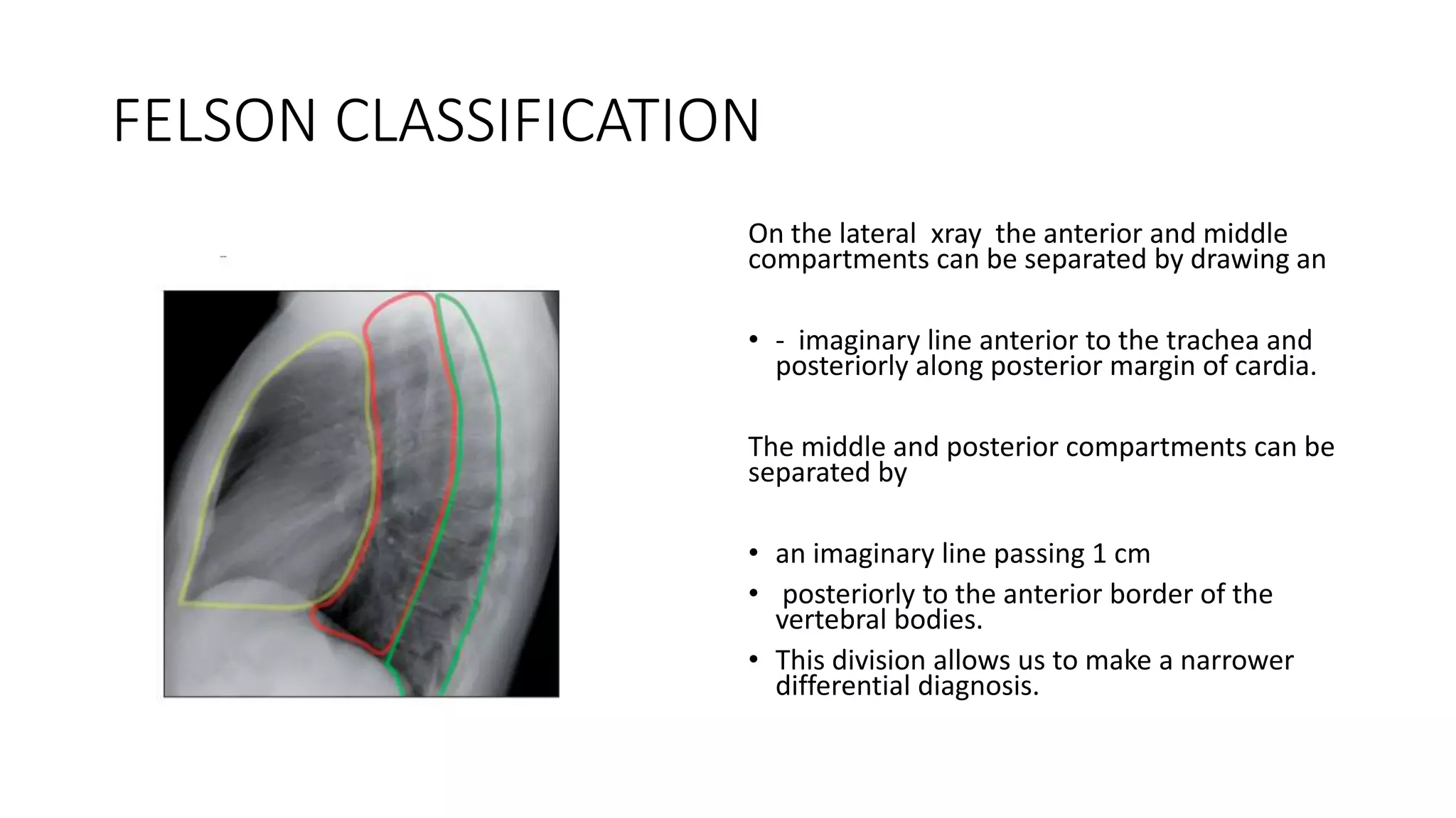
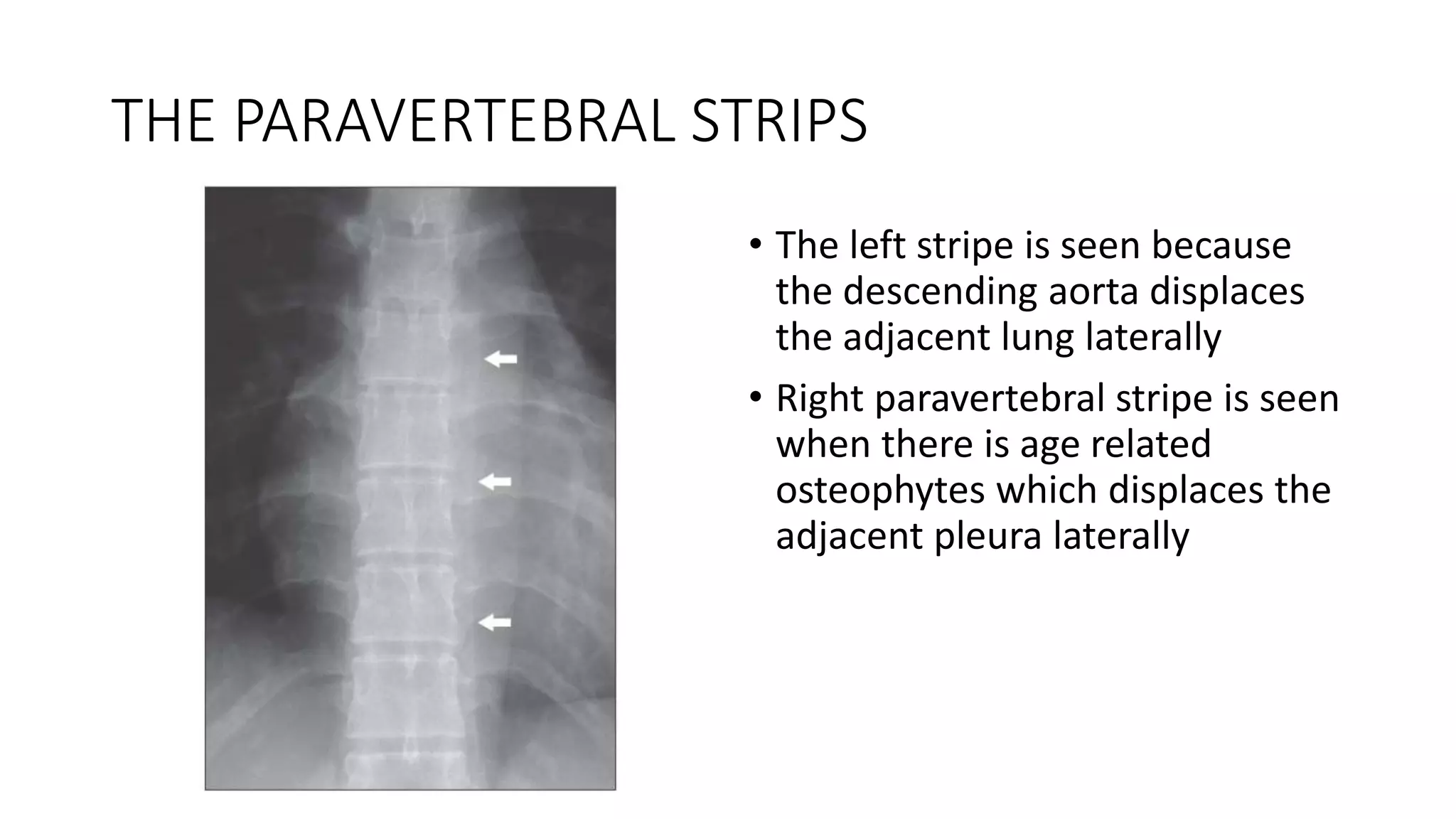
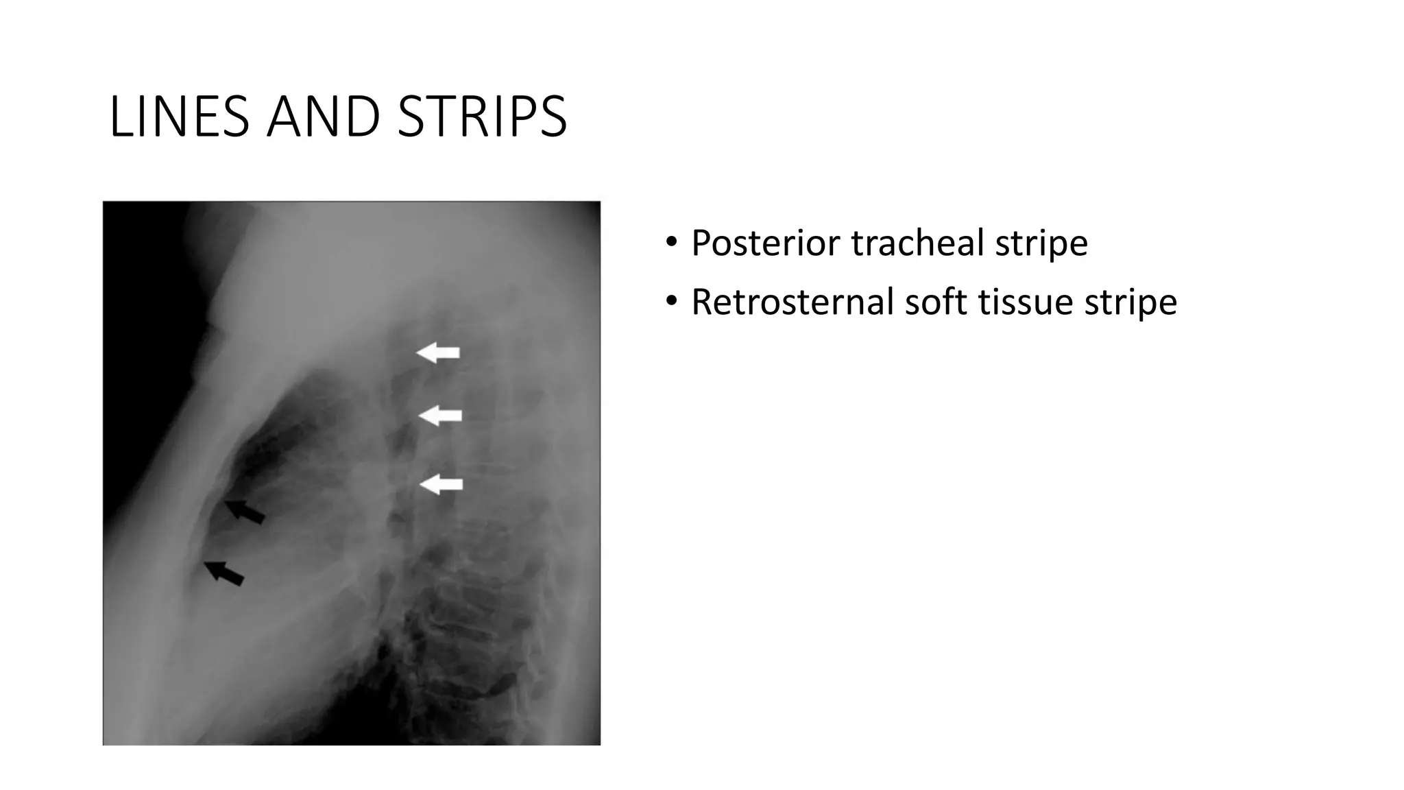
Students, digital devices and success - Andreas Schleicher - 27 May 2024..pptx
MEDIASTINUM.pptx
1.
2. INTRODUCTION
• The mediastinum is situated between the pleurae covering the medial
aspects of the right and left lungs. Its borders are the thoracic inlet
(superiorly), the diaphragm (inferiorly), the parietal pleurae
(laterally), the vertebral column (posteriorly) and the sternum
(anteriorly).
4. SUTTON CLASSIFICATION
Divisions :
• Anterior - lies in front of anterior
pericardium and trachea
• Middle – with in the pericardial
cavity including trachea
• Posterior – behind the posterior
pericardium and trachea
5. FELSON CLASSIFICATION
On the lateral xray the anterior and middle
compartments can be separated by drawing an
• - imaginary line anterior to the trachea and
posteriorly along posterior margin of cardia.
The middle and posterior compartments can be
separated by
• an imaginary line passing 1 cm
• posteriorly to the anterior border of the
vertebral bodies.
• This division allows us to make a narrower
differential diagnosis.
9. FAT COLLECTIONS
• Visible pads of fat are present at the right and / or left cardiophrenic
angles in some patients
• If a large fat collection is present it may:
• Cause part of the well-defined margin of the heart to be blurred.
• Mimic a mass lesion.
10.
11. INTERFACE LINES AND STRIPS
• THE RIGHT PARATRACHEAL STRIPE
• THE PARAVERTEBRAL STRIPES
• AZYGO-OESOPHAGEAL LINE
• ANTERIOR JUNCTION LINE
• POSTERIOR JUNCTION LINE
• AORTO-PULMONARY STRIPE
13. THE RIGHT PARATRACHEAL STRIPE
• Forms where the right lung
abuts the right side of the
trachea
• The thickness should not
exceed 2.5mm
14. THE PARAVERTEBRAL STRIPS
• The left stripe is seen because
the descending aorta displaces
the adjacent lung laterally
• Right paravertebral stripe is seen
when there is age related
osteophytes which displaces the
adjacent pleura laterally
15. AZYGO-OESOPHAGEAL LINE
• Formed where the right lung
abuts the right side of the
oesophagus and the azygos vein
• Extends below the aortic arch to
the diaphragm
16. ANTERIOR JUNCTION LINE
• Formed where the two lungs
abut each other anteriorly below
the level of the manubrium
• The line is made of 4 layers of
pleura
17. POSTERIOR JUNCTION LINE
• Formed where the two lungs
abut each other posteriorly
• Extends from above the clavicle
to the level of aortic arch
• Made up of 4 layers of pleura
20. FAT COLLECTIONS
• CARDIAC INCISURA:
The apex of the heart and
adjacent epicardial fat intrudes
into the left hemithorax and
displaces the most infero-medial
and anterior aspect of the left
lung. This often produces a
shadow which can simulate a
mass lesion