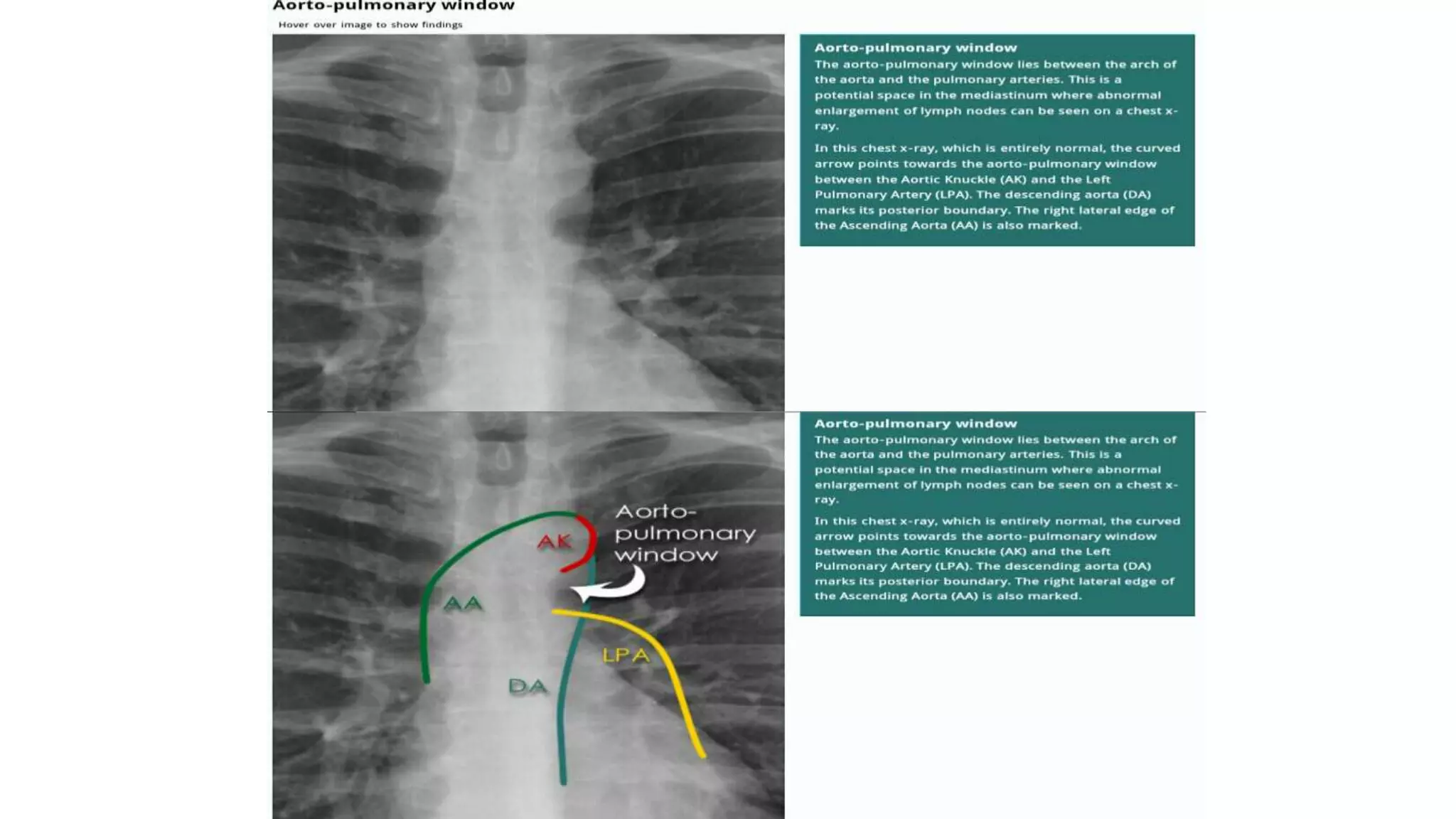
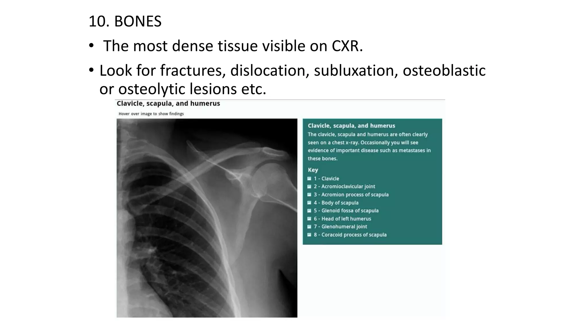
SlideShare a Scribd company logo
Chest X ray Basics-1
Presentation by:
Dr. Nishant Gupta
• Chest X-Ray is one of the most frequently requested hospital investigations.
• It is readily available and inexpensive in comparison to other imaging studies.
• The basic interpretation is of utmost importance in answering several clinical questions
at hand.
• It is an important tool to complement both history and initial clinical examination.
Basic Details on a Chest Xray
A. Patient details
• Name of the patient
• Age
• Date
B. Quality
• Image quality influences interpretation
• Quality is influenced by radiographic technique and patient factors.
• First determine if the clinical question can be answered.
• Check the image for – Projection, rotation, inspiration, penetration and artefacts.
CXR projection
Look to see if the film is antero-posterior (AP) or postero-anterior (PA) view
• With an AP view the X-ray beam is in front the patient and the X-Ray placed at the back,
and the other way round for PA.
• The standard CXR is PA but many emergency CXRs are AP.
• The CXR projection has an important bearing on the interpretation of the structures.
Chest Radiography: Basic Principles
Blackest
air- absorbs least Radiation
fat
soft tissue
calcium
bone
X-ray contrast
metal-absorbs max Radiation
Whitest
Maximum X-Ray
Transmission
(least dense tissue)
Maximum X-Ray
Absorption
(densest tissue)
 X-ray photon: Absorbed / scattered / transmitted
 X-ray absorption depends on:
• Beam energy (constant)
• Tissue density
Rotation
• Identify the medial ends of the clavicles and select one of the thoracic vertebra spinous processes that falls
between them.
• The medial ends of the clavicles should be equidistant from the spinous process, if that’s not the case then the
X-Ray is rotated.
Does rotation matter ?
• If the patient is rotated then interpretation may become difficult. Firstly, it may be difficult to know if the trachea
is deviated to one side by a disease process. It also becomes difficult to comment accurately on the heart size.
Changes in lung density due to asymmetry of overlying soft-tissue may be incorrectly interpreted as lung disease.
Rotation and heart size
• Heart size can be assessed accurately with a well-aligned posterior-anterior
(PA) chest X-ray. If the patient is rotated to their left, then the heart may
appear enlarged. If the patient is rotated to their right, then heart size may be
underestimated.
• Thickness of soft tissues of the chest, such as breast tissue, is altered by
rotation. This may give the misleading impression of pathology in the lungs.
• While reading out the chest X ray if the distance between the either of the
medial clavicle and spinous process is reduced, the rotation of that side is to
be mentioned in a PA view chest Xray.
Orientation
• Identify the left/right markings
• Identify the anatomical structures, erect/supine.
• Do not always assume that the heart will always be on the left
because certain pathologies can result with mediastinal shift,
dextrocardia can also be a possibility.
• You do not have to solely rely on just the CXR markings.
Right Rotation
12
Inspiration (Degree of inspiration)
• To judge the degree of inspiration, count the number of ribs above the diaphragm.
• The midpoint of the right hemi-diaphragm should be between the 5th and 7th ribs anteriorly.(slide 13 )
• The anterior end of the 6th rib should be above the diaphragm as should the posterior end of the 10th rib. ( slide
15)
• If more ribs are visible the patient is hyperinflated
• If fewer it indicates inadequate inspiration
• Poor inspiration will make the heart look larger, give appearance of basal shadowing and cause the trachea to
appear deviated to the right
Inspiratory Effort
Low Lung Volumes Full Inspiration
Image credit: Curry International Tuberculosis Center, University of California, San Francisco 9
Penetration
• To check the penetration, look at the lower part of the cardiac shadow
• The vertebral bodies should be barely visible through the cardiac shadow at this point.
• If they are clearly visible then the film is over penetrated and you may miss low density lesion.
• If you cannot see them at all then the film is under penetrated and the lung fields will appear falsely opaque
(white).
• The left hemidiaphragm should be visible to the edge of the spine
• When comparing X-Rays first determine if the level of penetration is similar.
Overexposure Proper Exposure
Image credit: Curry International Tuberculosis Center, University of California, San Francisco 11
Normal penetration
CHEST X-RAY ANATOMY
1. TRACHEA
• It should be central or slightly deviated to the right.
- In case of deviation decide if is due to rotation or pathology
• View the carina, angle should be between 60 –100 degrees.
• Because it contains air, it appears darker (blacker/radiolucent).
• Trachea normally narrows at the vocal cords (T3/T4)
2. HILAR STRUCTURES
• Also called lung root, consists of the major bronchi and
pulmonary vessels (veins/arteries).
• The hila are not symmetrical but consist of the same
basic structures.
• The lymph nodes are also present but no visible unless
abnormal.
3. LUNGS
• The lungs occupies the largest portion of the thoracic
cavity.
• The lungs are assessed and described by dividing them
into upper, middle and lower zones.
• The lung zones do not equate to lung lobes e.g. The lower
zone on the right consists of middle and lower lobes.
4. PLEURA AND PLEURAL SPACES
• The pleura are only visible when there is an abnormality
present.
• This can be due to pleural thickening and fluid or air
accumulating in the pleural spaces.
• Lung markings should reach the thoracic wall
5. COSTOPHRENIC ANGLE AND RECESS
• The costophrenic recesses are formed by
hemidiaphragms and chest wall.
• They contain the rim of the lung bases which lie over the
dome of each hemidiaphragm.
• These angles are known as the costophrenic angles.
• Costophrenic angles should form acute angles that are
sharp to the point.
6. HEMIDIAPHRAGM
7. HEART
• The heart lies more to the left of the thoracic cavity.
• The heart is assessed by means of the cardio-thoracic ratio
(CTR).
• CTR = Cardiac width : Thoracic width
• CTR > 50% is abnormal – PA view only
• The left hemidiaphragm should be visible behind the heart.
• The hemidiaphrams do not represent the lowest point of the
lungs.
8. THE MEDIASTINUM
• The mediastinum contains the heart and great vessels (Middle
mediatinum) and potential spaces in front of the heart (anterior
mediastinum), behind the heart (Posterior mediastinum) and
above the heart (superior mediastinum).
• These potential spaces are not defined on a normal CXR, but their
awareness can help in describing location of disease processes.
• There are several structures in the superior mediastinum that
should always be checked. These include aortic knuckle, aorto-
pulmonary window and the right para-tracheal stripe.
9. SOFT TISSUE
• Normal fat planes are clearly defined in the soft tissues.
• They appear as smooth layers of low density (black), between
layers of relatively dense (whiter) muscles.
• Irregular low density within soft tissues may be as a result of
tracking air as a result of injury to the airways or pleura.
• This is known as surgical emphysema and produces the
distinctive clinical sign of palpable subcutaneous ‘bubble
wrap’.
10. BONES
• The most dense tissue visible on CXR.
• Look for fractures, dislocation, subluxation, osteoblastic
or osteolytic lesions etc.
a. The CXR is an important tool to complement both history
and initial clinical examination.
b. Low density structures appear dark(black/radiolucent) and
high density are whitish (opaque).
c. Abnormalities need to be described in detail.
d. Identify the most striking abnormality first. However, once
you are done with this, it is vital to check the rest of the
image.
APPROACH TO CXR PATHOLOGY
Describing abnormalities
ABNORMAL CXRs
Chest X ray ppt.ppt
Chest X ray ppt.ppt
Chest X ray ppt.ppt
Chest X ray ppt.ppt

More Related Content

What's hot

Chest Radiology.ppt
Chest Radiology.pptChest Radiology.ppt
Chest Radiology.ppt
Maheen Fatima
 
Collapse and consolidation Lung Radiology
Collapse and consolidation Lung RadiologyCollapse and consolidation Lung Radiology
Collapse and consolidation Lung RadiologyNeelam Ashar
 
Interpretation of Chest X-Ray PPT
Interpretation of Chest X-Ray PPTInterpretation of Chest X-Ray PPT
Interpretation of Chest X-Ray PPT
drmainuddin
 
Chest X-ray Interpretation
Chest X-ray Interpretation Chest X-ray Interpretation
Chest X-ray Interpretation
Sarfraz Saleemi
 
Collapse- RADIOLOGY
Collapse- RADIOLOGYCollapse- RADIOLOGY
Collapse- RADIOLOGYNavdeep Shah
 
Chest imaging
Chest imagingChest imaging
Chest imaging
JANARDANCNMC
 
Basic CXR Interpretation_Diagnostic Radiography
Basic CXR Interpretation_Diagnostic RadiographyBasic CXR Interpretation_Diagnostic Radiography
Basic CXR Interpretation_Diagnostic Radiography
Imhotep Virtual Medical School
 
CXR Interpretation for Med Students
CXR Interpretation for Med StudentsCXR Interpretation for Med Students
CXR Interpretation for Med Students
ejheffernan
 
The Normal Chest X-ray
The Normal Chest X-rayThe Normal Chest X-ray
The Normal Chest X-raykaphoury
 
Normal chest x ray- Radiology Basics
Normal chest x  ray- Radiology BasicsNormal chest x  ray- Radiology Basics
Normal chest x ray- Radiology Basics
Sandeep Awal
 
Chest x ray - basics
Chest x ray - basicsChest x ray - basics
Chest x ray - basics
Rikin Hasnani
 
Chest Xray
Chest XrayChest Xray
Chest Xray
Hassan Tarig
 
Adult Lines and Tubes in Radiology
Adult Lines and Tubes in RadiologyAdult Lines and Tubes in Radiology
Adult Lines and Tubes in Radiology
Brian Wells, MD, MS, MPH
 
Pneumothorax case based
Pneumothorax case basedPneumothorax case based
Pneumothorax case based
Siruhan Ali
 
basics of chest X- ray interpretation
basics of chest X- ray interpretationbasics of chest X- ray interpretation
basics of chest X- ray interpretation
Maha Yousif
 
Radiological signs in chest medicine Part 1
Radiological signs in chest medicine Part 1Radiological signs in chest medicine Part 1
Radiological signs in chest medicine Part 1Gamal Agmy
 
Interpretation of chest CT
Interpretation of chest CTInterpretation of chest CT
Interpretation of chest CT
Sakiru Isa
 
Presentation1, ultrasound examination of the chest.
Presentation1, ultrasound examination of the chest.Presentation1, ultrasound examination of the chest.
Presentation1, ultrasound examination of the chest.
Abdellah Nazeer
 
Diagnosis of heart diseases from chest x ray
Diagnosis of heart diseases from chest x rayDiagnosis of heart diseases from chest x ray
Diagnosis of heart diseases from chest x ray
Ramachandra Barik
 

What's hot (20)

Chest Radiology.ppt
Chest Radiology.pptChest Radiology.ppt
Chest Radiology.ppt
 
Collapse and consolidation Lung Radiology
Collapse and consolidation Lung RadiologyCollapse and consolidation Lung Radiology
Collapse and consolidation Lung Radiology
 
Interpretation of Chest X-Ray PPT
Interpretation of Chest X-Ray PPTInterpretation of Chest X-Ray PPT
Interpretation of Chest X-Ray PPT
 
Chest X-ray Interpretation
Chest X-ray Interpretation Chest X-ray Interpretation
Chest X-ray Interpretation
 
Collapse- RADIOLOGY
Collapse- RADIOLOGYCollapse- RADIOLOGY
Collapse- RADIOLOGY
 
Chest imaging
Chest imagingChest imaging
Chest imaging
 
Basic CXR Interpretation_Diagnostic Radiography
Basic CXR Interpretation_Diagnostic RadiographyBasic CXR Interpretation_Diagnostic Radiography
Basic CXR Interpretation_Diagnostic Radiography
 
CXR Interpretation for Med Students
CXR Interpretation for Med StudentsCXR Interpretation for Med Students
CXR Interpretation for Med Students
 
The Normal Chest X-ray
The Normal Chest X-rayThe Normal Chest X-ray
The Normal Chest X-ray
 
Normal chest x ray- Radiology Basics
Normal chest x  ray- Radiology BasicsNormal chest x  ray- Radiology Basics
Normal chest x ray- Radiology Basics
 
Chest x ray - basics
Chest x ray - basicsChest x ray - basics
Chest x ray - basics
 
Chest Xray
Chest XrayChest Xray
Chest Xray
 
Adult Lines and Tubes in Radiology
Adult Lines and Tubes in RadiologyAdult Lines and Tubes in Radiology
Adult Lines and Tubes in Radiology
 
Pneumothorax case based
Pneumothorax case basedPneumothorax case based
Pneumothorax case based
 
1 the normal cxr
1 the normal cxr1 the normal cxr
1 the normal cxr
 
basics of chest X- ray interpretation
basics of chest X- ray interpretationbasics of chest X- ray interpretation
basics of chest X- ray interpretation
 
Radiological signs in chest medicine Part 1
Radiological signs in chest medicine Part 1Radiological signs in chest medicine Part 1
Radiological signs in chest medicine Part 1
 
Interpretation of chest CT
Interpretation of chest CTInterpretation of chest CT
Interpretation of chest CT
 
Presentation1, ultrasound examination of the chest.
Presentation1, ultrasound examination of the chest.Presentation1, ultrasound examination of the chest.
Presentation1, ultrasound examination of the chest.
 
Diagnosis of heart diseases from chest x ray
Diagnosis of heart diseases from chest x rayDiagnosis of heart diseases from chest x ray
Diagnosis of heart diseases from chest x ray
 

Similar to Chest X ray ppt.ppt

chest-x-ray.zp162335.ppt
chest-x-ray.zp162335.pptchest-x-ray.zp162335.ppt
chest-x-ray.zp162335.ppt
GowrishankarPotturi
 
How to read a Chest X Ray film (Radiograph).pptx
How to read a Chest X Ray film (Radiograph).pptxHow to read a Chest X Ray film (Radiograph).pptx
How to read a Chest X Ray film (Radiograph).pptx
Suresh Managutti
 
Chest x-ray.zp162335 (1)
Chest x-ray.zp162335 (1)Chest x-ray.zp162335 (1)
Chest x-ray.zp162335 (1)
AndrFares
 
chest-x-ray.zp162335.ppt
chest-x-ray.zp162335.pptchest-x-ray.zp162335.ppt
chest-x-ray.zp162335.ppt
nishantgupta867402
 
Chest x-ray.zp162335
Chest x-ray.zp162335Chest x-ray.zp162335
Chest x-ray.zp162335
Dr. Nitish kumar
 
chest-x-ray.zp162335.pptx
chest-x-ray.zp162335.pptxchest-x-ray.zp162335.pptx
chest-x-ray.zp162335.pptx
HamdiAlaqal
 
Chest x ray interpretation
Chest x ray interpretationChest x ray interpretation
Chest x ray interpretation
Kamal Sharma
 
chest-x-ray.pptx
chest-x-ray.pptxchest-x-ray.pptx
chest-x-ray.pptx
VasanthakohilaMuthuk
 
Normal chest x ray and collapse
Normal chest x ray and collapseNormal chest x ray and collapse
Normal chest x ray and collapse
Aabid Rahiman
 
Chest X-ray & Interpretation.pptx
Chest X-ray & Interpretation.pptxChest X-ray & Interpretation.pptx
Chest X-ray & Interpretation.pptx
Ashish yadav
 
Approach to cxr.pptx
Approach to cxr.pptxApproach to cxr.pptx
Approach to cxr.pptx
MohammadMamunuzzaman2
 
Learn Chest X-Ray With Its Normal Positioning & Radio-Anatomy
Learn Chest X-Ray With Its Normal Positioning & Radio-AnatomyLearn Chest X-Ray With Its Normal Positioning & Radio-Anatomy
Learn Chest X-Ray With Its Normal Positioning & Radio-Anatomy
Dr.Santosh Atreya
 
Chest X rays.pptx
Chest X rays.pptxChest X rays.pptx
Chest X rays.pptx
AkashJain123345
 
X RAY DETERMINATION AND EVALUATION.pptx
X RAY DETERMINATION AND EVALUATION.pptxX RAY DETERMINATION AND EVALUATION.pptx
X RAY DETERMINATION AND EVALUATION.pptx
ShoaibKhatik3
 
Chest x rays BY Dr Anoop K R
Chest x rays BY Dr Anoop K RChest x rays BY Dr Anoop K R
Chest x rays BY Dr Anoop K R
anoop k r
 
Chest radiology
Chest radiologyChest radiology
Chest radiology
Dr Vaziri
 
Normal chest x ray
Normal chest x rayNormal chest x ray
Normal chest x ray
KaustubhMohite4
 
Chest x ray anatomy - how to interpret chest x-ray (2)
Chest x ray anatomy - how to interpret chest x-ray (2)Chest x ray anatomy - how to interpret chest x-ray (2)
Chest x ray anatomy - how to interpret chest x-ray (2)
Yusuf Shieba Elhamd
 
pathology related topics and its complete focus on every aspect
pathology related topics and its complete focus on every aspectpathology related topics and its complete focus on every aspect
pathology related topics and its complete focus on every aspect
Nausheen57
 

Similar to Chest X ray ppt.ppt (20)

chest-x-ray.zp162335.ppt
chest-x-ray.zp162335.pptchest-x-ray.zp162335.ppt
chest-x-ray.zp162335.ppt
 
How to read a Chest X Ray film (Radiograph).pptx
How to read a Chest X Ray film (Radiograph).pptxHow to read a Chest X Ray film (Radiograph).pptx
How to read a Chest X Ray film (Radiograph).pptx
 
Chest x-ray.zp162335 (1)
Chest x-ray.zp162335 (1)Chest x-ray.zp162335 (1)
Chest x-ray.zp162335 (1)
 
chest-x-ray.zp162335.ppt
chest-x-ray.zp162335.pptchest-x-ray.zp162335.ppt
chest-x-ray.zp162335.ppt
 
Chest x-ray.zp162335
Chest x-ray.zp162335Chest x-ray.zp162335
Chest x-ray.zp162335
 
chest-x-ray.zp162335.pptx
chest-x-ray.zp162335.pptxchest-x-ray.zp162335.pptx
chest-x-ray.zp162335.pptx
 
Chest x ray interpretation
Chest x ray interpretationChest x ray interpretation
Chest x ray interpretation
 
chest-x-ray.pptx
chest-x-ray.pptxchest-x-ray.pptx
chest-x-ray.pptx
 
Normal chest x ray and collapse
Normal chest x ray and collapseNormal chest x ray and collapse
Normal chest x ray and collapse
 
Chest X-ray & Interpretation.pptx
Chest X-ray & Interpretation.pptxChest X-ray & Interpretation.pptx
Chest X-ray & Interpretation.pptx
 
Approach to cxr.pptx
Approach to cxr.pptxApproach to cxr.pptx
Approach to cxr.pptx
 
Learn Chest X-Ray With Its Normal Positioning & Radio-Anatomy
Learn Chest X-Ray With Its Normal Positioning & Radio-AnatomyLearn Chest X-Ray With Its Normal Positioning & Radio-Anatomy
Learn Chest X-Ray With Its Normal Positioning & Radio-Anatomy
 
Lung y3 2018 19 tl
Lung y3 2018 19 tlLung y3 2018 19 tl
Lung y3 2018 19 tl
 
Chest X rays.pptx
Chest X rays.pptxChest X rays.pptx
Chest X rays.pptx
 
X RAY DETERMINATION AND EVALUATION.pptx
X RAY DETERMINATION AND EVALUATION.pptxX RAY DETERMINATION AND EVALUATION.pptx
X RAY DETERMINATION AND EVALUATION.pptx
 
Chest x rays BY Dr Anoop K R
Chest x rays BY Dr Anoop K RChest x rays BY Dr Anoop K R
Chest x rays BY Dr Anoop K R
 
Chest radiology
Chest radiologyChest radiology
Chest radiology
 
Normal chest x ray
Normal chest x rayNormal chest x ray
Normal chest x ray
 
Chest x ray anatomy - how to interpret chest x-ray (2)
Chest x ray anatomy - how to interpret chest x-ray (2)Chest x ray anatomy - how to interpret chest x-ray (2)
Chest x ray anatomy - how to interpret chest x-ray (2)
 
pathology related topics and its complete focus on every aspect
pathology related topics and its complete focus on every aspectpathology related topics and its complete focus on every aspect
pathology related topics and its complete focus on every aspect
 

Recently uploaded

How to Give Better Lectures: Some Tips for Doctors
How to Give Better Lectures: Some Tips for DoctorsHow to Give Better Lectures: Some Tips for Doctors
How to Give Better Lectures: Some Tips for Doctors
LanceCatedral
 
Prix Galien International 2024 Forum Program
Prix Galien International 2024 Forum ProgramPrix Galien International 2024 Forum Program
Prix Galien International 2024 Forum Program
Levi Shapiro
 
Alcohol_Dr. Jeenal Mistry MD Pharmacology.pdf
Alcohol_Dr. Jeenal Mistry MD Pharmacology.pdfAlcohol_Dr. Jeenal Mistry MD Pharmacology.pdf
Alcohol_Dr. Jeenal Mistry MD Pharmacology.pdf
Dr Jeenal Mistry
 
BENIGN PROSTATIC HYPERPLASIA.BPH. BPHpdf
BENIGN PROSTATIC HYPERPLASIA.BPH. BPHpdfBENIGN PROSTATIC HYPERPLASIA.BPH. BPHpdf
BENIGN PROSTATIC HYPERPLASIA.BPH. BPHpdf
DR SETH JOTHAM
 
Antiulcer drugs Advance Pharmacology .pptx
Antiulcer drugs Advance Pharmacology .pptxAntiulcer drugs Advance Pharmacology .pptx
Antiulcer drugs Advance Pharmacology .pptx
Rohit chaurpagar
 
Tom Selleck Health: A Comprehensive Look at the Iconic Actor’s Wellness Journey
Tom Selleck Health: A Comprehensive Look at the Iconic Actor’s Wellness JourneyTom Selleck Health: A Comprehensive Look at the Iconic Actor’s Wellness Journey
Tom Selleck Health: A Comprehensive Look at the Iconic Actor’s Wellness Journey
greendigital
 
ARTIFICIAL INTELLIGENCE IN HEALTHCARE.pdf
ARTIFICIAL INTELLIGENCE IN  HEALTHCARE.pdfARTIFICIAL INTELLIGENCE IN  HEALTHCARE.pdf
ARTIFICIAL INTELLIGENCE IN HEALTHCARE.pdf
Anujkumaranit
 
Physiology of Special Chemical Sensation of Taste
Physiology of Special Chemical Sensation of TastePhysiology of Special Chemical Sensation of Taste
Physiology of Special Chemical Sensation of Taste
MedicoseAcademics
 
Charaka Samhita Sutra Sthana 9 Chapter khuddakachatuspadadhyaya
Charaka Samhita Sutra Sthana 9 Chapter khuddakachatuspadadhyayaCharaka Samhita Sutra Sthana 9 Chapter khuddakachatuspadadhyaya
Charaka Samhita Sutra Sthana 9 Chapter khuddakachatuspadadhyaya
Dr KHALID B.M
 
Report Back from SGO 2024: What’s the Latest in Cervical Cancer?
Report Back from SGO 2024: What’s the Latest in Cervical Cancer?Report Back from SGO 2024: What’s the Latest in Cervical Cancer?
Report Back from SGO 2024: What’s the Latest in Cervical Cancer?
bkling
 
Are There Any Natural Remedies To Treat Syphilis.pdf
Are There Any Natural Remedies To Treat Syphilis.pdfAre There Any Natural Remedies To Treat Syphilis.pdf
Are There Any Natural Remedies To Treat Syphilis.pdf
Little Cross Family Clinic
 
Superficial & Deep Fascia of the NECK.pptx
Superficial & Deep Fascia of the NECK.pptxSuperficial & Deep Fascia of the NECK.pptx
Superficial & Deep Fascia of the NECK.pptx
Dr. Rabia Inam Gandapore
 
Pulmonary Thromboembolism - etilogy, types, medical- Surgical and nursing man...
Pulmonary Thromboembolism - etilogy, types, medical- Surgical and nursing man...Pulmonary Thromboembolism - etilogy, types, medical- Surgical and nursing man...
Pulmonary Thromboembolism - etilogy, types, medical- Surgical and nursing man...
VarunMahajani
 
Non-respiratory Functions of the Lungs.pdf
Non-respiratory Functions of the Lungs.pdfNon-respiratory Functions of the Lungs.pdf
Non-respiratory Functions of the Lungs.pdf
MedicoseAcademics
 
Ozempic: Preoperative Management of Patients on GLP-1 Receptor Agonists
Ozempic: Preoperative Management of Patients on GLP-1 Receptor Agonists  Ozempic: Preoperative Management of Patients on GLP-1 Receptor Agonists
Ozempic: Preoperative Management of Patients on GLP-1 Receptor Agonists
Saeid Safari
 
Surat @ℂall @Girls ꧁❤8527049040❤꧂@ℂall @Girls Service Vip Top Model Safe
Surat @ℂall @Girls ꧁❤8527049040❤꧂@ℂall @Girls Service Vip Top Model SafeSurat @ℂall @Girls ꧁❤8527049040❤꧂@ℂall @Girls Service Vip Top Model Safe
Surat @ℂall @Girls ꧁❤8527049040❤꧂@ℂall @Girls Service Vip Top Model Safe
Savita Shen $i11
 
HOT NEW PRODUCT! BIG SALES FAST SHIPPING NOW FROM CHINA!! EU KU DB BK substit...
HOT NEW PRODUCT! BIG SALES FAST SHIPPING NOW FROM CHINA!! EU KU DB BK substit...HOT NEW PRODUCT! BIG SALES FAST SHIPPING NOW FROM CHINA!! EU KU DB BK substit...
HOT NEW PRODUCT! BIG SALES FAST SHIPPING NOW FROM CHINA!! EU KU DB BK substit...
GL Anaacs
 
BRACHYTHERAPY OVERVIEW AND APPLICATORS
BRACHYTHERAPY OVERVIEW  AND  APPLICATORSBRACHYTHERAPY OVERVIEW  AND  APPLICATORS
BRACHYTHERAPY OVERVIEW AND APPLICATORS
Krishan Murari
 
Factory Supply Best Quality Pmk Oil CAS 28578–16–7 PMK Powder in Stock
Factory Supply Best Quality Pmk Oil CAS 28578–16–7 PMK Powder in StockFactory Supply Best Quality Pmk Oil CAS 28578–16–7 PMK Powder in Stock
Factory Supply Best Quality Pmk Oil CAS 28578–16–7 PMK Powder in Stock
rebeccabio
 
basicmodesofventilation2022-220313203758.pdf
basicmodesofventilation2022-220313203758.pdfbasicmodesofventilation2022-220313203758.pdf
basicmodesofventilation2022-220313203758.pdf
aljamhori teaching hospital
 

Recently uploaded (20)

How to Give Better Lectures: Some Tips for Doctors
How to Give Better Lectures: Some Tips for DoctorsHow to Give Better Lectures: Some Tips for Doctors
How to Give Better Lectures: Some Tips for Doctors
 
Prix Galien International 2024 Forum Program
Prix Galien International 2024 Forum ProgramPrix Galien International 2024 Forum Program
Prix Galien International 2024 Forum Program
 
Alcohol_Dr. Jeenal Mistry MD Pharmacology.pdf
Alcohol_Dr. Jeenal Mistry MD Pharmacology.pdfAlcohol_Dr. Jeenal Mistry MD Pharmacology.pdf
Alcohol_Dr. Jeenal Mistry MD Pharmacology.pdf
 
BENIGN PROSTATIC HYPERPLASIA.BPH. BPHpdf
BENIGN PROSTATIC HYPERPLASIA.BPH. BPHpdfBENIGN PROSTATIC HYPERPLASIA.BPH. BPHpdf
BENIGN PROSTATIC HYPERPLASIA.BPH. BPHpdf
 
Antiulcer drugs Advance Pharmacology .pptx
Antiulcer drugs Advance Pharmacology .pptxAntiulcer drugs Advance Pharmacology .pptx
Antiulcer drugs Advance Pharmacology .pptx
 
Tom Selleck Health: A Comprehensive Look at the Iconic Actor’s Wellness Journey
Tom Selleck Health: A Comprehensive Look at the Iconic Actor’s Wellness JourneyTom Selleck Health: A Comprehensive Look at the Iconic Actor’s Wellness Journey
Tom Selleck Health: A Comprehensive Look at the Iconic Actor’s Wellness Journey
 
ARTIFICIAL INTELLIGENCE IN HEALTHCARE.pdf
ARTIFICIAL INTELLIGENCE IN  HEALTHCARE.pdfARTIFICIAL INTELLIGENCE IN  HEALTHCARE.pdf
ARTIFICIAL INTELLIGENCE IN HEALTHCARE.pdf
 
Physiology of Special Chemical Sensation of Taste
Physiology of Special Chemical Sensation of TastePhysiology of Special Chemical Sensation of Taste
Physiology of Special Chemical Sensation of Taste
 
Charaka Samhita Sutra Sthana 9 Chapter khuddakachatuspadadhyaya
Charaka Samhita Sutra Sthana 9 Chapter khuddakachatuspadadhyayaCharaka Samhita Sutra Sthana 9 Chapter khuddakachatuspadadhyaya
Charaka Samhita Sutra Sthana 9 Chapter khuddakachatuspadadhyaya
 
Report Back from SGO 2024: What’s the Latest in Cervical Cancer?
Report Back from SGO 2024: What’s the Latest in Cervical Cancer?Report Back from SGO 2024: What’s the Latest in Cervical Cancer?
Report Back from SGO 2024: What’s the Latest in Cervical Cancer?
 
Are There Any Natural Remedies To Treat Syphilis.pdf
Are There Any Natural Remedies To Treat Syphilis.pdfAre There Any Natural Remedies To Treat Syphilis.pdf
Are There Any Natural Remedies To Treat Syphilis.pdf
 
Superficial & Deep Fascia of the NECK.pptx
Superficial & Deep Fascia of the NECK.pptxSuperficial & Deep Fascia of the NECK.pptx
Superficial & Deep Fascia of the NECK.pptx
 
Pulmonary Thromboembolism - etilogy, types, medical- Surgical and nursing man...
Pulmonary Thromboembolism - etilogy, types, medical- Surgical and nursing man...Pulmonary Thromboembolism - etilogy, types, medical- Surgical and nursing man...
Pulmonary Thromboembolism - etilogy, types, medical- Surgical and nursing man...
 
Non-respiratory Functions of the Lungs.pdf
Non-respiratory Functions of the Lungs.pdfNon-respiratory Functions of the Lungs.pdf
Non-respiratory Functions of the Lungs.pdf
 
Ozempic: Preoperative Management of Patients on GLP-1 Receptor Agonists
Ozempic: Preoperative Management of Patients on GLP-1 Receptor Agonists  Ozempic: Preoperative Management of Patients on GLP-1 Receptor Agonists
Ozempic: Preoperative Management of Patients on GLP-1 Receptor Agonists
 
Surat @ℂall @Girls ꧁❤8527049040❤꧂@ℂall @Girls Service Vip Top Model Safe
Surat @ℂall @Girls ꧁❤8527049040❤꧂@ℂall @Girls Service Vip Top Model SafeSurat @ℂall @Girls ꧁❤8527049040❤꧂@ℂall @Girls Service Vip Top Model Safe
Surat @ℂall @Girls ꧁❤8527049040❤꧂@ℂall @Girls Service Vip Top Model Safe
 
HOT NEW PRODUCT! BIG SALES FAST SHIPPING NOW FROM CHINA!! EU KU DB BK substit...
HOT NEW PRODUCT! BIG SALES FAST SHIPPING NOW FROM CHINA!! EU KU DB BK substit...HOT NEW PRODUCT! BIG SALES FAST SHIPPING NOW FROM CHINA!! EU KU DB BK substit...
HOT NEW PRODUCT! BIG SALES FAST SHIPPING NOW FROM CHINA!! EU KU DB BK substit...
 
BRACHYTHERAPY OVERVIEW AND APPLICATORS
BRACHYTHERAPY OVERVIEW  AND  APPLICATORSBRACHYTHERAPY OVERVIEW  AND  APPLICATORS
BRACHYTHERAPY OVERVIEW AND APPLICATORS
 
Factory Supply Best Quality Pmk Oil CAS 28578–16–7 PMK Powder in Stock
Factory Supply Best Quality Pmk Oil CAS 28578–16–7 PMK Powder in StockFactory Supply Best Quality Pmk Oil CAS 28578–16–7 PMK Powder in Stock
Factory Supply Best Quality Pmk Oil CAS 28578–16–7 PMK Powder in Stock
 
basicmodesofventilation2022-220313203758.pdf
basicmodesofventilation2022-220313203758.pdfbasicmodesofventilation2022-220313203758.pdf
basicmodesofventilation2022-220313203758.pdf
 

Chest X ray ppt.ppt

  • 1. Chest X ray Basics-1 Presentation by: Dr. Nishant Gupta
  • 2. • Chest X-Ray is one of the most frequently requested hospital investigations. • It is readily available and inexpensive in comparison to other imaging studies. • The basic interpretation is of utmost importance in answering several clinical questions at hand. • It is an important tool to complement both history and initial clinical examination.
  • 3. Basic Details on a Chest Xray A. Patient details • Name of the patient • Age • Date B. Quality • Image quality influences interpretation • Quality is influenced by radiographic technique and patient factors. • First determine if the clinical question can be answered. • Check the image for – Projection, rotation, inspiration, penetration and artefacts.
  • 4. CXR projection Look to see if the film is antero-posterior (AP) or postero-anterior (PA) view • With an AP view the X-ray beam is in front the patient and the X-Ray placed at the back, and the other way round for PA. • The standard CXR is PA but many emergency CXRs are AP. • The CXR projection has an important bearing on the interpretation of the structures.
  • 5. Chest Radiography: Basic Principles Blackest air- absorbs least Radiation fat soft tissue calcium bone X-ray contrast metal-absorbs max Radiation Whitest Maximum X-Ray Transmission (least dense tissue) Maximum X-Ray Absorption (densest tissue)  X-ray photon: Absorbed / scattered / transmitted  X-ray absorption depends on: • Beam energy (constant) • Tissue density
  • 6.
  • 7.
  • 8.
  • 9. Rotation • Identify the medial ends of the clavicles and select one of the thoracic vertebra spinous processes that falls between them. • The medial ends of the clavicles should be equidistant from the spinous process, if that’s not the case then the X-Ray is rotated. Does rotation matter ? • If the patient is rotated then interpretation may become difficult. Firstly, it may be difficult to know if the trachea is deviated to one side by a disease process. It also becomes difficult to comment accurately on the heart size. Changes in lung density due to asymmetry of overlying soft-tissue may be incorrectly interpreted as lung disease.
  • 10. Rotation and heart size • Heart size can be assessed accurately with a well-aligned posterior-anterior (PA) chest X-ray. If the patient is rotated to their left, then the heart may appear enlarged. If the patient is rotated to their right, then heart size may be underestimated. • Thickness of soft tissues of the chest, such as breast tissue, is altered by rotation. This may give the misleading impression of pathology in the lungs. • While reading out the chest X ray if the distance between the either of the medial clavicle and spinous process is reduced, the rotation of that side is to be mentioned in a PA view chest Xray.
  • 11.
  • 12. Orientation • Identify the left/right markings • Identify the anatomical structures, erect/supine. • Do not always assume that the heart will always be on the left because certain pathologies can result with mediastinal shift, dextrocardia can also be a possibility. • You do not have to solely rely on just the CXR markings.
  • 14. Inspiration (Degree of inspiration) • To judge the degree of inspiration, count the number of ribs above the diaphragm. • The midpoint of the right hemi-diaphragm should be between the 5th and 7th ribs anteriorly.(slide 13 ) • The anterior end of the 6th rib should be above the diaphragm as should the posterior end of the 10th rib. ( slide 15) • If more ribs are visible the patient is hyperinflated • If fewer it indicates inadequate inspiration • Poor inspiration will make the heart look larger, give appearance of basal shadowing and cause the trachea to appear deviated to the right
  • 15.
  • 16.
  • 17.
  • 18. Inspiratory Effort Low Lung Volumes Full Inspiration Image credit: Curry International Tuberculosis Center, University of California, San Francisco 9
  • 19. Penetration • To check the penetration, look at the lower part of the cardiac shadow • The vertebral bodies should be barely visible through the cardiac shadow at this point. • If they are clearly visible then the film is over penetrated and you may miss low density lesion. • If you cannot see them at all then the film is under penetrated and the lung fields will appear falsely opaque (white). • The left hemidiaphragm should be visible to the edge of the spine • When comparing X-Rays first determine if the level of penetration is similar.
  • 20. Overexposure Proper Exposure Image credit: Curry International Tuberculosis Center, University of California, San Francisco 11
  • 23. 1. TRACHEA • It should be central or slightly deviated to the right. - In case of deviation decide if is due to rotation or pathology • View the carina, angle should be between 60 –100 degrees. • Because it contains air, it appears darker (blacker/radiolucent). • Trachea normally narrows at the vocal cords (T3/T4)
  • 24. 2. HILAR STRUCTURES • Also called lung root, consists of the major bronchi and pulmonary vessels (veins/arteries). • The hila are not symmetrical but consist of the same basic structures. • The lymph nodes are also present but no visible unless abnormal.
  • 25.
  • 26. 3. LUNGS • The lungs occupies the largest portion of the thoracic cavity. • The lungs are assessed and described by dividing them into upper, middle and lower zones. • The lung zones do not equate to lung lobes e.g. The lower zone on the right consists of middle and lower lobes.
  • 27. 4. PLEURA AND PLEURAL SPACES • The pleura are only visible when there is an abnormality present. • This can be due to pleural thickening and fluid or air accumulating in the pleural spaces. • Lung markings should reach the thoracic wall
  • 28.
  • 29. 5. COSTOPHRENIC ANGLE AND RECESS • The costophrenic recesses are formed by hemidiaphragms and chest wall. • They contain the rim of the lung bases which lie over the dome of each hemidiaphragm. • These angles are known as the costophrenic angles. • Costophrenic angles should form acute angles that are sharp to the point.
  • 30.
  • 32.
  • 33. 7. HEART • The heart lies more to the left of the thoracic cavity. • The heart is assessed by means of the cardio-thoracic ratio (CTR). • CTR = Cardiac width : Thoracic width • CTR > 50% is abnormal – PA view only • The left hemidiaphragm should be visible behind the heart. • The hemidiaphrams do not represent the lowest point of the lungs.
  • 34.
  • 35.
  • 36. 8. THE MEDIASTINUM • The mediastinum contains the heart and great vessels (Middle mediatinum) and potential spaces in front of the heart (anterior mediastinum), behind the heart (Posterior mediastinum) and above the heart (superior mediastinum). • These potential spaces are not defined on a normal CXR, but their awareness can help in describing location of disease processes. • There are several structures in the superior mediastinum that should always be checked. These include aortic knuckle, aorto- pulmonary window and the right para-tracheal stripe.
  • 37.
  • 38.
  • 39.
  • 40. 9. SOFT TISSUE • Normal fat planes are clearly defined in the soft tissues. • They appear as smooth layers of low density (black), between layers of relatively dense (whiter) muscles. • Irregular low density within soft tissues may be as a result of tracking air as a result of injury to the airways or pleura. • This is known as surgical emphysema and produces the distinctive clinical sign of palpable subcutaneous ‘bubble wrap’.
  • 41.
  • 42. 10. BONES • The most dense tissue visible on CXR. • Look for fractures, dislocation, subluxation, osteoblastic or osteolytic lesions etc.
  • 43. a. The CXR is an important tool to complement both history and initial clinical examination. b. Low density structures appear dark(black/radiolucent) and high density are whitish (opaque). c. Abnormalities need to be described in detail. d. Identify the most striking abnormality first. However, once you are done with this, it is vital to check the rest of the image. APPROACH TO CXR PATHOLOGY