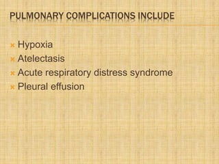
DR RIKIN HASNANI
Pulmonary complication
in Pancreatitis
INTRODUCTION
 Acute pancreatitis is an acute inflammatory
process of the pancreas in its severe form, is
complicated by the development of multi-
organ dysfunction syndrome with a mortality
of 15%-20%.
 Amongst the systemic complications,
pulmonary complications are the most
frequent and potentially the most serious.
RANSON’S CRITERIA
If the score ≥ 3, severe pancreatitis likely.
If the score < 3, severe pancreatitis is unlikely
PULMONARY COMPLICATIONS INCLUDE
 Hypoxia
 Atelectasis
 Acute respiratory distress syndrome
 Pleural effusion
PATHOPHYSIOLOGY OF ACUTE PANCREATITIS
Acute pancreatitis evolves in three phases
 Initial phase is characterized by
intrapancreatic digestive enzyme activation
and acinar cell injury which involves the first
few hours of acute Pancreatitis.
 The second phase characterized by an
intrapancreatic inflammatory reaction and
varying degrees of acinar cell necrosis, it
evolves in approximately 12-72 h.
 Finally, the third phase which involves the
rest of the progression of Acute Pancreatitis
is characterized by further progression of the
pancreatic injury and the appearance of
extrapancreatic changes including SIRS and
ARDS.
 It is during phase three that pulmonary insult
occurs
 Pulmonary complication are divided into
three stages
 Stage 1: Hypoxemia with no radiological
abnormalities
 Stage 2 : Hypoxemia with radiological
abnormalities
 Stage 3 : ARDS
STAGE 1: HYPOXEMIA WITH NO RADIOLOGICAL
ABNORMALITIES
 Tachypnea, mild respiratory alkalosis, and
hypoxemia are seen in almost two thirds of
patients with Acute Pancreatitis during the
first 2d of admission to the hospital.
 In these patients, physical examination is
essentially normal, chest radiographs rarely
demonstrate abnormalities.
 Imrie and co-workers showed that 45% of
patients have severe arterial hypoxemia
(PaO2 < 60mmHg) and indicated that a
PaO2 of < 52.5 mmHg is associated with a
mortality of more than 30%.
 The major cause of hypoxia is ventilation and
perfusion mismatch which results in a right to
left intrapulmonary shunting of up to 30% of
cardiac output.
 In two studies, Imrie and coworkers
correlated the overall mortality with the
degree of hypoxemia. They found that
patients with arterial pO2 below 70 mmHg
have a mortality of 3% to 5.9% and when the
arterial pO2 decreases below 60 mmHg the
mortality rate increases to about 14%.
STAGE 2 : HYPOXEMIA WITH RADIOLOGICAL
ABNORMALITIES
Radiological abnormalities seen are
 1- Pleural effusion(4%-17%)
 2- Atelactasis or Pulmonary infiltrates (15%)
 3- Pulmonary Edema(8%-50%)
PLEURAL EFFUSION
 Presence of pleural effusion is currently
considered an indication of severe
pancreatitis and not just a marker of the
disease.
 The pleural effusions are
Left sided in 68% ,
Bilateral in 22% ,
Right-sided in 10%, of cases.
PATHOPHYSIOLOGY OF PL. EFFUSION IN AC.
PANCREATITIS
 Various pathogenic mechanisms have been proposed
to explain the development of pleural effusions
associated with acute pancreatitis:
 (1) increased permeability of lymphatics and fluid leak
caused by pancreatic enzymes that may diffuse from
the peritoneal side to the thoracic side of the
diaphragm,
 (2) impaired lymphatic drainage of pleural exudate
caused by obstruction of lymphatic vessels by the
high enzymatic content of the pleural fluid, and
 (3) increased permeability of diaphragmatic
capillaries caused by the inflammatory process in the
adjacent pancreas.
CLINICAL FEATURES
 In patients with pleural effusion associated
with acute pancreatitis, symptoms are
primarily abdominal (pain,nausea and
vomiting);
 occasionally, respiratory symptoms(pleuritic
pain and dyspnea) may also be present.
 Pleural effusions in Acute Pancreatitis are
usually small, occasionally hemorrhagic , and
 are characterized by
 High amylase (up to 30 times greater than
corresponding serum value, usually >1000 IU/l)
 Protein (> 30 gm/L),
 Fluid LDH / serum LDH > 0.6 .
 WBC count vary from 1,000 to 50,000
cells/mm3.
MANAGEMENT
 Pleural effusions associated with acute
pancreatitis usually are self-limited and
resolve when the pancreatic inflammation
decreases without requiring therapeutic
drainage.
 If the pleural effusion does not resolve within
2 weeks after treatment for the pancreatic
disease, the possibility of a pancreatic
abscess or a pseudocyst should be
considered.
CHRONIC EFFUSIONS
 Chronic pleural effusions are usually associated
with chronic relapsing pancreatitis and
pancreatic pseudocyst.
 Chronic effusions are large, may occupy the
entire hemithorax,and reaccumulate rapidly
after thoracentesis.
 Mechanism of chronic effusion is a
pancreaticopleural fistula, a direct
communication between the pancreas and the
pleural space
 Pt with chronic effusion presents with respiratory
symptoms – dyspnoea, chest pain with minimal or no
abdominal symptoms.
 Patients with chronic pancreatic pleural effusions
should be treated initially with nasogastric tube
placement, no oral intake, parenteral nutrition, and
repeated therapeutic thoracenteses.
 Inhibition of pancreatic secretion with octreotide has
been shown to be useful in promoting the closure of
pancreaticopleural fistulas in some cases.
 Surgery is indicated if pleural fluid continues to
accumulate after 2-3 weeks of medical therapy.
ATELACTASIS
 Consolidation of lung tissue and atelectasis
are frequent radiological observations.
 Atelactasis is due to a decrease in the
production of pulmonary surfactant and
diaphragm muscle dysfunction.
 IL-1 and TNF-α plays avital role in diaphragm
muscle dysfunction.
ARDS
 Of the patients who develop Acute
pancreatitis , 15% to 20% develop ARDS
with an associated mortality of 56%.
 ARDS usually manifests itself between two to
seven days following the onset of Acute
pancreatitis, but may have a much more
rapid course.
 ARDS accounts for 50%-90% of all deaths
from pancreatitis
 Clinical features include severe dyspnea and
extreme hypoxemia refractory to a high
inspired oxygen concentration.
 Multilobar pulmonary infiltrates in patients
with previous normal radiographs and a
relatively normal pulmonary capillary wedge
pressure (< 18 mmHg) are noted.
 PaO2/FiO2 is usually <200
PATHOPHYSIOLOGY OF ARDS IN PANCREATITIS
 The pathophysiology of ARDS in Acute
pancreatitis is poorly understood.
 Actions of pancreatic enzymes as well as
inflammatory mediators released as a result
of pancreatic injury play a key role in the
pulmonary complications.
INFLAMMATORY MEDIATORS IN RESPIRATORY
INSUFFICIENCY
 1 Phospholipase A2
 2 Trypsin
 3 FFA/Lipoprotein lipase
 4 Complement activation (C5a)
 5 MIF
 6 IL-6, IL-8, IL-1β
 7 NO
 8 TNF-α
 9 ICAM-1
 10 β-2-integrin (CD11b/CD18)
 11 Trypsin
 12 NF-κB
 13 Substance P
 Activated trypsin: It is a possible source for
the cause of pulmonary insufficiency. Trypsin
causes damage to the pulmonary
vasculature and increases endothelial
permeability
 Trypsin is capable of activating different
complement factors directly, which can
stimulate cytolysis and chemotactic
leukocytes.
 Phospholipase A :PLA2, which is activated
by trypsin in the duodenum, is known for its
ability to remove fatty acids from
phospholipids.
 The basic reason for pulmonary insufficiency
and ARDS in Acute pancreatitis is due to the
destruction of the surfactant.
 One of the main components of surfactant is
phospholipid -
dipalmitoylphosphatidylcholine, which is a
perfect substrate for PLA2.
 Büchler et al showed that there is a strong
correlation between serum-activated PLA2 and
pulmonary insufficiency.
 In their prospective study, patients with
pulmonary failure demonstrated a notably higher
catalytic PLA2 activity during the first week of
Acute pancreatitis than patients without arterial
hypoxemia.
 They also showed that it was isotype II of PLA2
which is more specific to pulmonary
involvement.
 Platelet activating factor:PAF stimulates
PMN white cells and regulates the interaction
between PMN cells and endothelial cells,
facilitating migration of activated white cells
into tissue spaces. PAF is a structural
component of membrane lipids and is
released upon the action of PLA2
 Free fatty acid:The free fatty acids released
from triglycerides (TG), have been shown to
damage capillary alveolar wall membranes.
 TNF-α:TNF-α activates neutrophils and
increases lung damage levels at higher
concentration in the lung.
 Role of other substances like IL 8, IL10,NO,
Macrophage migration inhibitory factor,in
Acute pancreatitis are being studied.
MANAGEMET OF ACUTE PANCREATITIS
 NPO
 Fluid Resuscitation
 Octreotide 100-200mcg /sc/tid
 Management of oxygenation NIV or MV
 Management of Metabolic complications
 Lexipafant – PAF antagonist in stage 3 trials
decreases mortality if used at onset of
disease.
 Emerging drugs:
 CCK receptor antagonist: loxiglumide
 Prostaglandins: PGE1
 Platelet activating factor (PAF) antagonist
 TNF monoclonal antibody: Infliximab
THANK YOU

Lung in pancreatitis

  • 1.
    DR RIKIN HASNANI Pulmonarycomplication in Pancreatitis
  • 2.
    INTRODUCTION  Acute pancreatitisis an acute inflammatory process of the pancreas in its severe form, is complicated by the development of multi- organ dysfunction syndrome with a mortality of 15%-20%.  Amongst the systemic complications, pulmonary complications are the most frequent and potentially the most serious.
  • 3.
    RANSON’S CRITERIA If thescore ≥ 3, severe pancreatitis likely. If the score < 3, severe pancreatitis is unlikely
  • 4.
    PULMONARY COMPLICATIONS INCLUDE Hypoxia  Atelectasis  Acute respiratory distress syndrome  Pleural effusion
  • 5.
    PATHOPHYSIOLOGY OF ACUTEPANCREATITIS Acute pancreatitis evolves in three phases  Initial phase is characterized by intrapancreatic digestive enzyme activation and acinar cell injury which involves the first few hours of acute Pancreatitis.  The second phase characterized by an intrapancreatic inflammatory reaction and varying degrees of acinar cell necrosis, it evolves in approximately 12-72 h.
  • 6.
     Finally, thethird phase which involves the rest of the progression of Acute Pancreatitis is characterized by further progression of the pancreatic injury and the appearance of extrapancreatic changes including SIRS and ARDS.  It is during phase three that pulmonary insult occurs
  • 7.
     Pulmonary complicationare divided into three stages  Stage 1: Hypoxemia with no radiological abnormalities  Stage 2 : Hypoxemia with radiological abnormalities  Stage 3 : ARDS
  • 8.
    STAGE 1: HYPOXEMIAWITH NO RADIOLOGICAL ABNORMALITIES  Tachypnea, mild respiratory alkalosis, and hypoxemia are seen in almost two thirds of patients with Acute Pancreatitis during the first 2d of admission to the hospital.  In these patients, physical examination is essentially normal, chest radiographs rarely demonstrate abnormalities.
  • 9.
     Imrie andco-workers showed that 45% of patients have severe arterial hypoxemia (PaO2 < 60mmHg) and indicated that a PaO2 of < 52.5 mmHg is associated with a mortality of more than 30%.  The major cause of hypoxia is ventilation and perfusion mismatch which results in a right to left intrapulmonary shunting of up to 30% of cardiac output.
  • 10.
     In twostudies, Imrie and coworkers correlated the overall mortality with the degree of hypoxemia. They found that patients with arterial pO2 below 70 mmHg have a mortality of 3% to 5.9% and when the arterial pO2 decreases below 60 mmHg the mortality rate increases to about 14%.
  • 11.
    STAGE 2 :HYPOXEMIA WITH RADIOLOGICAL ABNORMALITIES Radiological abnormalities seen are  1- Pleural effusion(4%-17%)  2- Atelactasis or Pulmonary infiltrates (15%)  3- Pulmonary Edema(8%-50%)
  • 12.
    PLEURAL EFFUSION  Presenceof pleural effusion is currently considered an indication of severe pancreatitis and not just a marker of the disease.  The pleural effusions are Left sided in 68% , Bilateral in 22% , Right-sided in 10%, of cases.
  • 13.
    PATHOPHYSIOLOGY OF PL.EFFUSION IN AC. PANCREATITIS  Various pathogenic mechanisms have been proposed to explain the development of pleural effusions associated with acute pancreatitis:  (1) increased permeability of lymphatics and fluid leak caused by pancreatic enzymes that may diffuse from the peritoneal side to the thoracic side of the diaphragm,  (2) impaired lymphatic drainage of pleural exudate caused by obstruction of lymphatic vessels by the high enzymatic content of the pleural fluid, and  (3) increased permeability of diaphragmatic capillaries caused by the inflammatory process in the adjacent pancreas.
  • 14.
    CLINICAL FEATURES  Inpatients with pleural effusion associated with acute pancreatitis, symptoms are primarily abdominal (pain,nausea and vomiting);  occasionally, respiratory symptoms(pleuritic pain and dyspnea) may also be present.
  • 15.
     Pleural effusionsin Acute Pancreatitis are usually small, occasionally hemorrhagic , and  are characterized by  High amylase (up to 30 times greater than corresponding serum value, usually >1000 IU/l)  Protein (> 30 gm/L),  Fluid LDH / serum LDH > 0.6 .  WBC count vary from 1,000 to 50,000 cells/mm3.
  • 16.
    MANAGEMENT  Pleural effusionsassociated with acute pancreatitis usually are self-limited and resolve when the pancreatic inflammation decreases without requiring therapeutic drainage.  If the pleural effusion does not resolve within 2 weeks after treatment for the pancreatic disease, the possibility of a pancreatic abscess or a pseudocyst should be considered.
  • 17.
    CHRONIC EFFUSIONS  Chronicpleural effusions are usually associated with chronic relapsing pancreatitis and pancreatic pseudocyst.  Chronic effusions are large, may occupy the entire hemithorax,and reaccumulate rapidly after thoracentesis.  Mechanism of chronic effusion is a pancreaticopleural fistula, a direct communication between the pancreas and the pleural space
  • 18.
     Pt withchronic effusion presents with respiratory symptoms – dyspnoea, chest pain with minimal or no abdominal symptoms.  Patients with chronic pancreatic pleural effusions should be treated initially with nasogastric tube placement, no oral intake, parenteral nutrition, and repeated therapeutic thoracenteses.  Inhibition of pancreatic secretion with octreotide has been shown to be useful in promoting the closure of pancreaticopleural fistulas in some cases.  Surgery is indicated if pleural fluid continues to accumulate after 2-3 weeks of medical therapy.
  • 19.
    ATELACTASIS  Consolidation oflung tissue and atelectasis are frequent radiological observations.  Atelactasis is due to a decrease in the production of pulmonary surfactant and diaphragm muscle dysfunction.  IL-1 and TNF-α plays avital role in diaphragm muscle dysfunction.
  • 20.
    ARDS  Of thepatients who develop Acute pancreatitis , 15% to 20% develop ARDS with an associated mortality of 56%.  ARDS usually manifests itself between two to seven days following the onset of Acute pancreatitis, but may have a much more rapid course.  ARDS accounts for 50%-90% of all deaths from pancreatitis
  • 21.
     Clinical featuresinclude severe dyspnea and extreme hypoxemia refractory to a high inspired oxygen concentration.  Multilobar pulmonary infiltrates in patients with previous normal radiographs and a relatively normal pulmonary capillary wedge pressure (< 18 mmHg) are noted.  PaO2/FiO2 is usually <200
  • 22.
    PATHOPHYSIOLOGY OF ARDSIN PANCREATITIS  The pathophysiology of ARDS in Acute pancreatitis is poorly understood.  Actions of pancreatic enzymes as well as inflammatory mediators released as a result of pancreatic injury play a key role in the pulmonary complications.
  • 23.
    INFLAMMATORY MEDIATORS INRESPIRATORY INSUFFICIENCY  1 Phospholipase A2  2 Trypsin  3 FFA/Lipoprotein lipase  4 Complement activation (C5a)  5 MIF  6 IL-6, IL-8, IL-1β  7 NO  8 TNF-α  9 ICAM-1  10 β-2-integrin (CD11b/CD18)  11 Trypsin  12 NF-κB  13 Substance P
  • 24.
     Activated trypsin:It is a possible source for the cause of pulmonary insufficiency. Trypsin causes damage to the pulmonary vasculature and increases endothelial permeability  Trypsin is capable of activating different complement factors directly, which can stimulate cytolysis and chemotactic leukocytes.
  • 25.
     Phospholipase A:PLA2, which is activated by trypsin in the duodenum, is known for its ability to remove fatty acids from phospholipids.  The basic reason for pulmonary insufficiency and ARDS in Acute pancreatitis is due to the destruction of the surfactant.  One of the main components of surfactant is phospholipid - dipalmitoylphosphatidylcholine, which is a perfect substrate for PLA2.
  • 26.
     Büchler etal showed that there is a strong correlation between serum-activated PLA2 and pulmonary insufficiency.  In their prospective study, patients with pulmonary failure demonstrated a notably higher catalytic PLA2 activity during the first week of Acute pancreatitis than patients without arterial hypoxemia.  They also showed that it was isotype II of PLA2 which is more specific to pulmonary involvement.
  • 27.
     Platelet activatingfactor:PAF stimulates PMN white cells and regulates the interaction between PMN cells and endothelial cells, facilitating migration of activated white cells into tissue spaces. PAF is a structural component of membrane lipids and is released upon the action of PLA2
  • 28.
     Free fattyacid:The free fatty acids released from triglycerides (TG), have been shown to damage capillary alveolar wall membranes.  TNF-α:TNF-α activates neutrophils and increases lung damage levels at higher concentration in the lung.  Role of other substances like IL 8, IL10,NO, Macrophage migration inhibitory factor,in Acute pancreatitis are being studied.
  • 29.
    MANAGEMET OF ACUTEPANCREATITIS  NPO  Fluid Resuscitation  Octreotide 100-200mcg /sc/tid  Management of oxygenation NIV or MV  Management of Metabolic complications  Lexipafant – PAF antagonist in stage 3 trials decreases mortality if used at onset of disease.
  • 30.
     Emerging drugs: CCK receptor antagonist: loxiglumide  Prostaglandins: PGE1  Platelet activating factor (PAF) antagonist  TNF monoclonal antibody: Infliximab
  • 31.