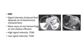
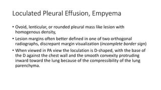
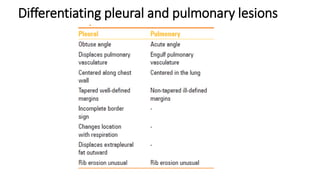
Downloaded 144 times

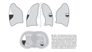
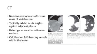

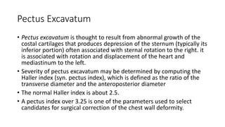
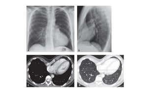
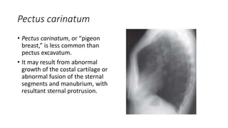

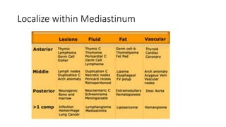
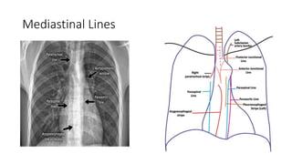
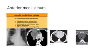

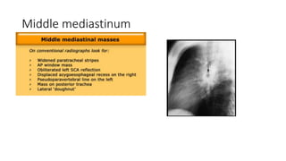
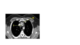
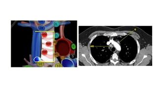

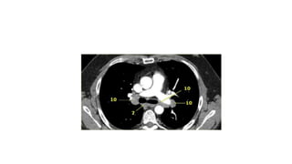


This document discusses computed tomography (CT) imaging findings of various chest diseases, including pleural diseases, chest wall diseases, and mediastinal diseases. It describes how CT can be used to identify and characterize pleural effusions, pleural thickening, asbestos-related pleural disease, and tumors of the pleura. It also discusses chest wall abnormalities such as pectus excavatum, pectus carinatum, and Poland syndrome. Finally, it provides guidance on using CT findings to localize diseases within the mediastinum and differentiate various mediastinal abnormalities.


![PERI-PROSTHETIC FRACTURE NAIL-PLATE CONSTRUCT [NPC].pptx](https://cdn.slidesharecdn.com/ss_thumbnails/drarunkumardrmohamedashrafperiprostheticfrasturenail-plateconstructnpc-260209164459-7e9d15a1-thumbnail.jpg?width=640&height=640&fit=bounds)

