Submit Search
Upload
The radiology assistant chest x ray - basic interpretation
•
26 likes
•
3,334 views
kosar kamal
Follow
basic chest interpretation
Read less
Read more
Health & Medicine
Report
Share
Report
Share
1 of 56
Download now
Download to read offline
Recommended
Signs in Chest Xray
Signs in Chest Xray
Archana Koshy
Presentation1, interpretation of x ray of the abdomen.
Presentation1, interpretation of x ray of the abdomen.
Abdellah Nazeer
Radiology: Chest Imaging
Radiology: Chest Imaging
SCGH ED CME
Paediatric chest imaging
Paediatric chest imaging
Sidra Afzal
Abdominal X ray
Abdominal X ray
Richin Koshy
Presentation1.pptx. interpretation of x ray chest.
Presentation1.pptx. interpretation of x ray chest.
Abdellah Nazeer
Abdominal CT scan made easy
Abdominal CT scan made easy
Masrur Akbar Khan
Imaging of the large bowel
Imaging of the large bowel
Archana Koshy
Chest x ray - basics
Chest x ray - basics
Rikin Hasnani
Interpretation of Chest X-Ray PPT
Interpretation of Chest X-Ray PPT
drmainuddin
RAH Med 4 MHU - Chest Xray 1
RAH Med 4 MHU - Chest Xray 1
Luke Oakden-Rayner
Presentation1.pptx, imaging of the urinary system.
Presentation1.pptx, imaging of the urinary system.
Abdellah Nazeer
Radiological features of pneumonia
Radiological features of pneumonia
Department of Health & Family Welfare, Government of West Bengal
Imaging in Skull base
Imaging in Skull base
Rakesh Ca
Radiology ----Classical Signs in GIT Dr. Muhammad Bin Zulfiqar
Radiology ----Classical Signs in GIT Dr. Muhammad Bin Zulfiqar
Dr. Muhammad Bin Zulfiqar
Chest x ray
Chest x ray
Dr,saket Jain
Pneumomediastinum
Pneumomediastinum
Gamal Agmy
Presentation1.pptx, radiological signs in thoracic radiology.
Presentation1.pptx, radiological signs in thoracic radiology.
Abdellah Nazeer
Chest x ray basic interpretation
Chest x ray basic interpretation
Vikram Patil
Intracranial ultrasound
Intracranial ultrasound
Dr. Mohit Goel
Ultrasound of the urinary tract - Renal infections
Ultrasound of the urinary tract - Renal infections
Samir Haffar
Congenital cystic adenomatoid malformation
Congenital cystic adenomatoid malformation
Sneha Volvoikar
Radiology of nose and paranasal sinuses
Radiology of nose and paranasal sinuses
Andrea R Salins
Pediatric radiology
Pediatric radiology
Brian Wells, MD, MS, MPH
Ultrasound of acute & chronic cholecystitis
Ultrasound of acute & chronic cholecystitis
Samir Haffar
radiology anatomy of chest.ppt
radiology anatomy of chest.ppt
ranjitharadhakrishna3
Abdominal xray images
Abdominal xray images
Le Jang
Interpretation of chest xray ppt
Interpretation of chest xray ppt
Rithwik Karumuri
The radiology assistant chest x ray - lung disease
The radiology assistant chest x ray - lung disease
kosar kamal
Chest xray
Chest xray
Abino David
More Related Content
What's hot
Chest x ray - basics
Chest x ray - basics
Rikin Hasnani
Interpretation of Chest X-Ray PPT
Interpretation of Chest X-Ray PPT
drmainuddin
RAH Med 4 MHU - Chest Xray 1
RAH Med 4 MHU - Chest Xray 1
Luke Oakden-Rayner
Presentation1.pptx, imaging of the urinary system.
Presentation1.pptx, imaging of the urinary system.
Abdellah Nazeer
Radiological features of pneumonia
Radiological features of pneumonia
Department of Health & Family Welfare, Government of West Bengal
Imaging in Skull base
Imaging in Skull base
Rakesh Ca
Radiology ----Classical Signs in GIT Dr. Muhammad Bin Zulfiqar
Radiology ----Classical Signs in GIT Dr. Muhammad Bin Zulfiqar
Dr. Muhammad Bin Zulfiqar
Chest x ray
Chest x ray
Dr,saket Jain
Pneumomediastinum
Pneumomediastinum
Gamal Agmy
Presentation1.pptx, radiological signs in thoracic radiology.
Presentation1.pptx, radiological signs in thoracic radiology.
Abdellah Nazeer
Chest x ray basic interpretation
Chest x ray basic interpretation
Vikram Patil
Intracranial ultrasound
Intracranial ultrasound
Dr. Mohit Goel
Ultrasound of the urinary tract - Renal infections
Ultrasound of the urinary tract - Renal infections
Samir Haffar
Congenital cystic adenomatoid malformation
Congenital cystic adenomatoid malformation
Sneha Volvoikar
Radiology of nose and paranasal sinuses
Radiology of nose and paranasal sinuses
Andrea R Salins
Pediatric radiology
Pediatric radiology
Brian Wells, MD, MS, MPH
Ultrasound of acute & chronic cholecystitis
Ultrasound of acute & chronic cholecystitis
Samir Haffar
radiology anatomy of chest.ppt
radiology anatomy of chest.ppt
ranjitharadhakrishna3
Abdominal xray images
Abdominal xray images
Le Jang
Interpretation of chest xray ppt
Interpretation of chest xray ppt
Rithwik Karumuri
What's hot
(20)
Chest x ray - basics
Chest x ray - basics
Interpretation of Chest X-Ray PPT
Interpretation of Chest X-Ray PPT
RAH Med 4 MHU - Chest Xray 1
RAH Med 4 MHU - Chest Xray 1
Presentation1.pptx, imaging of the urinary system.
Presentation1.pptx, imaging of the urinary system.
Radiological features of pneumonia
Radiological features of pneumonia
Imaging in Skull base
Imaging in Skull base
Radiology ----Classical Signs in GIT Dr. Muhammad Bin Zulfiqar
Radiology ----Classical Signs in GIT Dr. Muhammad Bin Zulfiqar
Chest x ray
Chest x ray
Pneumomediastinum
Pneumomediastinum
Presentation1.pptx, radiological signs in thoracic radiology.
Presentation1.pptx, radiological signs in thoracic radiology.
Chest x ray basic interpretation
Chest x ray basic interpretation
Intracranial ultrasound
Intracranial ultrasound
Ultrasound of the urinary tract - Renal infections
Ultrasound of the urinary tract - Renal infections
Congenital cystic adenomatoid malformation
Congenital cystic adenomatoid malformation
Radiology of nose and paranasal sinuses
Radiology of nose and paranasal sinuses
Pediatric radiology
Pediatric radiology
Ultrasound of acute & chronic cholecystitis
Ultrasound of acute & chronic cholecystitis
radiology anatomy of chest.ppt
radiology anatomy of chest.ppt
Abdominal xray images
Abdominal xray images
Interpretation of chest xray ppt
Interpretation of chest xray ppt
Viewers also liked
The radiology assistant chest x ray - lung disease
The radiology assistant chest x ray - lung disease
kosar kamal
Chest xray
Chest xray
Abino David
A case of SLE polyserositis & pneumonitis
A case of SLE polyserositis & pneumonitis
Stanley Medical College, Department of Medicine
Pulmonary manifestations of lupus
Pulmonary manifestations of lupus
mohmeet
Pericardial effusion
Pericardial effusion
Todd Peterson
Sepsis
Sepsis
軒名 林
Surviving Sepsis Guidelines 2012
Surviving Sepsis Guidelines 2012
Sun Yai-Cheng
Systemic lupus
Systemic lupus
Siva Nanda Reddy
андрология 1
андрология 1
imad almasri
Professional Persona Project - John Patino
Professional Persona Project - John Patino
John Patino
Pertemuan ke 1 pengenalan komputer
Pertemuan ke 1 pengenalan komputer
Harry-antho Putheraa
How to revolutionise an industry with ZeroVM
How to revolutionise an industry with ZeroVM
Jakub Krajcovic
Professional Persona Project - John Patino
Professional Persona Project - John Patino
John Patino
андрология 2
андрология 2
imad almasri
Food art
Food art
Elpida Adalopoulou
soon
soon
chloekaiyun
Place and community based education - Prof Greg Smith
Place and community based education - Prof Greg Smith
Meg Collis
Openstack SAGE-AU
Openstack SAGE-AU
Jakub Krajcovic
อุปกรณ์พื้นฐานคอมพิวเตอร์
อุปกรณ์พื้นฐานคอมพิวเตอร์
BoMz Zilch
Conventions of a Thriller
Conventions of a Thriller
charlottemedia13
Viewers also liked
(20)
The radiology assistant chest x ray - lung disease
The radiology assistant chest x ray - lung disease
Chest xray
Chest xray
A case of SLE polyserositis & pneumonitis
A case of SLE polyserositis & pneumonitis
Pulmonary manifestations of lupus
Pulmonary manifestations of lupus
Pericardial effusion
Pericardial effusion
Sepsis
Sepsis
Surviving Sepsis Guidelines 2012
Surviving Sepsis Guidelines 2012
Systemic lupus
Systemic lupus
андрология 1
андрология 1
Professional Persona Project - John Patino
Professional Persona Project - John Patino
Pertemuan ke 1 pengenalan komputer
Pertemuan ke 1 pengenalan komputer
How to revolutionise an industry with ZeroVM
How to revolutionise an industry with ZeroVM
Professional Persona Project - John Patino
Professional Persona Project - John Patino
андрология 2
андрология 2
Food art
Food art
soon
soon
Place and community based education - Prof Greg Smith
Place and community based education - Prof Greg Smith
Openstack SAGE-AU
Openstack SAGE-AU
อุปกรณ์พื้นฐานคอมพิวเตอร์
อุปกรณ์พื้นฐานคอมพิวเตอร์
Conventions of a Thriller
Conventions of a Thriller
Similar to The radiology assistant chest x ray - basic interpretation
Cxr new
Cxr new
LPS Institute of Cardiology Kanpur UP India
GROUP 6B PEDIATRIC CHEST AND ABDOMEN XRAY TECHNIQUE, PITFALLS.pptx
GROUP 6B PEDIATRIC CHEST AND ABDOMEN XRAY TECHNIQUE, PITFALLS.pptx
YasinKidiri
Fetal MRI
Fetal MRI
Patty G.
AAMD Presentation
AAMD Presentation
Lee Culp, M.S., C.M.D, R.T.(T.)
Myo protect
Myo protect
Sandeep Jose K
Engaging with data
Engaging with data
Nuffield Trust
CHEST XRAY RDG 303.pptx
CHEST XRAY RDG 303.pptx
EmmanuelOluseyi1
Interventional Radiography
Interventional Radiography
Jessica Myers
Imagen Torácica
Imagen Torácica
Cesar Rosenberg González
Intensivist-delivered echo: dangerous in the wrong hands? - Price
Intensivist-delivered echo: dangerous in the wrong hands? - Price
intensivecaresociety
07 radiology in surgery tutorial hajhamad m msu
07 radiology in surgery tutorial hajhamad m msu
Mohammed M. H. Hajhamad
Journal club presentation (chest tube) _.pptx dr shakil
Journal club presentation (chest tube) _.pptx dr shakil
Shakil Ahmad
C.V Hany
C.V Hany
Hany Kerbalou
9241541792 eng
9241541792 eng
Nurmaniar Majid
Echocardiographic anatomy in the fetus 2009 pg
Echocardiographic anatomy in the fetus 2009 pg
Nguyen Phong Trung
2020 fun with thoracostomy's
2020 fun with thoracostomy's
Robert Cole
Protocol
Protocol
Sahana Subramani
Thyroid Cancer
Thyroid Cancer
fitango
Damage Control Symposium 24th Nov 2014
Damage Control Symposium 24th Nov 2014
Alan Batt
WE Ladd - Seminar - EN - 2018
WE Ladd - Seminar - EN - 2018
Oguz Kizilkaya
Similar to The radiology assistant chest x ray - basic interpretation
(20)
Cxr new
Cxr new
GROUP 6B PEDIATRIC CHEST AND ABDOMEN XRAY TECHNIQUE, PITFALLS.pptx
GROUP 6B PEDIATRIC CHEST AND ABDOMEN XRAY TECHNIQUE, PITFALLS.pptx
Fetal MRI
Fetal MRI
AAMD Presentation
AAMD Presentation
Myo protect
Myo protect
Engaging with data
Engaging with data
CHEST XRAY RDG 303.pptx
CHEST XRAY RDG 303.pptx
Interventional Radiography
Interventional Radiography
Imagen Torácica
Imagen Torácica
Intensivist-delivered echo: dangerous in the wrong hands? - Price
Intensivist-delivered echo: dangerous in the wrong hands? - Price
07 radiology in surgery tutorial hajhamad m msu
07 radiology in surgery tutorial hajhamad m msu
Journal club presentation (chest tube) _.pptx dr shakil
Journal club presentation (chest tube) _.pptx dr shakil
C.V Hany
C.V Hany
9241541792 eng
9241541792 eng
Echocardiographic anatomy in the fetus 2009 pg
Echocardiographic anatomy in the fetus 2009 pg
2020 fun with thoracostomy's
2020 fun with thoracostomy's
Protocol
Protocol
Thyroid Cancer
Thyroid Cancer
Damage Control Symposium 24th Nov 2014
Damage Control Symposium 24th Nov 2014
WE Ladd - Seminar - EN - 2018
WE Ladd - Seminar - EN - 2018
More from kosar kamal
Upper limb radiography
Upper limb radiography
kosar kamal
Final exam practical
Final exam practical
kosar kamal
GIT
GIT
kosar kamal
Chest
Chest
kosar kamal
neuroradiology quiz module
neuroradiology quiz module
kosar kamal
Lecture 7
Lecture 7
kosar kamal
Lectures 6
Lectures 6
kosar kamal
Chest radiology essentials mediastinal masses
Chest radiology essentials mediastinal masses
kosar kamal
Chest radiology essentials signs
Chest radiology essentials signs
kosar kamal
basic approach for CXR interpretation
basic approach for CXR interpretation
kosar kamal
Quiz 2
Quiz 2
kosar kamal
Second and third trimester emergencies
Second and third trimester emergencies
kosar kamal
First trimester ultrasound
First trimester ultrasound
kosar kamal
Extended focused abdominal assesment in trauma patients
Extended focused abdominal assesment in trauma patients
kosar kamal
More from kosar kamal
(14)
Upper limb radiography
Upper limb radiography
Final exam practical
Final exam practical
GIT
GIT
Chest
Chest
neuroradiology quiz module
neuroradiology quiz module
Lecture 7
Lecture 7
Lectures 6
Lectures 6
Chest radiology essentials mediastinal masses
Chest radiology essentials mediastinal masses
Chest radiology essentials signs
Chest radiology essentials signs
basic approach for CXR interpretation
basic approach for CXR interpretation
Quiz 2
Quiz 2
Second and third trimester emergencies
Second and third trimester emergencies
First trimester ultrasound
First trimester ultrasound
Extended focused abdominal assesment in trauma patients
Extended focused abdominal assesment in trauma patients
Recently uploaded
Low Rate Call Girls Ambattur Anika 8250192130 Independent Escort Service Amba...
Low Rate Call Girls Ambattur Anika 8250192130 Independent Escort Service Amba...
narwatsonia7
Call Girl Bangalore Nandini 7001305949 Independent Escort Service Bangalore
Call Girl Bangalore Nandini 7001305949 Independent Escort Service Bangalore
narwatsonia7
Artifacts in Nuclear Medicine with Identifying and resolving artifacts.
Artifacts in Nuclear Medicine with Identifying and resolving artifacts.
MiadAlsulami
CALL ON ➥9907093804 🔝 Call Girls Hadapsar ( Pune) Girls Service
CALL ON ➥9907093804 🔝 Call Girls Hadapsar ( Pune) Girls Service
Miss joya
High Profile Call Girls Jaipur Vani 8445551418 Independent Escort Service Jaipur
High Profile Call Girls Jaipur Vani 8445551418 Independent Escort Service Jaipur
parulsinha
Low Rate Call Girls Pune Esha 9907093804 Short 1500 Night 6000 Best call girl...
Low Rate Call Girls Pune Esha 9907093804 Short 1500 Night 6000 Best call girl...
Miss joya
Hi,Fi Call Girl In Mysore Road - 7001305949 | 24x7 Service Available Near Me
Hi,Fi Call Girl In Mysore Road - 7001305949 | 24x7 Service Available Near Me
narwatsonia7
VIP Call Girls Indore Kirti 💚😋 9256729539 🚀 Indore Escorts
VIP Call Girls Indore Kirti 💚😋 9256729539 🚀 Indore Escorts
aditipandeya
sauth delhi call girls in Bhajanpura 🔝 9953056974 🔝 escort Service
sauth delhi call girls in Bhajanpura 🔝 9953056974 🔝 escort Service
9953056974 Low Rate Call Girls In Saket, Delhi NCR
VIP Call Girls Pune Vrinda 9907093804 Short 1500 Night 6000 Best call girls S...
VIP Call Girls Pune Vrinda 9907093804 Short 1500 Night 6000 Best call girls S...
Miss joya
Call Girls Service In Shyam Nagar Whatsapp 8445551418 Independent Escort Service
Call Girls Service In Shyam Nagar Whatsapp 8445551418 Independent Escort Service
parulsinha
Russian Call Girls in Bangalore Manisha 7001305949 Independent Escort Service...
Russian Call Girls in Bangalore Manisha 7001305949 Independent Escort Service...
narwatsonia7
Sonagachi Call Girls Services 9907093804 @24x7 High Class Babes Here Call Now
Sonagachi Call Girls Services 9907093804 @24x7 High Class Babes Here Call Now
Riya Pathan
VIP Call Girls Tirunelveli Aaradhya 8250192130 Independent Escort Service Tir...
VIP Call Girls Tirunelveli Aaradhya 8250192130 Independent Escort Service Tir...
narwatsonia7
Call Girls In Andheri East Call 9920874524 Book Hot And Sexy Girls
Call Girls In Andheri East Call 9920874524 Book Hot And Sexy Girls
nehamumbai
Call Girls Doddaballapur Road Just Call 7001305949 Top Class Call Girl Servic...
Call Girls Doddaballapur Road Just Call 7001305949 Top Class Call Girl Servic...
narwatsonia7
Housewife Call Girls Hoskote | 7001305949 At Low Cost Cash Payment Booking
Housewife Call Girls Hoskote | 7001305949 At Low Cost Cash Payment Booking
narwatsonia7
Kesar Bagh Call Girl Price 9548273370 , Lucknow Call Girls Service
Kesar Bagh Call Girl Price 9548273370 , Lucknow Call Girls Service
makika9823
Call Girls Service Surat Samaira ❤️🍑 8250192130 👄 Independent Escort Service ...
Call Girls Service Surat Samaira ❤️🍑 8250192130 👄 Independent Escort Service ...
CALL GIRLS
Call Girls Service Jaipur Grishma WhatsApp ❤8445551418 VIP Call Girls Jaipur
Call Girls Service Jaipur Grishma WhatsApp ❤8445551418 VIP Call Girls Jaipur
parulsinha
Recently uploaded
(20)
Low Rate Call Girls Ambattur Anika 8250192130 Independent Escort Service Amba...
Low Rate Call Girls Ambattur Anika 8250192130 Independent Escort Service Amba...
Call Girl Bangalore Nandini 7001305949 Independent Escort Service Bangalore
Call Girl Bangalore Nandini 7001305949 Independent Escort Service Bangalore
Artifacts in Nuclear Medicine with Identifying and resolving artifacts.
Artifacts in Nuclear Medicine with Identifying and resolving artifacts.
CALL ON ➥9907093804 🔝 Call Girls Hadapsar ( Pune) Girls Service
CALL ON ➥9907093804 🔝 Call Girls Hadapsar ( Pune) Girls Service
High Profile Call Girls Jaipur Vani 8445551418 Independent Escort Service Jaipur
High Profile Call Girls Jaipur Vani 8445551418 Independent Escort Service Jaipur
Low Rate Call Girls Pune Esha 9907093804 Short 1500 Night 6000 Best call girl...
Low Rate Call Girls Pune Esha 9907093804 Short 1500 Night 6000 Best call girl...
Hi,Fi Call Girl In Mysore Road - 7001305949 | 24x7 Service Available Near Me
Hi,Fi Call Girl In Mysore Road - 7001305949 | 24x7 Service Available Near Me
VIP Call Girls Indore Kirti 💚😋 9256729539 🚀 Indore Escorts
VIP Call Girls Indore Kirti 💚😋 9256729539 🚀 Indore Escorts
sauth delhi call girls in Bhajanpura 🔝 9953056974 🔝 escort Service
sauth delhi call girls in Bhajanpura 🔝 9953056974 🔝 escort Service
VIP Call Girls Pune Vrinda 9907093804 Short 1500 Night 6000 Best call girls S...
VIP Call Girls Pune Vrinda 9907093804 Short 1500 Night 6000 Best call girls S...
Call Girls Service In Shyam Nagar Whatsapp 8445551418 Independent Escort Service
Call Girls Service In Shyam Nagar Whatsapp 8445551418 Independent Escort Service
Russian Call Girls in Bangalore Manisha 7001305949 Independent Escort Service...
Russian Call Girls in Bangalore Manisha 7001305949 Independent Escort Service...
Sonagachi Call Girls Services 9907093804 @24x7 High Class Babes Here Call Now
Sonagachi Call Girls Services 9907093804 @24x7 High Class Babes Here Call Now
VIP Call Girls Tirunelveli Aaradhya 8250192130 Independent Escort Service Tir...
VIP Call Girls Tirunelveli Aaradhya 8250192130 Independent Escort Service Tir...
Call Girls In Andheri East Call 9920874524 Book Hot And Sexy Girls
Call Girls In Andheri East Call 9920874524 Book Hot And Sexy Girls
Call Girls Doddaballapur Road Just Call 7001305949 Top Class Call Girl Servic...
Call Girls Doddaballapur Road Just Call 7001305949 Top Class Call Girl Servic...
Housewife Call Girls Hoskote | 7001305949 At Low Cost Cash Payment Booking
Housewife Call Girls Hoskote | 7001305949 At Low Cost Cash Payment Booking
Kesar Bagh Call Girl Price 9548273370 , Lucknow Call Girls Service
Kesar Bagh Call Girl Price 9548273370 , Lucknow Call Girls Service
Call Girls Service Surat Samaira ❤️🍑 8250192130 👄 Independent Escort Service ...
Call Girls Service Surat Samaira ❤️🍑 8250192130 👄 Independent Escort Service ...
Call Girls Service Jaipur Grishma WhatsApp ❤8445551418 VIP Call Girls Jaipur
Call Girls Service Jaipur Grishma WhatsApp ❤8445551418 VIP Call Girls Jaipur
The radiology assistant chest x ray - basic interpretation
1.
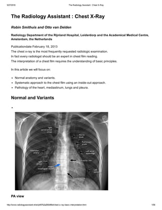
5/27/2016 The Radiology Assistant : Chest XRay http://www.radiologyassistant.nl/en/p497b2a265d96d/chestxraybasicinterpretation.html 1/56 The Radiology Assistant : Chest XRay Robin Smithuis and Otto van Delden Radiology Department of the Rijnland Hospital, Leiderdorp and the Academical Medical Centre, Amsterdam, the Netherlands Publicationdate February 18, 2013 The chest xray is the most frequently requested radiologic examination. In fact every radiologst should be an expert in chest film reading. The interpretation of a chest film requires the understanding of basic principles. In this article we will focus on: Normal anatomy and variants. Systematic approach to the chest film using an insideout approach. Pathology of the heart, mediastinum, lungs and pleura. Normal and Variants PA view
2.
5/27/2016 The Radiology Assistant : Chest XRay http://www.radiologyassistant.nl/en/p497b2a265d96d/chestxraybasicinterpretation.html 2/56 On the PA chestfilm it is important to examine all the areas where the lung borders the diaphragm, the heart and other mediastinal structures. At these borders lungsoft tissue interfaces are seen resulting in a: Line or stripe for instance the right para tracheal stripe. Silhouette for instance the normal silhouette of the aortic knob or left ventricle These lines and silhouettes are useful localizers of disease, because they can be displaced or obscured with loss of the normal silhouette. This is called the silhouette sign, which we will discuss later. The paraspinal line may be displaced by a paravertebral abscess, hemorrhage due to a fracture or extravertebral extension of a neoplasm. Widening of the paratracheal line (> 23mm) may be due to lymphadenopathy, pleural thickening, hemorrhage or fluid overload and heart failure. Displacement of the paraaortic line can be due to elongation of the aorta, aneurysm, dissection and rupture. The anterior and posterior junction lines are formed where the upper lobes join anteriorly and posteriorly. These are usely not well seen and we will not discuss them. An important mediastinallung interface to look for is the azygoesophageal line or recess (arrow). Azygoesophageal recess. The blue arrow indicates the paraaortic line. The azygoesophageal recess is the region inferior to the level of the azygos vein arch in which the right lung forms an interface with the mediastinum between the heart anteriorly and vertebral column posteriorly.
3.
5/27/2016 The Radiology Assistant : Chest XRay http://www.radiologyassistant.nl/en/p497b2a265d96d/chestxraybasicinterpretation.html 3/56 It is bordered on the left by the esophagus. Deviation of the azygoesophageal line is caused by (5): Hiatal hernia Esophageal disease Left atrial enlargement Subcarinal lymphadenopathy Bronchogenic cyst Notice the deviation of the azygoesophageal line on the PAfilm. It is caused by a hiatal hernia. Vena azygos lobe A common normal variant is the azygos lobe. The azygos lobe is created when a laterally displaced azygos vein makes a deep fissure in the upper
4.
5/27/2016 The Radiology Assistant : Chest XRay http://www.radiologyassistant.nl/en/p497b2a265d96d/chestxraybasicinterpretation.html 4/56 part of the lung. On a chest film it is seen as a fine line that crosses the apex of the right lung. Here another patient with an azygos lobe. The azygos vein is seen as a thick structure within the azygos fissure. In some patients an extra joint is seen in the anterior part of the first rib at the point where the bone meets the calcified cartilageneous part (arrow). This may simulate a lung mass.
5.
5/27/2016 The Radiology Assistant : Chest XRay http://www.radiologyassistant.nl/en/p497b2a265d96d/chestxraybasicinterpretation.html 5/56 Pectus excavatum In patients with a pectus excavatum the right heart border can be illdefined, but this is normal. It produces a silhouette sign and thus simulating a consolidation or atelectasis of the right middle lobe. The lateral view is helpful in such cases. Pectus excavatum is a congenital deformity of the ribs and the sternum producing a concave appearance of the anterior chest wall. Lateral view On a normal lateral view the contours of the heart are visible and the IVC is seen entering the right atrium. The retrosternal space should be radiolucent, since it only contains air. Any radiopacity in this area is suspective of a proces in the anterior mediastinum or upper lobes of the lung.
6.
5/27/2016 The Radiology Assistant : Chest XRay http://www.radiologyassistant.nl/en/p497b2a265d96d/chestxraybasicinterpretation.html 6/56 As you go from superior to inferior over the vertebral bodies they should get darker, because usually there will be less soft tissue and more radiolucent lung tissue (red arrow). If this is not the case, look carefully for pathology in the lower lobes. The contours of the left and right diaphragm should be visible. The right diaphragm should be visible all the way to the anterior chest wall (red arrow). Actually we see the interface between the air in the lungs and the soft tissue structures in the abdomen. The left diaphragm can only be seen to a point where it borders the heart (blue arrow). Here the interface is lost, since the heart has the same density as the structures below the diaphragm. The left main pulmonary artery (in purple) passes over the left main bronchus and is higher than the right pulmonary artery (in blue) which passes in front of the right main bronchus.
7.
5/27/2016 The Radiology Assistant : Chest XRay http://www.radiologyassistant.nl/en/p497b2a265d96d/chestxraybasicinterpretation.html 7/56 Once you know how the normal hilar structures look like on a lateral view, it is easier to detect abnormalities. In this case on the PAview there is hilar enlargement. On the PAview it is not clear whether this is due to dilated vessels or enlarged lymph nodes. On the lateral view there are round structures in areas where you don't expect any vessels. So we can conclude that we are dealing with enlarged lymph nodes. This patient has sarcoidosis. Notice also the widening of the paratracheal line (or stripe) as a result of enlarged lymph nodes. On the lateral view spondylosis may mimick a lung mass. Any density in the area of the vertebral bodies should lead you to the PAfilm to look for spondylosis, which is usually located on the right side (arrows). On the left side the formation of osteophytes is hampered by the pulsations of the aorta.
8.
5/27/2016 The Radiology Assistant : Chest XRay http://www.radiologyassistant.nl/en/p497b2a265d96d/chestxraybasicinterpretation.html 8/56 On the PAview the superior mediastinum is widened. The lateral view is helpful in this case because it demonstrates a density in the retrosternal space. Now the differential diagnosis is limited to a mass in the anterior mediastinum (4 T's). This was a Hodgkins lymphoma. A common incidental finding in adults is a Bochdalek hernia, which is due to a congenital defect in the posterior diaphragm (arrows). In most cases it only contains retroperitoneal fat and is asymptomatic, but occasionally it may contain abdominal organs. Large hernias are sometimes seen in neonates and can be complicated by pulmonary hypoplasia. A hernia of Morgagni is also a congenital diaphragmatic hernia, but is less common. It is located anteriorly.
9.
5/27/2016 The Radiology Assistant : Chest XRay http://www.radiologyassistant.nl/en/p497b2a265d96d/chestxraybasicinterpretation.html 9/56 Systematic Approach Whenever you review a chest xray, always use a systematic approach. We use an insideout approach from central to peripheral. First the heart figure is evaluated, followed by mediastinum and hili. Subsequently the lungs, lungborders and finally the chest wall and abdomen are examined. You have to know the normal anatomy and variants. Find subtle abnormalities by using the sihouette sign and mediastinal lines. Once you see an abnormality use a pattern approach to come up with the most likely diagnosis and differential diagnosis. Old films It is extremely important to always compare with old films, as we will demonstrate in this case.
10.
5/27/2016 The Radiology Assistant : Chest XRay http://www.radiologyassistant.nl/en/p497b2a265d96d/chestxraybasicinterpretation.html 10/56 Actually someone said that the most important radiograph is the old film, since it gives you so much information. For instance a lung mass, which hasn't changed in many years is not a lung cancer. First study the chest films. Then continue. Based on the CXR that you just saw, you could have made the diagnosis of congestive heart failure, but the findings are very subtle. However once you compare it to the old film, things become more obvious and you will be much more confident in your diagnosis: 1. The size of the heart is slightly increased compared to the old film. 2. The pulmonary vessels are slightly increased in diameter indicating increased pulmonary pressure. 3. There are subtle interstitial markings as a result of interstitial edema. 4. There is pleural fluid bilaterally. Notice that the inferior border of the lower lobes has changed in position. All these findings indicate the presence of heart failure.
11.
5/27/2016 The Radiology Assistant : Chest XRay http://www.radiologyassistant.nl/en/p497b2a265d96d/chestxraybasicinterpretation.html 11/56 Silhouette sign in a consolidation located in the lingula (blue arrow). The silhouette of the left heart border will still be visible in a consolidation in the left lower lobe (red arrow). Silhouette sign This is a very important sign. It enables us to find subtle pathology and to locate it within the chest. The loss of the normal silhouette of a structure is called the silhouette sign. Here an example to explain the silhouette sign: The heart is located anteriorly in the chest and it is bordered by the lingula of the left lung. The difference in density between the heart and the air in the lung enables us to see the silhouette of the left ventricle. When there is something in the lingula with the same 'water density' as the heart, the normal silhouette will be lost (blue arrow). When there is a pneumonia in the left lower lobe, which is located more posteriorly in the chest, the left ventricle will still be bordered by air in the lingula and we will still see the silhouette of the heart (red arrow). The PAfilm shows a silhouette sign of the left heart border.
12.
5/27/2016 The Radiology Assistant : Chest XRay http://www.radiologyassistant.nl/en/p497b2a265d96d/chestxraybasicinterpretation.html 12/56 Even without looking at the lateral film, we know, that the pathology must be located anteriorly in the left lung. This was a consolidation due to a pneumonia caused by Sterptococcus pneumoniae. Here we see a consolidation which is located in the left lower lobe. There is a normal silhouette of the left heart border. On this lateral film there is too much density over the lower part of the spine. By only looking at the interfaces of the left and right diaphragm on the lateral film, it is possible to tell on which side the pathology is located. First study the lateral film. Then continue.
13.
5/27/2016 The Radiology Assistant : Chest XRay http://www.radiologyassistant.nl/en/p497b2a265d96d/chestxraybasicinterpretation.html 13/56 On a normal lateral chest film the silhouette of the left diaphragm 2 can be seen from posterior up to where it is bordered by the heart, which has the same density (blue arrow). One should be able to follow the contour of the right diaphragm 1 from posterior all the way to anterior, because it is only bordered by the lung. Here we cannot follow the contour of the right diaphragm all the way to posterior, which indicates that there is something of waterdensity in the right lower lobe (red arrow). On the PAfilm there is a normal silhouette of the heart border, so the pathology is not in the anterior part of the chest, which we already suspected by studying the lateral view. Why do we still see the silhouette of the right diaphragm on the PAfilm? What we see is actually the highest point of the right diaphragm, which is anterior to the pneumonia in the right lower lobe. The pneumonia does not border the highest point of the diaphragm.
14.
5/27/2016 The Radiology Assistant : Chest XRay http://www.radiologyassistant.nl/en/p497b2a265d96d/chestxraybasicinterpretation.html 14/56 Hidden areas There are some areas that need special attention, because pathology in these areas can easily be overlooked: apical zones hilar zones retrocardial zone zone below the dome of diaphragm These areas are also known as the hidden areas. Notice that there is quite some lung volume below the dome of the diaphragm, which will need your attention (arrow).
15.
5/27/2016 The Radiology Assistant : Chest XRay http://www.radiologyassistant.nl/en/p497b2a265d96d/chestxraybasicinterpretation.html 15/56 Here an example of a large lesion in the right lower lobe, which is difficult to detect on the PAfilm, unless when you give special attention to the hidden areas. Click on the image for an enlarged view. Here a pneumonia which was hidden in the right lower lobe mainly below the level of the dome of the diaphragm (red arrow). Notice the increase in density on the lateral film in the lower vertebral region. You may have to enlarge the image to get a better view.
16.
5/27/2016 The Radiology Assistant : Chest XRay http://www.radiologyassistant.nl/en/p497b2a265d96d/chestxraybasicinterpretation.html 16/56 First study the CXR. Notice the subtle increased density in the area behind the heart that needs special attention (blue arrow). This was a lower lobe pneumonia. First study the CXR. We know that in some cases there is an extra joint in the anterior part of the first rib which may simulate a mass. However this is also a hidden area where it can be difficult to detect a mass. In this case a small lung cancer is seen behind the left first rib. Notice that is is also seen on the lateral view in the retrosternal area. Continue with the PETCT.
17.
5/27/2016 The Radiology Assistant : Chest XRay http://www.radiologyassistant.nl/en/p497b2a265d96d/chestxraybasicinterpretation.html 17/56 The PETCT demonstrates the tumor (arrow) which has already spread to the bone and liver. The diagnosis was made by a biopsy of an osteeolytic metastasis in the iliac bone. First study the CXRs. There is a subtle consolidation in the left lower lobe in the hidden area behind the heart. Again there is increased density over the lower vertrebral region. Heart and Pericardium
18.
5/27/2016 The Radiology Assistant : Chest XRay http://www.radiologyassistant.nl/en/p497b2a265d96d/chestxraybasicinterpretation.html 18/56 On a chest film only the outer contours of the heart are seen. In many cases we can only tell whether the heart figure is normal or enlarged and it will be difficult to say anything about the different heart compartments. However it can be helpful to know where the different compartments are situated. Left Atrium Most posterior structure. Receives blood from the pulmonary veins that run almost horizontally towards the left atrium. Left atrial appendage (in purple) can sometimes be seen as a small outpouching just below the pulmonary trunk. Enlargement of the left atrium results on the PAview in outpouching of the upper heart contour on the right and an obtuse angle between the right and left main bronchus. On the lateral view bulging of the upper posterior contour will be seen. Right Atrium Receives blood from the inferior and superior vena cava. Enlargement will cause an outpouching of the right heart contour. Left Ventricle
19.
5/27/2016 The Radiology Assistant : Chest XRay http://www.radiologyassistant.nl/en/p497b2a265d96d/chestxraybasicinterpretation.html 19/56 Situated to the left and posteriorly to the right ventricle. Enlargement will result on the PAview in an increase of the heart size to the left and on the lateral view in bulging of the lower posterior contour. Right Ventricle Most anterior structure and is situated behind the sternum. Enlargement will result on the PAview in an increase of the heart size to the left and can finally result in the left heart border being formed by the right ventricle. Left Atrium The upper posterior border of the heart is formed by the left atrium. Enlargement will result in bulging of the upper posterior contour Left Ventricle Forms the lower posterior border. Enlargement will displace the contour more posteriorly. Right Ventricle The lower retrosternal space is filled by the right ventricle. Enlargement of the right ventricle will result in more superior filling of this retrosternal space.
20.
5/27/2016 The Radiology Assistant : Chest XRay http://www.radiologyassistant.nl/en/p497b2a265d96d/chestxraybasicinterpretation.html 20/56 Left Atrium enlargement This is a patient with longstanding mitral valve disease and mitral valve replacement. Extreme dilatation of the left atrium has resulted in bulging of the contours (blue and black arrows). Right ventricle enlargement First study the PA and lateral chest film and then continue reading. On these chest films the heart is extremely dilated. Notice that it is especially the right ventricle that is dilated. This is well seen on the lateral film (yellow arrow). There is a small aortic knob (blue arrow), while the pulmonary trunk and the right lower pulmonary artery are dilated.
21.
5/27/2016 The Radiology Assistant : Chest XRay http://www.radiologyassistant.nl/en/p497b2a265d96d/chestxraybasicinterpretation.html 21/56 All these findings are probably the result of a lefttoright shunt with subsequent development of pulmonary hypertension. The location of the cardiac valves is best determined on the lateral radiograph. A line is drawn on the lateral radiograph from the carina to the cardiac apex. The pulmonic and aortic valves generally sit above this line and the tricuspid and mitral valves sit below this line (4). On this lateral view you can get a good impression of the enlargement of the left atrium. Cardiac incisura
22.
5/27/2016 The Radiology Assistant : Chest XRay http://www.radiologyassistant.nl/en/p497b2a265d96d/chestxraybasicinterpretation.html 22/56 Click image to enlarge. On the right side of the chest the lung will lie against the anterior chest wall. On the left however the inferior part of the lung may not reach the anterior chest wall, since the heart or pericardial fat or effusion is situated there. This causes a density on the anteroinferior side on the lateral view which can have many forms. It is a normal finding, which can be seen on many chest xrays and should not be mistaken for pathology in the lingula or middle lobe. The explanation for the cardiac incisura is seen on this CTimage. At the level of the inferior part of the heart we can appreciate that the lower lobe of the right lung is seen more anteriorly compared to the left lower lobe.
23.
5/27/2016 The Radiology Assistant : Chest XRay http://www.radiologyassistant.nl/en/p497b2a265d96d/chestxraybasicinterpretation.html 23/56 Pacemaker There are different types of cardiac pacemakers. Here we see a pacemaker with one lead in the right atrium and another in the right ventricle. A third lead is seen, which is guided through the coronary sinus towards the left ventricle. This is done in patients with asynchrone ventricular contractions. Pacing both ventricles at the same time will lead to synchrone contractions and a better cardiac output. Pericardial effusion Whenever we encounter a large heart figure, we should always be aware of the possibility of pericardial effusion simulating a large heart. On the chest xray it looks as if this patient has a dilated heart while on the CT it is clear, that it is the pericardial effusion that is responsible for the enlarged heart figure.
24.
5/27/2016 The Radiology Assistant : Chest XRay http://www.radiologyassistant.nl/en/p497b2a265d96d/chestxraybasicinterpretation.html 24/56 Especially in patients who had recent cardiac surgery an enlargement of the heart figure can indicate pericardial bleeding. This patient had a change in the heart configuration and pericardial bleeding was suspected. Ultrasound demonstrated only a minimal pericardial effusion. Continue with the CT. There is a large pericardial effusion, which is located posteriorly to the left ventricle (blue arrow). The left ventricle id filled with contrast and is compressed (red arrow). At surgery a large hematoma in the posterior part of the pericardium was found.
25.
5/27/2016 The Radiology Assistant : Chest XRay http://www.radiologyassistant.nl/en/p497b2a265d96d/chestxraybasicinterpretation.html 25/56 Notice that on the anterior side there is only a minimal collection of pericardial fluid, which explains why the ultrasound examination underestimated the amount of pericardial fluid. Here another patient who had valvereplacement. Notice the large heart size. There is redistribution of the pulmonary vessels which indicates heart failure. Continue with the CT. The CTimage shows a large pericardial effusion. Always compare these postoperative chest films with the preoperative ones.
26.
5/27/2016 The Radiology Assistant : Chest XRay http://www.radiologyassistant.nl/en/p497b2a265d96d/chestxraybasicinterpretation.html 26/56 Calcifications Detection of calcifications within the heart is quite common. The most common are coronary artery calcifications and valve calcifications. Here we see pericardial calcifications which can be associated with constrictive pericarditis. In this case there are calcifications that look like pericardial calcifications, but these are myocardial calcifications in an infarcted area of the left ventricle. Notice that they follow the contour of the left ventricle.
27.
5/27/2016 The Radiology Assistant : Chest XRay http://www.radiologyassistant.nl/en/p497b2a265d96d/chestxraybasicinterpretation.html 27/56 Pericardial fatpad Pericardial fat depositions are common. Sometimes a large fat pad can be seen (figure). Necrosis of the fat pad has pathologic features similar to fat necrosis in epiploic appendagitis. It is an uncommon benign condition, that manifests as acute pleuritic chest pain in previously healthy persons (10). Pericardial cyst Pericardial cysts are connected to the pericardium and usually contain clear fluid. The majority of pericardial cysts arise in the anterior cardiophrenic angle, more frequently on the rightside, but they can be seen as high as the pericardial recesses at the level of the proximal aorta and pulmonary arteries (11). Most patients are asymptomatic. On the chest xray it seems as if there is a elevated left hemidiaphragm.
28.
5/27/2016 The Radiology Assistant : Chest XRay http://www.radiologyassistant.nl/en/p497b2a265d96d/chestxraybasicinterpretation.html 28/56 On CT however there is a cyst connected to the pericardium. Hili The normal hilar shadow is for 99% composed of vessels pulmonary arteries and to a lesser extent veins (1). The vessel margins are smooth and the vessels have branches. The left hilum should never be lower than the right hilum. The left pulmonary artery runs over the left main bronchus, while the right pulmonary artery runs in front of the right main bronchus, which is usually lower in position than the left main bronchus. Hence the left hilum is higher than the right. Only in a minority of cases the right hilus is at the same level as the left, but never higher.
29.
5/27/2016 The Radiology Assistant : Chest XRay http://www.radiologyassistant.nl/en/p497b2a265d96d/chestxraybasicinterpretation.html 29/56 In this illustration the lower lobe arteries are coloured blue because they contain oxygenpoor blood. They have a more vertical orientation, while the pulmonary veins run more horizontally towards the left atrium, which is located below the level of the main pulmonary arteries. Both pulmonary arteries and veins can be identified on a lateral view and should not be mistaken for lymphadenopathy. Sometimes the pulmonary veins can be very prominent. The left main pulmonary artery passes over the left main bronchus and is higher than the right pulmonary artery which passes in front of the right main bronchus. These images are thick slab sagittal reconstructions of a chestct to get a better view of the hilar structures.
30.
5/27/2016 The Radiology Assistant : Chest XRay http://www.radiologyassistant.nl/en/p497b2a265d96d/chestxraybasicinterpretation.html 30/56 The lower lobe pulmonary arteries extend inferiorly from the hilum. They are described as little fingers, because each has the size of a little finger (1). On the right side the little finger will be visible in 94% of normal CXRs and on the left side in 62% of normals (1). Study the CXR of a 70year old male who fell from the stairs and has severe pain on the right flank.. Notice on the PAfilm the absence of the little finger on the right and on the lateral view the increased density over the lower vertebral column. What is your diagnosis?
31.
5/27/2016 The Radiology Assistant : Chest XRay http://www.radiologyassistant.nl/en/p497b2a265d96d/chestxraybasicinterpretation.html 31/56 There is a right lower lobe atelectasis. Notice the abnormal right border of the heart. The right interlobar artery is not visible, because it is not surrounded by aerated lung but by the collapsed lower lobe, which is adjacent to the right atrium. On a followup chest film the atelectasis has resolved. We assume that the atelectasis was a result of posttraumatic poor ventilation with mucus plugging. Notice the reappearance of the right little finger (red arrow) and the normal right heart border (blue arrow). Hilar enlargement
32.
5/27/2016 The Radiology Assistant : Chest XRay http://www.radiologyassistant.nl/en/p497b2a265d96d/chestxraybasicinterpretation.html 32/56 The table summarizes the causes of hilar enlargement. Normal hili are: Normal in position left higher than right Equal density Normal branching vessels Enlargement of the hili is usually due to lymphadenopathy or enlarged vessels. In this case there is an enlarged hilar shadow on both sides. This could be the result of enlarged vessels or enlarged lymph nodes. A very helpful finding in this case is the mass on the right of the trachea. This is known as the 123 sign in sarcoidosis, i.e. enlargement of left hilum, right hilum and paratracheal.
33.
5/27/2016 The Radiology Assistant : Chest XRay http://www.radiologyassistant.nl/en/p497b2a265d96d/chestxraybasicinterpretation.html 33/56 Here some more examples of sarcoidosis. Click to enlarge. 1. Lymphadenopathy and groundglass appearance of the lungs 2. Lymphadenopathy, 123 sign 3. Bulky lymphadenopathy 4. 123 sign 5. Nodular lung pattern, no lymphadenopathy 6. Hilar and paratracheal lymphadenopathy Mediastinum Mediastinal masses are discussed in more detail in Mediastinal masses. Here is just a brief overview.
34.
5/27/2016 The Radiology Assistant : Chest XRay http://www.radiologyassistant.nl/en/p497b2a265d96d/chestxraybasicinterpretation.html 34/56 The mediastinum can be divided into an anterior, middle and posterior compartment, each with it's own pathology. Mediastinal lines Mediastinal lines or stripes are interfaces between the soft tissue of mediastinal structures and the lung. Displacement of these lines is helpful in finding mediastinal pathology, as we have discussed above.
35.
5/27/2016 The Radiology Assistant : Chest XRay http://www.radiologyassistant.nl/en/p497b2a265d96d/chestxraybasicinterpretation.html 35/56 Azygoesophageal recess The most important mediastinal line to look for is the azygoesophageal line, which borders the azygoesophageal recess. This line is visible on most frontal CXRs. The causes of displacement of this line are summarized in the table. A hiatal hernia is the most common cause of displacement of the azygoesophageal line. Notice the air within the hernia on the lateral view.
36.
5/27/2016 The Radiology Assistant : Chest XRay http://www.radiologyassistant.nl/en/p497b2a265d96d/chestxraybasicinterpretation.html 36/56 Another common cause of displacement of the azygoesophageal line is subcarinal lymphadenopathy. Notice the displacement of the upper part of the azygoesophageal line on the chest xray in the area below the carina. This is the result of massive lymphadenopathy in the subcarinal region (station 7). There are also nodes on the right of the trachea displacing the right paratracheal line. On the PET we can appreciate the massive lymphadenopathy far better than on the CXR. There are also lymphomas in the neck. this is an important finding, since these nodes are accessible for biopsy. Continue with images of CT and ultrasound.
37.
5/27/2016 The Radiology Assistant : Chest XRay http://www.radiologyassistant.nl/en/p497b2a265d96d/chestxraybasicinterpretation.html 37/56 Here we see a CTimage. The azygoesophageal recess is displaced by lymph nodes that compress the left atrium. The final diagnosis of small cel lungcancer was made through a biopsy of a lymphnode in the neck. First study the chest xray. Then continue reading. Notice the following: 1. There is displacement of the azygoesophageal line both superiorly an inferiorly. 2. There is an airfluid level (arrow). Combined with the above this must be a dilated esophagus with residual fluid. The final diagnosis was achalasia. 3. The density on the left in the region of the lingula is the result from prior aspiration pneumonia.
38.
5/27/2016 The Radiology Assistant : Chest XRay http://www.radiologyassistant.nl/en/p497b2a265d96d/chestxraybasicinterpretation.html 38/56 Here we have a prior CXR of this patient. The APfilm shows a right paratracheal mass. The azygoesophageal recess is not identified, because it is displaced and parallels the border of the right atrium. The large round density in the left lung is the result of aspiration. Notice the massive dilatation of the esophagus on the CT. Aortopulmonary window The aortopulmonary window is the interface below the aorta and above the pulmonary trunk and is concave or straight laterally.
39.
5/27/2016 The Radiology Assistant : Chest XRay http://www.radiologyassistant.nl/en/p497b2a265d96d/chestxraybasicinterpretation.html 39/56 Here the APwindow is convex laterally due to a mass that fills the retrosternal space on the lateral view. On the CTimages a mass in the anterior mediastinum is seen. Final diagnosis: Hodgkins lymphoma. The PET better demonstrates the extent of the lymphnode metastases in this patient. Final diagnosis: small cell lungcarcinoma. Lungs
40.
5/27/2016 The Radiology Assistant : Chest XRay http://www.radiologyassistant.nl/en/p497b2a265d96d/chestxraybasicinterpretation.html 40/56 Lung abnormalities mostly present as areas of increased density, which can be divided into the following patterns: 1. Consolidation 2. Atelectasis 3. Nodule or mass solitary or multiple 4. Interstitial Less frequently areas of decreased density are seen as in emphysema or lungcysts. These lungpatterns will discussed in more detail in an article that will be published soon: Chest XRay Lung disease.
41.
5/27/2016 The Radiology Assistant : Chest XRay http://www.radiologyassistant.nl/en/p497b2a265d96d/chestxraybasicinterpretation.html 41/56 Consolidation Atelectasis
42.
5/27/2016 The Radiology Assistant : Chest XRay http://www.radiologyassistant.nl/en/p497b2a265d96d/chestxraybasicinterpretation.html 42/56 Interstitial pattern Click on the table to enlarge. Interstitial lung diseases are discussed here. Pleura Pleural fluid It takes about 200300 ml of fluid before it comes visible on an CXR (figure). About 5 liters of pleural fluid are present when there is total opacification of the hemithorax.
43.
5/27/2016 The Radiology Assistant : Chest XRay http://www.radiologyassistant.nl/en/p497b2a265d96d/chestxraybasicinterpretation.html 43/56 Total opacification of the right hemithorax in a patient with pleuritis carcinomatosa on both sides. On the right there is only some air visible in the major bronchi creating an air bronchogram within the compressed lung. Pleural fluid may become encysted. Here we see fluid entrapped within the fissure. This can sometimes give the impression of a mass and is called 'vanishing tumor'.
44.
5/27/2016 The Radiology Assistant : Chest XRay http://www.radiologyassistant.nl/en/p497b2a265d96d/chestxraybasicinterpretation.html 44/56 Pneumothorax The table lists the most common causes of a pneumothorax. The other cystic lungdisease which causes pneumothorax is Langerhans cell histiocytosis (LCH) which is seen in smokers. Study the CXR. There are two important findings.
45.
5/27/2016 The Radiology Assistant : Chest XRay http://www.radiologyassistant.nl/en/p497b2a265d96d/chestxraybasicinterpretation.html 45/56 The retracted visceral pleura is seen (blue arrow) which indicates that there is a pneumothorax. There is a horizontal line visible (yellow arrow). Normally there are no straight lines in the human body unless when there is an airfluid level. This means that there is a hydropneumothorax. When a pneumothorax is small, this airfluid level can be the only key to the diagnosis of a pneumothorax.
46.
5/27/2016 The Radiology Assistant : Chest XRay http://www.radiologyassistant.nl/en/p497b2a265d96d/chestxraybasicinterpretation.html 46/56 Study the CXR. There are 3 important findings. Notice that the mediastinum is slightly displaced to the left. Does this mean that there is a tension pneumothorax? Do you have an idea about the cause of the pneumothorax?
47.
5/27/2016 The Radiology Assistant : Chest XRay http://www.radiologyassistant.nl/en/p497b2a265d96d/chestxraybasicinterpretation.html 47/56 There is a hydropneumothorax. Notice the airfluid level (blue arrow). The upper lobe is still attached to the chest wall by adhesions. Maybe this patient was treated for a prior pneumothorax. There is a lung cyst in the upper lobe (red arrow). So we can assume that the pneumothorax has something to do with a cystic lung disease. Since this patient is a woman, lymphangioleiomyomatosis (LAM) is a possible diagnosis. LAM is a rare lung disease that results in a proliferation of smooth muscle throughout the lungs resulting in the obstruction of small airways leading to pulmonary cyst formation and pneumothorax. LAM also occurs in patients who have tuberous sclerosis.
48.
5/27/2016 The Radiology Assistant : Chest XRay http://www.radiologyassistant.nl/en/p497b2a265d96d/chestxraybasicinterpretation.html 48/56 Study the CXR. What is your diagnosis? This is not a pneumothorax but a skin fold. The radiography was performed supine with a CR cassette inserted underneath the patient, which resulted in a skinfold. Notice that there are lung markings beyond the apparent pneumothorax. Here two CXRs of another patient with obvious skinfolds.
49.
5/27/2016 The Radiology Assistant : Chest XRay http://www.radiologyassistant.nl/en/p497b2a265d96d/chestxraybasicinterpretation.html 49/56 Recognition of a pneumothorax depends on the volume of air in the pleural space and the position of the body. On a supine radiograph a pneumothorax can be subtle and approximately 30% of pneumothoraces are undetected. A sign to look for is the 'deep sulcus sign'. It represents lucency of the lateral costophrenic angle extending toward the hypochondrium (Figure). The image is of a patient in the ICU who is on mechanical ventilation. There was an acute exacerbation of the dyspnoe. There is a deep sulcus sign on the left. Notice that the left hemidiaphragm is depressed. This is an important finding since it indicates a tension pneumothorax.
50.
5/27/2016 The Radiology Assistant : Chest XRay http://www.radiologyassistant.nl/en/p497b2a265d96d/chestxraybasicinterpretation.html 50/56 The image on the right is after insertion of an intercostal drain. Notice that the diaphragm has regained its normal appearance. Pleural opacities The table lists the most common causes of pleural opacities.
51.
5/27/2016 The Radiology Assistant : Chest XRay http://www.radiologyassistant.nl/en/p497b2a265d96d/chestxraybasicinterpretation.html 51/56 Pleural plaques The CXR shows multiple opacities. They have irregular shapes and do not look like a lung masses or consolidations. Some of these opacities are clearly bordering the chest wall (red arrows). All these findings indicate that we are dealing asbestos related pleural plaques. Asbestos related pleural plaques are usually: 1. bilateral and extensive. 2. covering the dome of the diaphragm.
52.
5/27/2016 The Radiology Assistant : Chest XRay http://www.radiologyassistant.nl/en/p497b2a265d96d/chestxraybasicinterpretation.html 52/56 Unilateral pleural calcifications are usually due to: infection (TB) empyema hemorrhagic Pleural hematoma These images are of a patient, who had a pleural opacity after a chest trauma. It was believed to be a hematoma and resolved spontaneously. Chest wall
53.
5/27/2016 The Radiology Assistant : Chest XRay http://www.radiologyassistant.nl/en/p497b2a265d96d/chestxraybasicinterpretation.html 53/56 Ribfractures The most common identified chest wall abnormalities are old ribfractures. The CXR shows many rib deformities due to old fracturees. When a rib fracture heals, the callus formation may create a masslike appearance (blue arrow). Sometimes a CT is necessary to differentiate a healing fracture from a lung mass.
54.
5/27/2016 The Radiology Assistant : Chest XRay http://www.radiologyassistant.nl/en/p497b2a265d96d/chestxraybasicinterpretation.html 54/56 Notice the large lung volume and the enlarged pulmonary vessels. Probably we are dealing with pulmonary arterial hypertension in a patient with COPD. The second most common chest wall abnormalities that we see on a CXR are metastases in vertebral bodies and ribs. Notice the expansile mass in the posterior rib on the right. Abdomen
55.
5/27/2016 The Radiology Assistant : Chest XRay http://www.radiologyassistant.nl/en/p497b2a265d96d/chestxraybasicinterpretation.html 55/56 The most obvious finding on this CXR is free air under the diaphragm. This finding indicates a bowel perforation, unless when the patient had recent abdominal surgery and there is still some air left in the abdomen, which can stay there for several days. There is another subtle finding in the left upper lobe. A subtle density projecting over the first rib hidden area proved to be a lungcarcinoma.
56.
5/27/2016 The Radiology Assistant : Chest XRay http://www.radiologyassistant.nl/en/p497b2a265d96d/chestxraybasicinterpretation.html 56/56 Here another patient with free abdominal air. Notice the very thin regular line which is the diaphragm (arrow). At first impression one might think that this is just some platelike atelectasis due to poor inspiration.
Download now