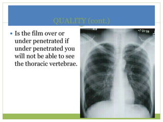
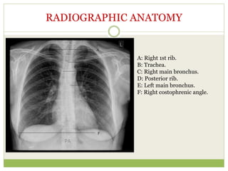
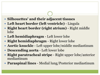
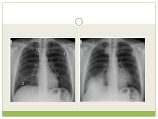
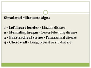
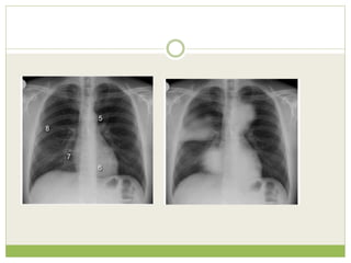
BASIC CHEST X-RAY INTERPRETATION
DR.S.SHRINUVASAN
ASSISTANT PROFESSOR,SLIMS
CONTENTS
๏‚— INTRODUCTION TO X- RAYS
๏‚— Radiographic views
๏‚— SYSTEMATIC APPROACH
๏‚— Radiographic anatomy
LUNGS/MEDIASTNUM
๏‚— Common lung conditions
X-rays- describe radiation which is part of the
spectrum which includes visible light, gamma
rays and cosmic radiation.
Unlike visible light, radiation passes through
stuff.
When you shine a beam of X-Ray at a person
and put a film on the other side of them a shadow
is produced of the inside of their body.
Different tissues in our body absorb X-rays at different
extents:
โ€ขBone- high absorption (white)
โ€ขTissue- somewhere in the middle absorption (grey)
โ€ขAir- low absorption (black)
PA VS AP
VIEWS
๏‚— Decubitus - useful for differentiating pleural
effusions from consolidation (e.g. pneumonia) and
loculated effusions from free fluid in the pleural
space.
๏‚— Lordotic view - used to visualize the apex of the
lung to pick-up abnormalities such as a Pancoast
tumour.
๏‚— Expiratory view - helpful for the diagnosis of
pneumothorax .
BE
SYSTEMATIC
Film Quality
๏‚— First determine is the film a PA or AP view.
All x-rays in the PICU are portable and are AP
view
IMAGE QUALITY
CHECK FOR ROTATION
๏‚ก Does the thoracic spine
align in the center of the
sternum and between
the clavicles?
๏‚ก Are the clavicles level?
Was film taken under full inspiration?
-10 posterior ribs should be visible.
A really good film will show anterior ribs too, there should
Be 6 to qualify as a good inspiratory film.
QUALITY (cont.)
๏‚— Is the film over or
under penetrated if
under penetrated you
will not be able to see
the thoracic vertebrae.
THE THORAX
๏‚— The thorax is divided into
TWO LATERAL COMPARTMENTS
each containing a lung and associated pleura
CENTRAL COMPARTMENT
the mediastinum, which contains
the other thoracic structures.
๏‚— The lung root contains:
๏‚— * Main bronchus.
๏‚— * Pulmonary artery.
๏‚— * Pulmonary vein.
๏‚— * Bronchial arteries.
๏‚— * Lymph nodes
RADIOGRAPHIC ANATOMY
A: Right 1st rib.
B: Trachea.
C: Right main bronchus.
D: Posterior rib.
E: Left main bronchus.
F: Right costophrenic angle.
LATERAL VIEW
CHECK THE HEART
๏‚— Size
๏‚— Shape
๏‚— Silhouette-margins should be sharp
๏‚— Diameter (>1/2 thoracic diameter is enlarged heart)
Remember: AP views make heart appear larger than it
actually is.
MEDIASTINUM
๏‚— The mediastinum is the central part of the thoracic
cavity which lies between thepleural sacs.
๏‚— It extends from the superior thoracic aperture to the
diaphragm and from the sternum and costal
cartilages to the thoracic vertebrae.
๏‚— An arbitrary line formed between the sternal angle to
the inferior border of T4 divides the mediastinum
into superior and inferior parts
๏‚— The inferior mediastinum is subdivided by
thepericardium into anterior, middle and posterior
parts. The heart and great vessels lie
within the middle mediastinum.
๏‚— The heart has a base, apex, three surfaces and four
borders. The base of the heart is located posteriorly
and formed mainly by the left atrium.
MEDIASTINUM
A: Aortic knuckle.
B: Superior vena cava (SVC).
C: Left pulmonary artery.
D: Right pulmonary artery.
E: Left heart border: left
ventricle.
F: Right heart border: right
atrium.
G: Inferior heart border: right
ventricle.
Cardiac Silhouette
1. R Atrium
2. R Ventricle
3. Apex of L Ventricle
4. Superior Vena Cava
5. Inferior Vena Cava
6. Tricuspid Valve
7. Pulmonary Valve
8. Pulmonary Trunk
9. R PA 10. L PA
The 'silhouette' sign
๏‚— The silhouette sign is a misnomer! It should be called
the 'loss of silhouette' sign. Normal adjacent
anatomical structures of differing densities form a
crisp 'silhouette,' or contour. Loss of a specific
contour can help determine the position of a disease
process.
๏‚— Silhouettes' and their adjacent tissues
๏‚— Left heart border (left ventricle) - Lingula
๏‚— Right heart border (right atrium) - Right middle
lobe
๏‚— Left hemidiaphragm - Left lower lobe
๏‚— Right hemidiaphragm - Right lower lobe
๏‚— Aortic knuckle - Left upper lobe/middle mediastinum
๏‚— Descending aorta - Left lower lobe
๏‚— Right paratracheal stripe - Right upper lobe/anterior
mediastinum
๏‚— Paraspinal lines - Medial lung/Posterior mediastinum
Simulated silhouette signs
1 - Left heart border - Lingula disease
2 - Hemidiaphragm - Lower lobe lung disease
3 - Paratracheal stripe - Paratracheal disease
4 - Chest wall - Lung, pleural or rib disease
5 - Aortic knuckle - Anterior mediastinal or left
upper lobe disease
6 - Paraspinal line - Posterior thorax disease
7 - Right heart border - Middle lobe disease
8 - Density above horizontal fissure* - Anterior
segment of the right upper lobe
Key points - Review areas
๏‚— Apices - Pneumothorax?
๏‚— Bones/soft-tissues - Fractures/density?
๏‚— Cardiac shadow- Consolidation/mass?
๏‚— Diaphragm - Pneumoperitoneum?
๏‚— Edge of the image - Unexpected findings?
๏‚— Mnemonic - ABCDE
REVIEW AREAS
OPACIFICATION OF A
HEMITHORAX
๏‚— Pleural effusion
๏‚— Consolidation
๏‚— Collapse
๏‚— Massive tumour
๏‚— Fibrothorax
๏‚— Combination of above lesions
Pneumonectomy
๏‚— Lung agenesis
PLEURAL EFFUSION
๏‚— Pleural effusion is excess fluid that accumulates
between the two pleural layers , the fluid-filled space
that surrounds the lungs.
๏‚— Excessive amounts of such fluid can impair
breathing by limiting the expansion of the lungs
during ventilation.
๏‚— Four types of fluids can accumulate in the pleural
space:
๏‚— Serous fluid (hydrothorax)
๏‚— Blood(haemothorax)
๏‚— Chyle (chylothorax)
๏‚— Pus(pyothorax or empyema)
๏‚— Conditions associated with transudative pleural effusions
๏‚— Congestive Heart Failure (CHF)
๏‚— Hepatic cirrhosis
๏‚— Hypoproteinemia
๏‚— Nephrotic syndrome
๏‚— Acute atelectasis
๏‚— Myxedema
๏‚— Peritoneal dialysis
๏‚— Meig's syndrome
๏‚— Obstructive uropathy
๏‚— Conditions associated with exudative pleural effusions
๏‚— Malignancy
๏‚— Infection
๏‚— Trauma
๏‚— Pulmonary infarction
๏‚— Pulmonary embolism
๏‚— Autoimmune disorders
๏‚— Pancreatitis
๏‚— Ruptured esophagus ( or Boerhaave's syndrome)
๏‚— Rheumatoid Pleurisy
๏‚— Drug-induced Lupus
๏‚— Tuberculosis
PNEUMONIA
๏‚— Pneumonia is an inflammatory condition of the
lungโ€”affecting primarily the microscopic air sacs
known as alveoli. It is usually caused by infection
with viruses or bacteria and less commonly other
microorganisms, certain drugs and other conditions
such as autoimmune diseases
COLLAPSE
๏‚— Atelectasis is defined as the collapse or closure of
the lung resulting in reduced or absent gas exchange.
It may affect part or all of one lung. It is a condition
where the alveoli are deflated, as distinct from
pulmonary consolidation
CAUSES OF HYPERTRANSRADIANT
๏‚— Mastectomy โ€” absent breast ยฑ absent pectoral muscle shadows.
๏‚— Poland's syndrome โ€” unilateral congenital absence of pectoral
๏‚— muscles ยฑ rib defects.
๏‚— PLEURA
PNEUMOTHORAX
๏‚— LUNG
๏‚ก EMPHYSEMA
๏‚ก UNILATERAL BULLAE โ€” vessels are absent rather than attenuated. May mimic
pneumothorax.
๏‚ก MACLEOD'S SYNDROME โ€” the late sequela of childhood bronchiolitis.
Small lung with small main and peripheral arteries. Air trapping occurs on
expiration
PNEUMOTHORAX
๏‚— A pneumothorax
is an abnormal
collection of air or
gas in the pleural
space that separates
the lung from the
chest wall and
which may interfere
with normal
breathing
๏‚— A Primary Pneumothorax is one that occurs without
an apparent cause and in the absence of significant lung
disease
๏‚— Secondary Pneumothorax occurs in the presence of
existing lung pathology.
๏‚— In a minority of cases, the amount of air in the chest
increases markedly when a one-way valve is formed by
an area of damaged tissue, leading to a Tension
Pneumothorax.
๏‚— This condition is a medical emergency that can cause
steadily worsening oxygen shortage and low blood
pressure. Unless reversed by effective treatment, these
sequelae can progress and cause death.
Chronic Obstructive Pulmonary Disease
PULMONARY EDEMA
๏‚— Pulmonary edema is fluid accumulation in the air
spaces and parenchyma of the lungs. It leads to
impaired gas exchange and may cause respiratory
failure.
๏‚— It is due to either failure of the left ventricle of the
heart to adequately remove blood from the
pulmonary circulation ("cardiogenic pulmonary
edema"), or an injury to the lung parenchyma or
vasculature of the lung ("noncardiogenic pulmonary
edema")
๏‚— The key findings of cardiogenic pulmonary edema
Kerley B lines (septal lines)
๏‚ก Seen at the lung bases, usually no more than 1 mm thick and 1 cm
long, perpendicular to the pleural surface
๏‚— Pleural effusions
๏‚ก Usually bilateral, frequently the right side being larger than the left
๏‚ก If unilateral, more often on the right
๏‚— Fluid in the fissures
๏‚ก Thickening of the major or minor fissure
๏‚— Peribronchial cuffing
๏‚ก Visualization of small doughnut-shaped rings representing fluid in
thickened bronchial walls
๏‚— Non-cardiogenic pulmonary edema
๏‚ก Bilateral, peripheral air space disease with air bronchograms
or central bat-wing pattern
๏‚ก Kerley B lines and pleural effusions are uncommon
๏‚ก Typically occurs 48 hours or more after the initial insult
๏‚ก Stabilizes at around five days and may take weeks to
completely clear
๏‚ก On CT
๏ƒท Gravity-dependent consolidation or ground glass opacification
๏ƒท Air bronchograms are common
The clinical question
Clinical information provided
๏‚— Recent increase in shortness of breath
๏‚— No fever or productive cough
๏‚— Left shoulder and arm pain
๏‚— Heavy smoker
๏‚— Weight loss
Findings
Left apical shadowing
Raised left hemidiaphragm
Increased extra-thoracic
soft tissue density (*) with
displacement of the
scapula on the left
(arrowheads) - compare
with right
๏‚— Interpretation in view of clinical details
๏‚— Cancer - Smoker with weight loss and left apical
consolidation/mass and no clinical features of
infection
๏‚— Phrenic nerve palsy - Increased shortness of
breath and raised left hemidiaphragm
๏‚— Brachial plexopathy - Arm pain and axillary soft
tissue swelling
THANK
YOU

BASIC CHEST X-RAY INTERPRETATION

  • 1.
    BASIC CHEST X-RAYINTERPRETATION DR.S.SHRINUVASAN ASSISTANT PROFESSOR,SLIMS
  • 2.
    CONTENTS ๏‚— INTRODUCTION TOX- RAYS ๏‚— Radiographic views ๏‚— SYSTEMATIC APPROACH ๏‚— Radiographic anatomy LUNGS/MEDIASTNUM ๏‚— Common lung conditions
  • 3.
    X-rays- describe radiationwhich is part of the spectrum which includes visible light, gamma rays and cosmic radiation. Unlike visible light, radiation passes through stuff. When you shine a beam of X-Ray at a person and put a film on the other side of them a shadow is produced of the inside of their body.
  • 4.
    Different tissues inour body absorb X-rays at different extents: โ€ขBone- high absorption (white) โ€ขTissue- somewhere in the middle absorption (grey) โ€ขAir- low absorption (black)
  • 5.
  • 7.
    VIEWS ๏‚— Decubitus -useful for differentiating pleural effusions from consolidation (e.g. pneumonia) and loculated effusions from free fluid in the pleural space. ๏‚— Lordotic view - used to visualize the apex of the lung to pick-up abnormalities such as a Pancoast tumour. ๏‚— Expiratory view - helpful for the diagnosis of pneumothorax .
  • 8.
  • 10.
    Film Quality ๏‚— Firstdetermine is the film a PA or AP view. All x-rays in the PICU are portable and are AP view
  • 11.
  • 12.
    CHECK FOR ROTATION ๏‚กDoes the thoracic spine align in the center of the sternum and between the clavicles? ๏‚ก Are the clavicles level?
  • 13.
    Was film takenunder full inspiration? -10 posterior ribs should be visible. A really good film will show anterior ribs too, there should Be 6 to qualify as a good inspiratory film.
  • 14.
    QUALITY (cont.) ๏‚— Isthe film over or under penetrated if under penetrated you will not be able to see the thoracic vertebrae.
  • 15.
    THE THORAX ๏‚— Thethorax is divided into TWO LATERAL COMPARTMENTS each containing a lung and associated pleura CENTRAL COMPARTMENT the mediastinum, which contains the other thoracic structures.
  • 16.
    ๏‚— The lungroot contains: ๏‚— * Main bronchus. ๏‚— * Pulmonary artery. ๏‚— * Pulmonary vein. ๏‚— * Bronchial arteries. ๏‚— * Lymph nodes
  • 17.
    RADIOGRAPHIC ANATOMY A: Right1st rib. B: Trachea. C: Right main bronchus. D: Posterior rib. E: Left main bronchus. F: Right costophrenic angle.
  • 18.
  • 19.
    CHECK THE HEART ๏‚—Size ๏‚— Shape ๏‚— Silhouette-margins should be sharp ๏‚— Diameter (>1/2 thoracic diameter is enlarged heart) Remember: AP views make heart appear larger than it actually is.
  • 21.
    MEDIASTINUM ๏‚— The mediastinumis the central part of the thoracic cavity which lies between thepleural sacs. ๏‚— It extends from the superior thoracic aperture to the diaphragm and from the sternum and costal cartilages to the thoracic vertebrae. ๏‚— An arbitrary line formed between the sternal angle to the inferior border of T4 divides the mediastinum into superior and inferior parts
  • 22.
    ๏‚— The inferiormediastinum is subdivided by thepericardium into anterior, middle and posterior parts. The heart and great vessels lie within the middle mediastinum. ๏‚— The heart has a base, apex, three surfaces and four borders. The base of the heart is located posteriorly and formed mainly by the left atrium.
  • 24.
    MEDIASTINUM A: Aortic knuckle. B:Superior vena cava (SVC). C: Left pulmonary artery. D: Right pulmonary artery. E: Left heart border: left ventricle. F: Right heart border: right atrium. G: Inferior heart border: right ventricle.
  • 25.
    Cardiac Silhouette 1. RAtrium 2. R Ventricle 3. Apex of L Ventricle 4. Superior Vena Cava 5. Inferior Vena Cava 6. Tricuspid Valve 7. Pulmonary Valve 8. Pulmonary Trunk 9. R PA 10. L PA
  • 26.
    The 'silhouette' sign ๏‚—The silhouette sign is a misnomer! It should be called the 'loss of silhouette' sign. Normal adjacent anatomical structures of differing densities form a crisp 'silhouette,' or contour. Loss of a specific contour can help determine the position of a disease process.
  • 27.
    ๏‚— Silhouettes' andtheir adjacent tissues ๏‚— Left heart border (left ventricle) - Lingula ๏‚— Right heart border (right atrium) - Right middle lobe ๏‚— Left hemidiaphragm - Left lower lobe ๏‚— Right hemidiaphragm - Right lower lobe ๏‚— Aortic knuckle - Left upper lobe/middle mediastinum ๏‚— Descending aorta - Left lower lobe ๏‚— Right paratracheal stripe - Right upper lobe/anterior mediastinum ๏‚— Paraspinal lines - Medial lung/Posterior mediastinum
  • 29.
    Simulated silhouette signs 1- Left heart border - Lingula disease 2 - Hemidiaphragm - Lower lobe lung disease 3 - Paratracheal stripe - Paratracheal disease 4 - Chest wall - Lung, pleural or rib disease
  • 31.
    5 - Aorticknuckle - Anterior mediastinal or left upper lobe disease 6 - Paraspinal line - Posterior thorax disease 7 - Right heart border - Middle lobe disease 8 - Density above horizontal fissure* - Anterior segment of the right upper lobe
  • 32.
    Key points -Review areas ๏‚— Apices - Pneumothorax? ๏‚— Bones/soft-tissues - Fractures/density? ๏‚— Cardiac shadow- Consolidation/mass? ๏‚— Diaphragm - Pneumoperitoneum? ๏‚— Edge of the image - Unexpected findings? ๏‚— Mnemonic - ABCDE
  • 33.
  • 37.
    OPACIFICATION OF A HEMITHORAX ๏‚—Pleural effusion ๏‚— Consolidation ๏‚— Collapse ๏‚— Massive tumour ๏‚— Fibrothorax ๏‚— Combination of above lesions Pneumonectomy ๏‚— Lung agenesis
  • 38.
    PLEURAL EFFUSION ๏‚— Pleuraleffusion is excess fluid that accumulates between the two pleural layers , the fluid-filled space that surrounds the lungs. ๏‚— Excessive amounts of such fluid can impair breathing by limiting the expansion of the lungs during ventilation.
  • 39.
    ๏‚— Four typesof fluids can accumulate in the pleural space: ๏‚— Serous fluid (hydrothorax) ๏‚— Blood(haemothorax) ๏‚— Chyle (chylothorax) ๏‚— Pus(pyothorax or empyema)
  • 43.
    ๏‚— Conditions associatedwith transudative pleural effusions ๏‚— Congestive Heart Failure (CHF) ๏‚— Hepatic cirrhosis ๏‚— Hypoproteinemia ๏‚— Nephrotic syndrome ๏‚— Acute atelectasis ๏‚— Myxedema ๏‚— Peritoneal dialysis ๏‚— Meig's syndrome ๏‚— Obstructive uropathy
  • 44.
    ๏‚— Conditions associatedwith exudative pleural effusions ๏‚— Malignancy ๏‚— Infection ๏‚— Trauma ๏‚— Pulmonary infarction ๏‚— Pulmonary embolism ๏‚— Autoimmune disorders ๏‚— Pancreatitis ๏‚— Ruptured esophagus ( or Boerhaave's syndrome) ๏‚— Rheumatoid Pleurisy ๏‚— Drug-induced Lupus ๏‚— Tuberculosis
  • 45.
  • 46.
    ๏‚— Pneumonia isan inflammatory condition of the lungโ€”affecting primarily the microscopic air sacs known as alveoli. It is usually caused by infection with viruses or bacteria and less commonly other microorganisms, certain drugs and other conditions such as autoimmune diseases
  • 48.
    COLLAPSE ๏‚— Atelectasis isdefined as the collapse or closure of the lung resulting in reduced or absent gas exchange. It may affect part or all of one lung. It is a condition where the alveoli are deflated, as distinct from pulmonary consolidation
  • 51.
    CAUSES OF HYPERTRANSRADIANT ๏‚—Mastectomy โ€” absent breast ยฑ absent pectoral muscle shadows. ๏‚— Poland's syndrome โ€” unilateral congenital absence of pectoral ๏‚— muscles ยฑ rib defects. ๏‚— PLEURA PNEUMOTHORAX ๏‚— LUNG ๏‚ก EMPHYSEMA ๏‚ก UNILATERAL BULLAE โ€” vessels are absent rather than attenuated. May mimic pneumothorax. ๏‚ก MACLEOD'S SYNDROME โ€” the late sequela of childhood bronchiolitis. Small lung with small main and peripheral arteries. Air trapping occurs on expiration
  • 52.
    PNEUMOTHORAX ๏‚— A pneumothorax isan abnormal collection of air or gas in the pleural space that separates the lung from the chest wall and which may interfere with normal breathing
  • 53.
    ๏‚— A PrimaryPneumothorax is one that occurs without an apparent cause and in the absence of significant lung disease ๏‚— Secondary Pneumothorax occurs in the presence of existing lung pathology. ๏‚— In a minority of cases, the amount of air in the chest increases markedly when a one-way valve is formed by an area of damaged tissue, leading to a Tension Pneumothorax. ๏‚— This condition is a medical emergency that can cause steadily worsening oxygen shortage and low blood pressure. Unless reversed by effective treatment, these sequelae can progress and cause death.
  • 56.
  • 61.
    PULMONARY EDEMA ๏‚— Pulmonaryedema is fluid accumulation in the air spaces and parenchyma of the lungs. It leads to impaired gas exchange and may cause respiratory failure. ๏‚— It is due to either failure of the left ventricle of the heart to adequately remove blood from the pulmonary circulation ("cardiogenic pulmonary edema"), or an injury to the lung parenchyma or vasculature of the lung ("noncardiogenic pulmonary edema")
  • 62.
    ๏‚— The keyfindings of cardiogenic pulmonary edema Kerley B lines (septal lines) ๏‚ก Seen at the lung bases, usually no more than 1 mm thick and 1 cm long, perpendicular to the pleural surface ๏‚— Pleural effusions ๏‚ก Usually bilateral, frequently the right side being larger than the left ๏‚ก If unilateral, more often on the right ๏‚— Fluid in the fissures ๏‚ก Thickening of the major or minor fissure ๏‚— Peribronchial cuffing ๏‚ก Visualization of small doughnut-shaped rings representing fluid in thickened bronchial walls
  • 64.
    ๏‚— Non-cardiogenic pulmonaryedema ๏‚ก Bilateral, peripheral air space disease with air bronchograms or central bat-wing pattern ๏‚ก Kerley B lines and pleural effusions are uncommon ๏‚ก Typically occurs 48 hours or more after the initial insult ๏‚ก Stabilizes at around five days and may take weeks to completely clear ๏‚ก On CT ๏ƒท Gravity-dependent consolidation or ground glass opacification ๏ƒท Air bronchograms are common
  • 66.
  • 67.
    Clinical information provided ๏‚—Recent increase in shortness of breath ๏‚— No fever or productive cough ๏‚— Left shoulder and arm pain ๏‚— Heavy smoker ๏‚— Weight loss
  • 68.
    Findings Left apical shadowing Raisedleft hemidiaphragm Increased extra-thoracic soft tissue density (*) with displacement of the scapula on the left (arrowheads) - compare with right
  • 69.
    ๏‚— Interpretation inview of clinical details ๏‚— Cancer - Smoker with weight loss and left apical consolidation/mass and no clinical features of infection ๏‚— Phrenic nerve palsy - Increased shortness of breath and raised left hemidiaphragm ๏‚— Brachial plexopathy - Arm pain and axillary soft tissue swelling
  • 70.

Editor's Notes

  • #7ย This is a PA film on the left compared with a AP supine film on the right.ย ย  The AP shows magnification of the heart and widening of the mediastinum.ย  Whenever possible the patient should be imaged in an upright PA position.ย  AP views are less useful and should be reserved for very ill patients who cannot stand erect. ย  This is a PA film on the left compared with a AP supine film on the right.ย ย  The AP shows magnification of the heart and widening of the mediastinum.ย  Whenever possible the patient should be imaged in an upright PA position.ย  AP views are less useful and should be reserved for very ill patients who cannot stand erect. ย 
  • #19ย Normal lateral film. Note the retrosternal and retrocardiac clear spaces (open arrows) and the increased translucency of the lower vertebrae. The axillary folds (straight black arrows) and scapulae (curved black arrows) overlie the lungs. The tracheal translucency is well seen (s
  • #43ย Massive pleural effusion with mediastinal shift to the left. (A) Chest radiograph and (B) CT coronal reconstruction. A massive effusion displaces the mediastinum to the left. CT shows the important pleural effusion together with the enhanced atelectatic left lung. Note also the depression of the right hemidiaphragm (arrows).
  • #58ย A lateral chest x-ray of a person with emphysema. Note the barrel chest and flat diaphragm.
  • #66ย Acute intra-alveolar pulmonary oedema with a bat's wing distribution.